


Home / Training / Manuals / Atlas of breast cancer early detection / Cases
Atlas of breast cancer early detection
Filter by language: English / Русский
Go back to the list of case studies
.png) Click on the pictures to magnify and display the legends
Click on the pictures to magnify and display the legends
| Case number: | 153 |
| Age: | 50 |
| Clinical presentation: | Postmenopausal woman with average risk of breast cancer presented with a right breast lump noticed 23 years earlier. Examination revealed a right breast lump (3 × 2 cm). |
Mammography:
 |  |
 |  |
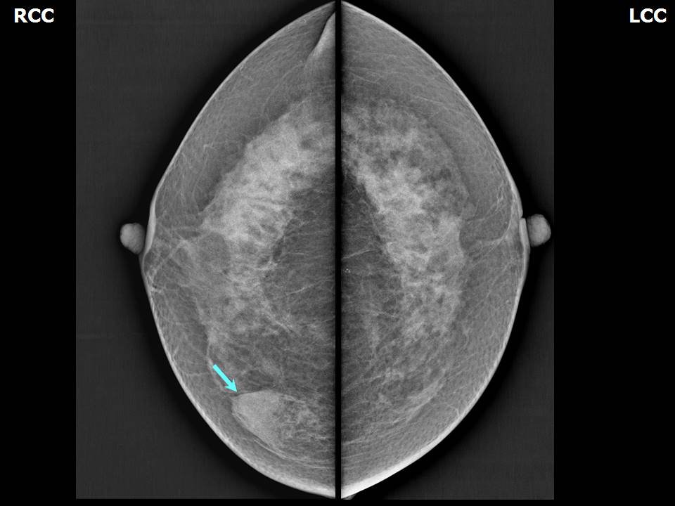
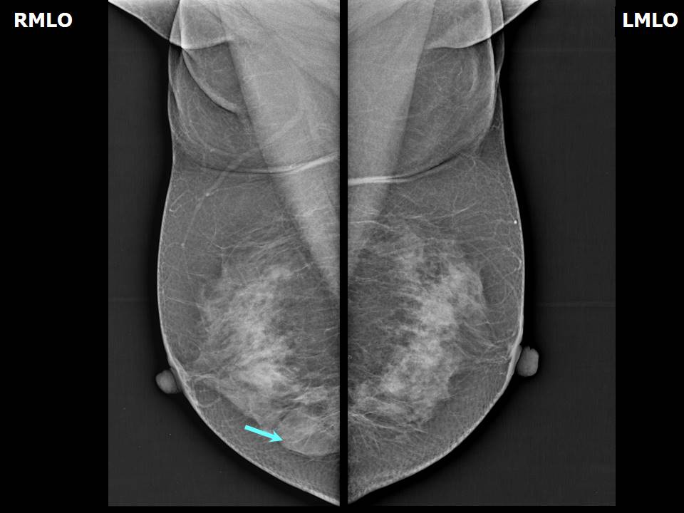
| Breast composition: | ACR category c (the breasts are heterogeneously dense, which may obscure small masses) | Mammography features: |
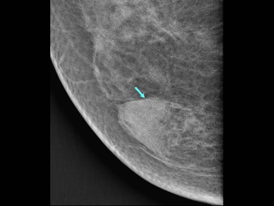
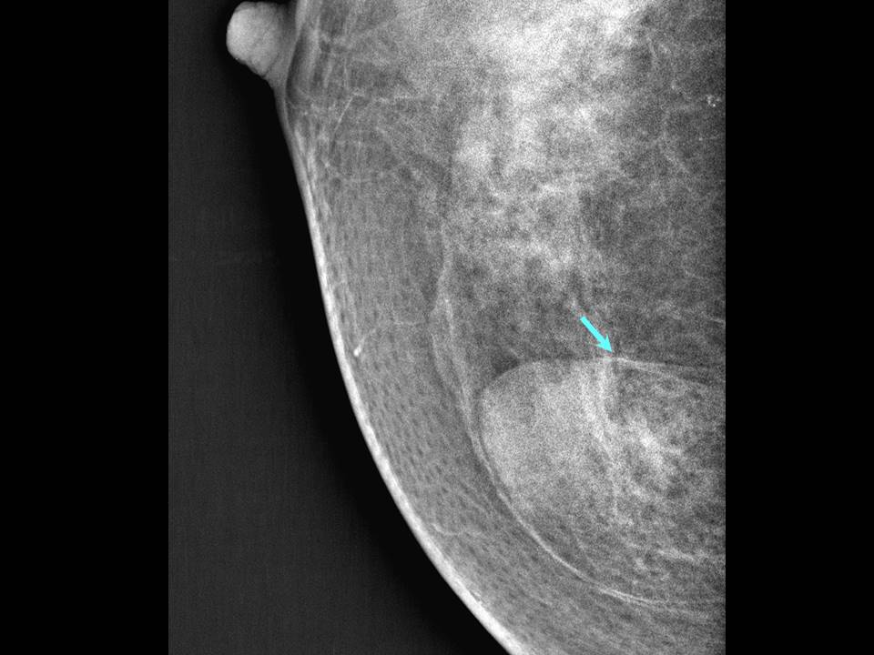
| ‣ Location of the lesion: | Right breast, inner quadrants at 3 oclock, middle and posterior thirds |
| ‣ Mass: | |
| • Number: | 1 |
| • Size: | 3.2 × 2.7 cm |
| • Shape: | Oval |
| • Margins: | Circumscribed |
| • Density: | Fat-containing |
| ‣ Calcifications: | |
| • Typically benign: | None |
| • Suspicious: | None |
| • Distribution: | None |
| ‣ Architectural distortion: | None |
| ‣ Asymmetry: | None |
| ‣ Intramammary node: | None |
| ‣ Skin lesion: | None |
| ‣ Solitary dilated duct: | None |
| ‣ Associated features: | None |
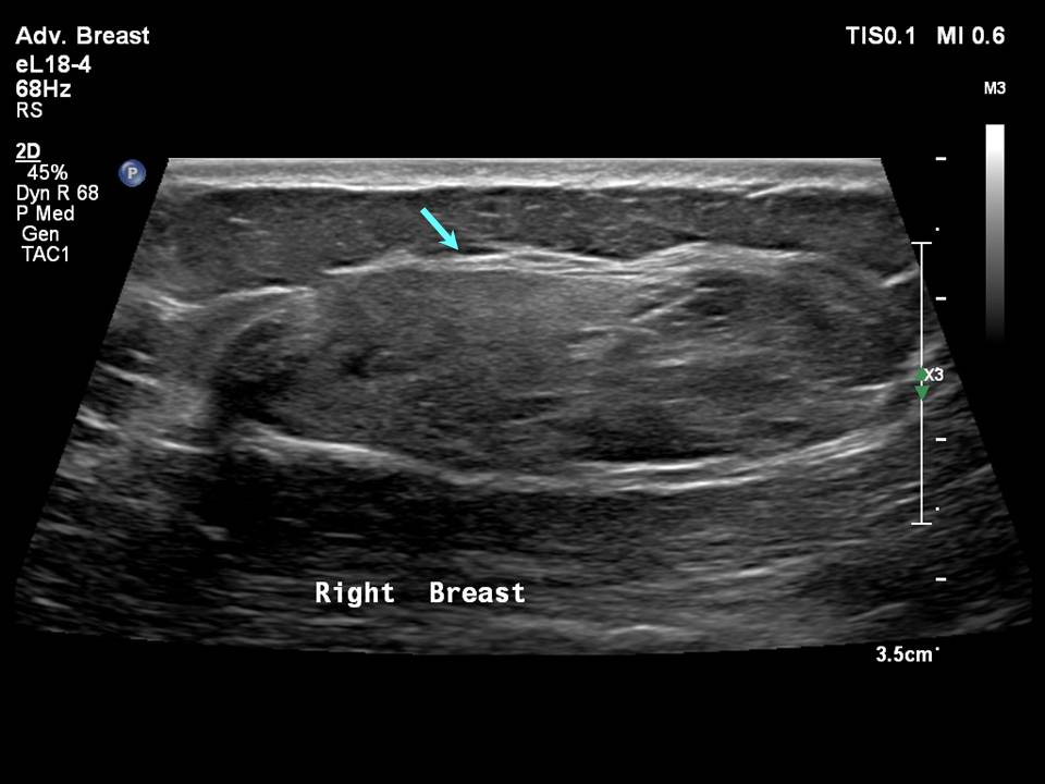
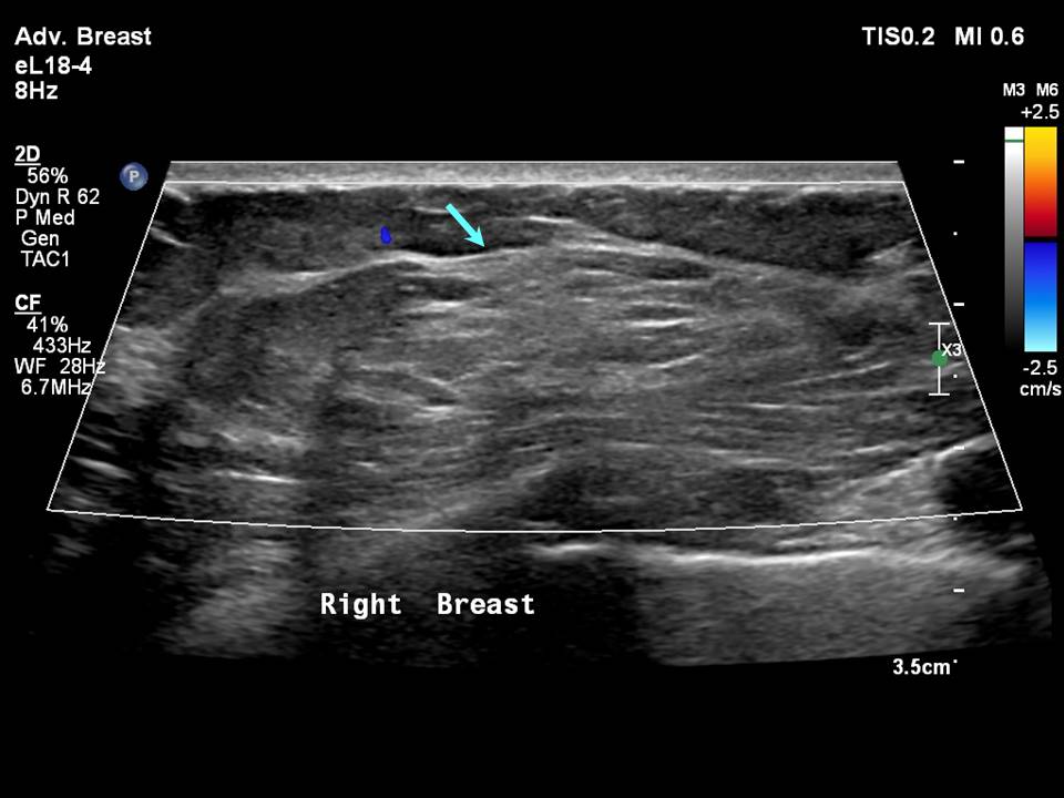
Ultrasound:
 |  |
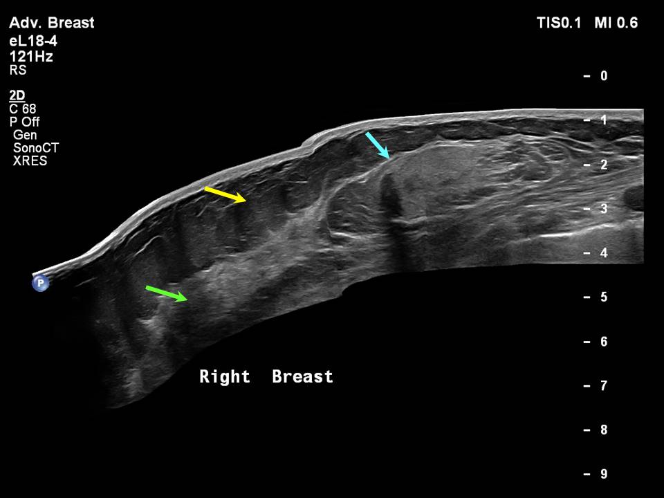 |
| Ultrasound features: Right breast, inner quadrants at 3 oclock, 7.0 cm from the nipple and at 1.5 cm skin depth | |
| ‣ Mass | |
| • Location: | Right breast, inner quadrants at 3 oclock, 7.0 cm from the nipple and at 1.5 cm skin depth |
| • Number: | 1 |
| • Size: | 4.0 × 1.5 cm |
| • Shape: | Oval |
| • Orientation: | Parallel |
| • Margins: | Circumscribed |
| • Echo pattern: | Heterogeneous |
| • Posterior features: | No posterior features |
| ‣ Calcifications: | None |
| ‣ Associated features: | None |
| ‣ Special cases: | None |
BI-RADS:
BI-RADS Category: 2 (benign)Further assessment:
Further assessment advised: Referral for cytologyCytology:
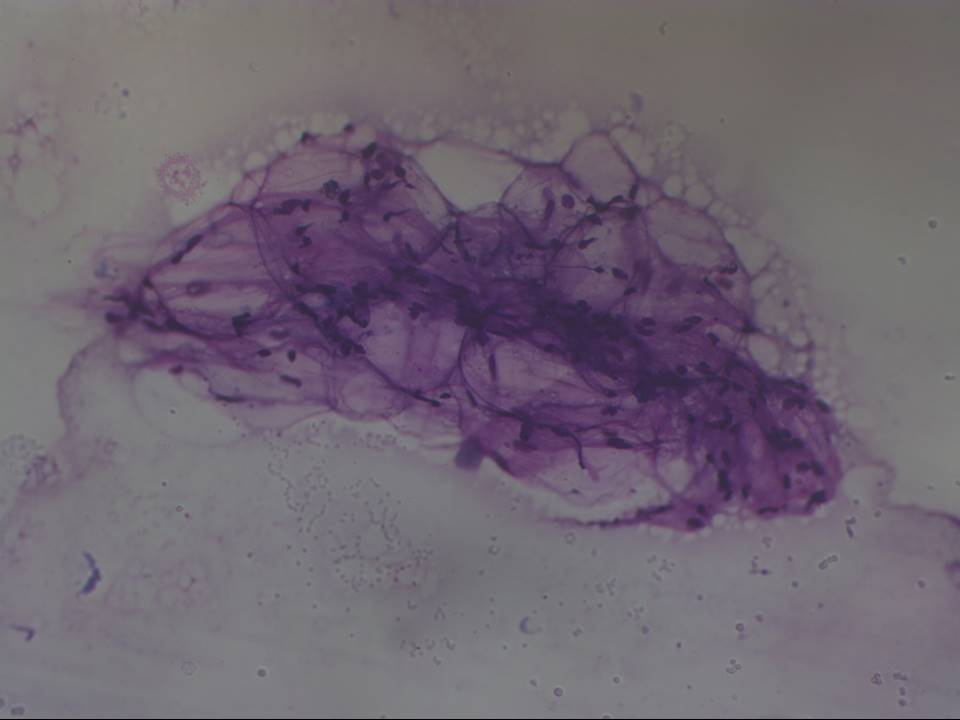 |
| Cytology features: | |
| ‣ Type of sample: | FNAC |
| ‣ Site of biopsy: | |
| • Laterality: | Right |
| • Quadrant: | Inner |
| • Localization technique: | Palpation |
| • Nature of aspirate: | Fatty aspirate |
| ‣ Cytological description: | Cytological smears showed many adipose tissue fragments. A few ductal myoepithelial cells and isolated naked nuclei were seen |
| ‣ Reporting category: | Benign |
| ‣ Diagnosis: | On correlating with CBE and imaging findings, consistent with hamartoma |
| ‣ Comments: | None |
Case summary:
| Postmenopausal woman presented with right breast lump. Diagnosed as hamartoma (fibroadenolipoma) BI-RADS 2 on imaging and as hamartoma on cytology. |
Learning points:
|