Home / Training / Manuals / Atlas of breast cancer early detection / Cases
Atlas of breast cancer early detection
Filter by language: English / Русский
Go back to the list of case studies
.png) Click on the pictures to magnify and display the legends
Click on the pictures to magnify and display the legends
| Case number: | 149 |
| Age: | 53 |
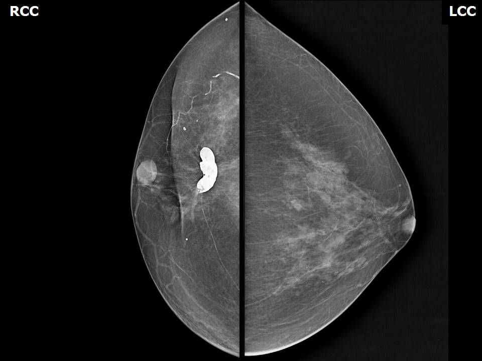
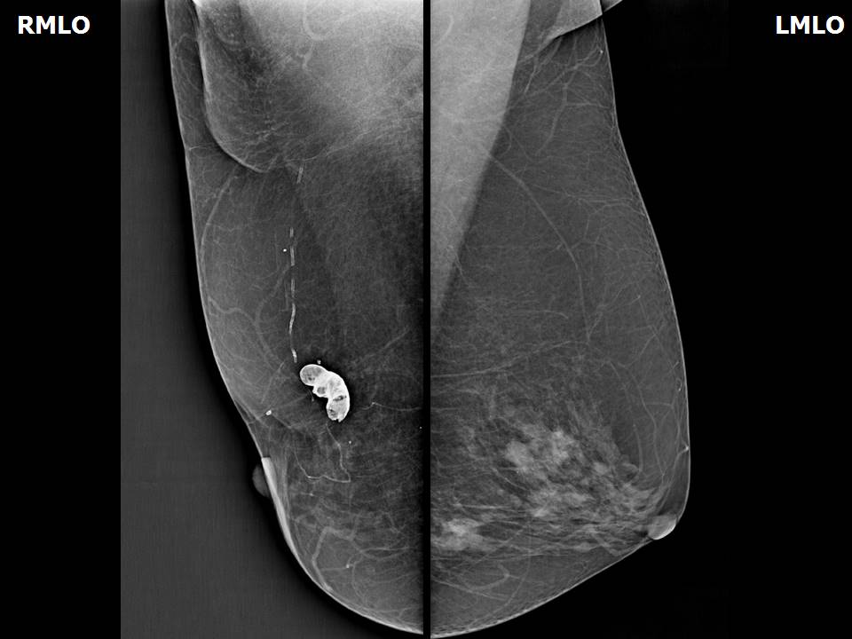
| Clinical presentation: | Postmenopausal woman with increased risk of developing breast cancer presented for postoperative surveillance. She had previously undergone breast-conserving surgery for invasive carcinoma of the right breast and also has family history of breast carcinoma. |
Mammography:
| Breast composition: | ACR category b (there are scattered areas of fibroglandular density) | Mammography features: |
| ‣ Location of the lesion: | Right breast, upper central quadrant along surgical scar |
| ‣ Mass: | |
| • Number: | 0 |
| • Size: | None |
| • Shape: | None |
| • Margins: | None |
| • Density: | None |
| ‣ Calcifications: | |
| • Typically benign: | Eggshell, typically benign with lucent centres along surgical scar |
| • Suspicious: | None |
| • Distribution: | None |
| ‣ Architectural distortion: | None |
| ‣ Asymmetry: | None |
| ‣ Intramammary node: | None |
| ‣ Skin lesion: | None |
| ‣ Solitary dilated duct: | None |
| ‣ Associated features: | None |
BI-RADS:
BI-RADS Category: 2 (benign)Case summary:
| Postmenopausal woman who had undergone breast-conserving surgery for right breast cancer on regular surveillance. Diagnosed as dystrophic calcification in proximity to the surgical scar, BI-RADS 2 on imaging. |
Learning points:
|


