


Home / Training / Manuals / Atlas of breast cancer early detection / Cases
Atlas of breast cancer early detection
Filter by language: English / Русский
Go back to the list of case studies
.png) Click on the pictures to magnify and display the legends
Click on the pictures to magnify and display the legends
| Case number: | 034 |
| Age: | 67 |
| Clinical presentation: | Postmenopausal woman with increased risk of developing breast cancer because of family history of uterine carcinoma presented with a left breast lump noticed 2 days ago. |
Mammography:
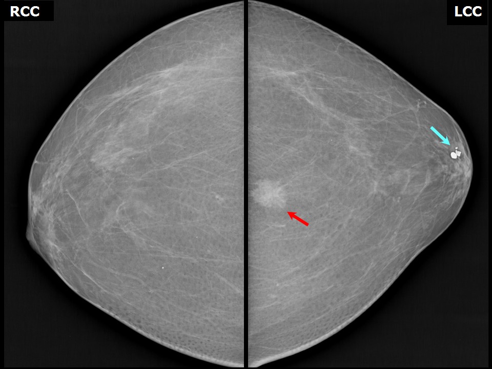 | 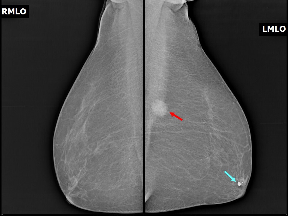 |
| Breast composition: | ACR category a (the breasts are almost entirely fatty) | Mammography features: |
| ‣ Location of the lesion: | Left breast, upper inner quadrant at 11 oclock, posterior third, 9.0 cm from the nipple and 5.0 cm from the skin |
| ‣ Mass: | |
| • Number: | 1 |
| • Size: | 2.5 × 2.4 × 2.0 cm |
| • Shape: | Irregular |
| • Margins: | Spiculated |
| • Density: | High |
| ‣ Calcifications: | |
| • Typically benign: | Round and coarse calcifications in the left subareolar region and round calcifications in the right breast posteriorly |
| • Suspicious: | None |
| • Distribution: | None |
| ‣ Architectural distortion: | None |
| ‣ Asymmetry: | None |
| ‣ Intramammary node: | None |
| ‣ Skin lesion: | None |
| ‣ Solitary dilated duct: | None |
| ‣ Associated features: | None |
Ultrasound:
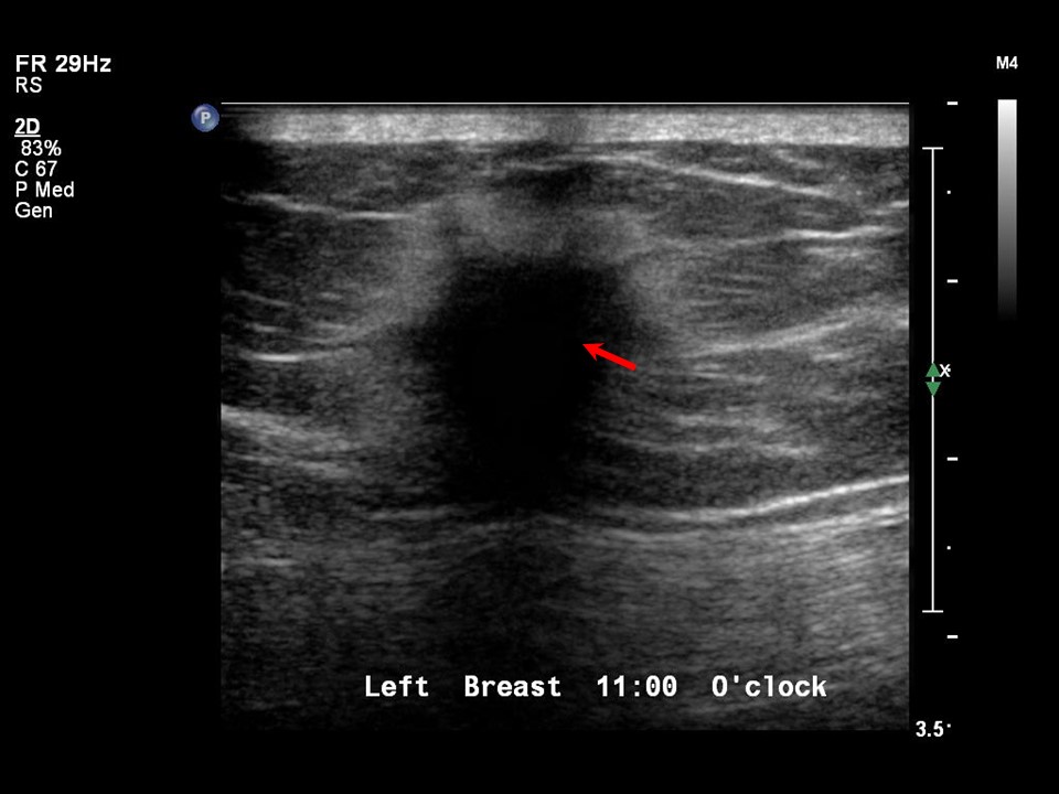 | 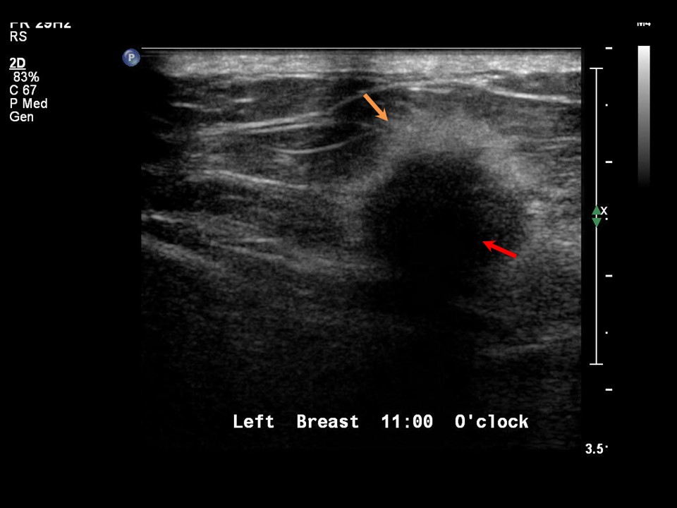 |
| Ultrasound features: Left breast, upper inner quadrant at 11 oclock position | |
| ‣ Mass | |
| • Location: | Left breast, upper inner quadrant at 11 oclock position |
| • Number: | 1 |
| • Size: | 2.0 × 1.6 cm |
| • Shape: | Irregular |
| • Orientation: | Not parallel |
| • Margins: | Spiculated |
| • Echo pattern: | Hypoechoic |
| • Posterior features: | Strong posterior shadowing |
| ‣ Calcifications: | None |
| ‣ Associated features: | None |
| ‣ Special cases: | None |
BI-RADS:
BI-RADS Category: 5 (highly suggestive of malignancy)Further assessment:
Further assessment advised: Referral for cytologyCytology:
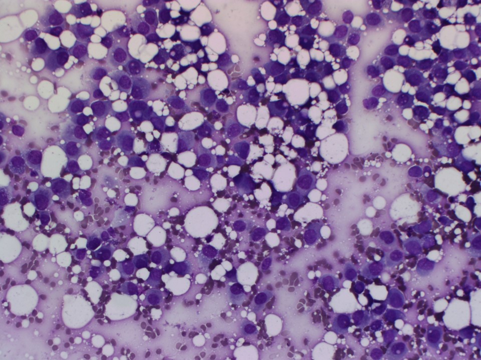 | 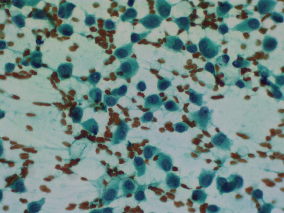 |
| Cytology features: | |
| ‣ Type of sample: | FNAC |
| ‣ Site of biopsy: | |
| • Laterality: | Left |
| • Quadrant: | Upper inner |
| • Localization technique: | Palpation |
| • Nature of aspirate: | 0.2 mL of whitish material |
| ‣ Cytological description: | Cellular smears reveal dyscohesive sheets and isolated malignant cells. Pleomorphic cells with hyperchromatic nuclei and moderate amount of cytoplasm present |
| ‣ Reporting category: | Malignant |
| ‣ Diagnosis: | Carcinoma |
| ‣ Comments: | None |
Histopathology:
Lumpectomy
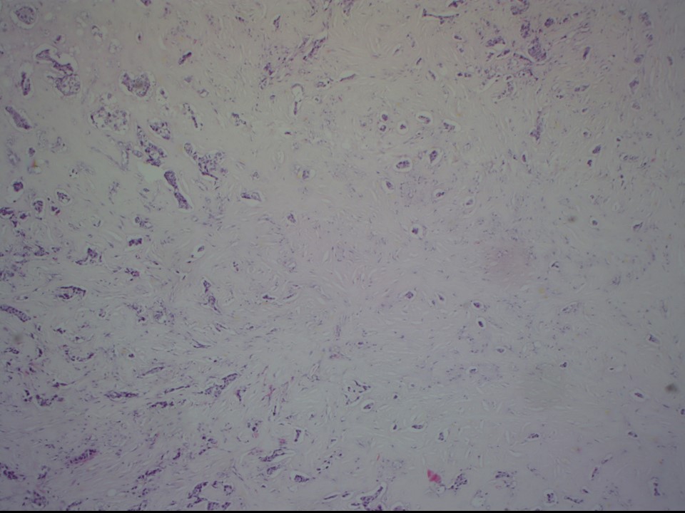 | 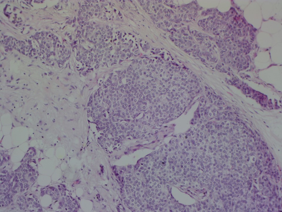 |
| Histopathology features: | |
| ‣ Specimen type: | Lumpectomy |
| ‣ Laterality: | Left |
| ‣ Macroscopy: | Lumpectomy specimen (7.0 × 6.0 × 3.5 cm) oriented with long suture laterally and short suture superiorly, with skin flap (5.0 × 2.0 cm). On serial sectioning, a very firm to hard, greyish white tumour (1.8 × 1.5 × 1.5 cm) with irregular borders is seen. It is located 1.5 cm from the skin (anterior margin), 0.2 cm from the posterior margin, 2.5 cm from the superior margin, 2.5 cm from the inferior margin, 2.0 cm from the medial margin, and 3.0 cm from the lateral margin |
| ‣ Histological type: | Invasive breast carcinoma of no special type |
| ‣ Histological grade: | Grade 3 (3 + 3 + 2 = 8) |
| ‣ Mitosis: | 14 |
| ‣ Maximum invasive tumour size: | 1.8 cm |
| ‣ Lymph node status: | 0/20 |
| ‣ Peritumoural lymphovascular invasion: | Present |
| ‣ DCIS/EIC: | Absent |
| ‣ Margins: | Free of tumour |
| ‣ Pathological stage: | pT1cN0 |
| ‣ Biomarkers: | |
| ‣ Comments: |
Case summary:
| Postmenopausal woman presented with a left breast lump. Diagnosed as left breast carcinoma with spiculated margins, BI-RADS 5 on imaging, as carcinoma left breast on cytology, and as invasive breast carcinoma of no special type, pT1cN0 on histopathology. |
Learning points:
|