


Home / Training / Manuals / Atlas of breast cancer early detection / Cases
Atlas of breast cancer early detection
Filter by language: English / Русский
Go back to the list of case studies
.png) Click on the pictures to magnify and display the legends
Click on the pictures to magnify and display the legends
| Case number: | 028 |
| Age: | 62 |
| Clinical presentation: | Postmenopausal woman with average risk of developing breast cancer presented with a left breast lump noticed 45 months ago. |
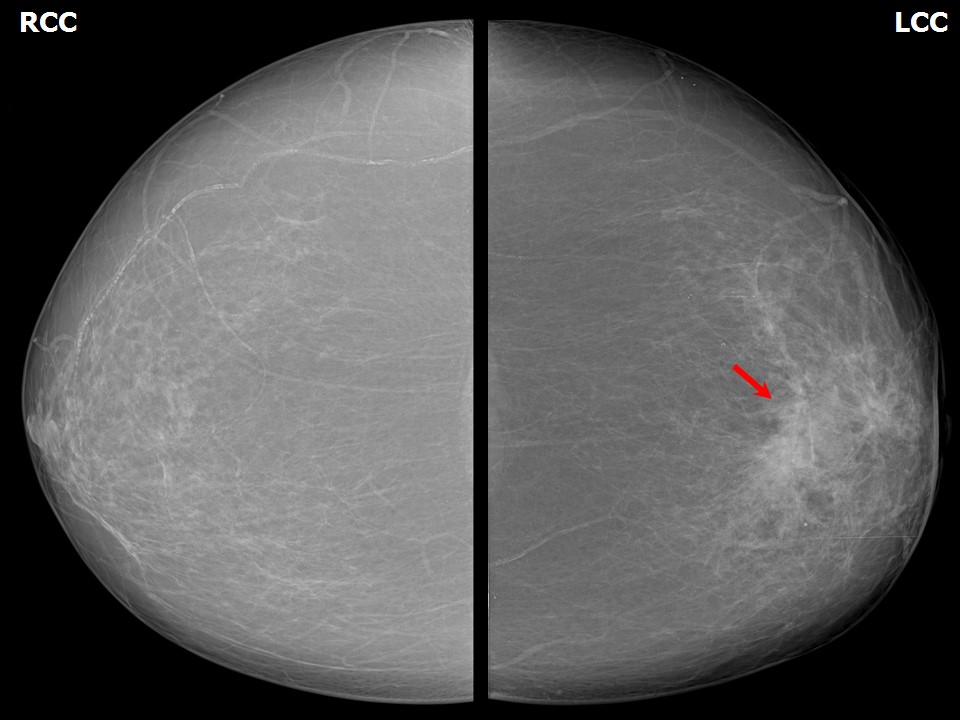
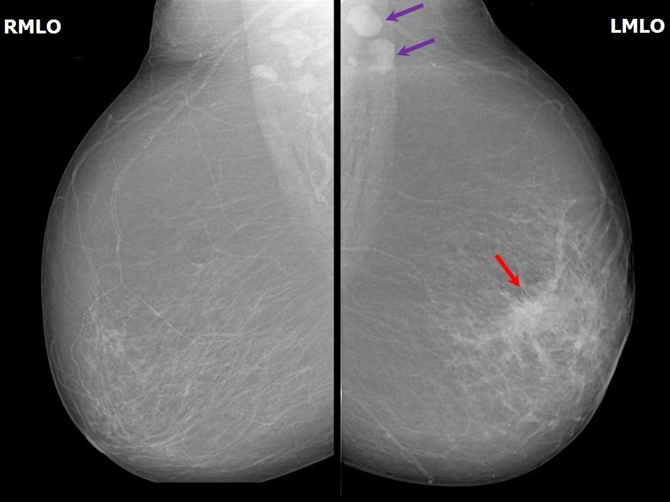
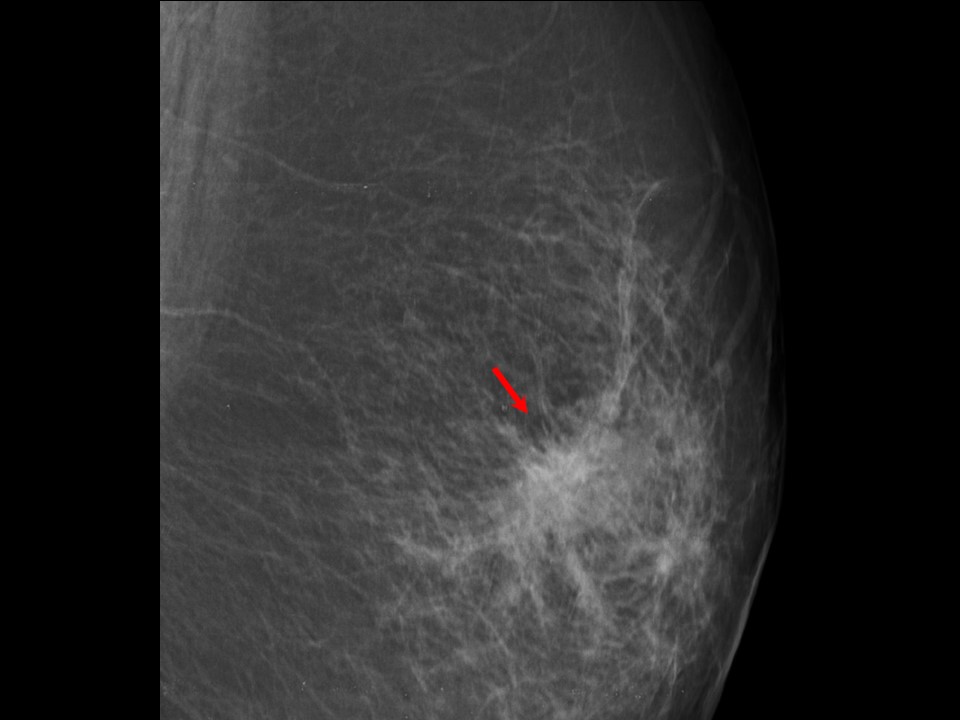
Mammography:
 |  |
 |
| Breast composition: | ACR category a (the breasts are almost entirely fatty) | Mammography features: |
| ‣ Location of the lesion: | Left breast, central portion of the breast, central zone, anterior third |
| ‣ Mass: | |
| • Number: | 1 |
| • Size: | None |
| • Shape: | Irregular |
| • Margins: | Spiculated |
| • Density: | High |
| ‣ Calcifications: | |
| • Typically benign: | None |
| • Suspicious: | None |
| • Distribution: | None |
| ‣ Architectural distortion: | Area of architectural distortion, 4.5 × 3.0 cm, with radiating linear shadows |
| ‣ Asymmetry: | Focal |
| ‣ Intramammary node: | None |
| ‣ Skin lesion: | None |
| ‣ Solitary dilated duct: | None |
| ‣ Associated features: | Axillary lymphadenopathy |
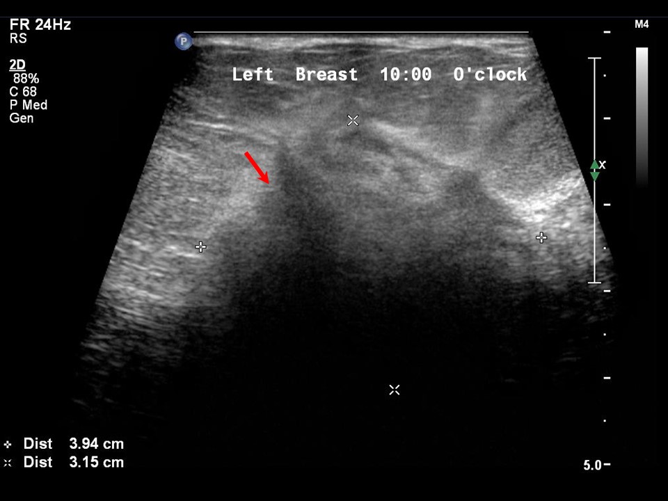
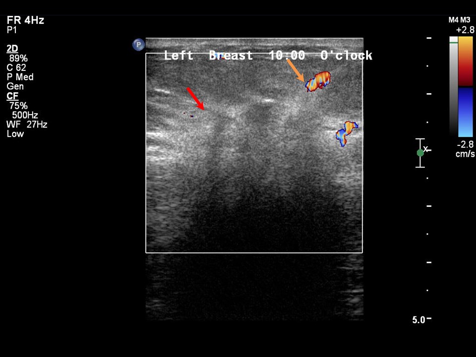
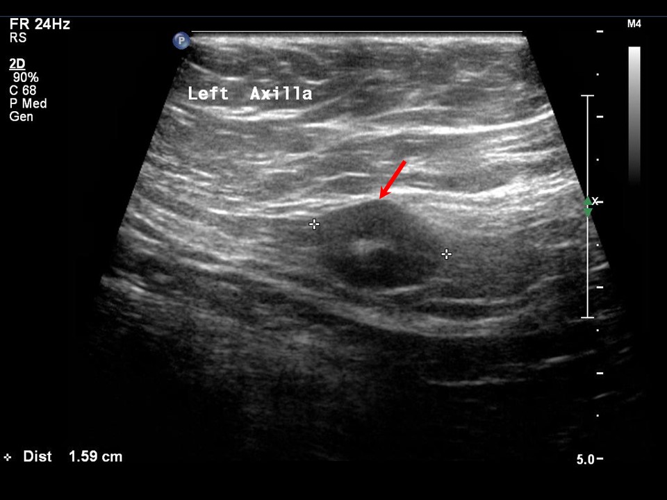
Ultrasound:
 |  |
 |
| Ultrasound features: Left breast, central portion of the breast | |
| ‣ Mass | |
| • Location: | Left breast, central portion of the breast |
| • Number: | 1 |
| • Size: | 4.0 × 3.2 cm |
| • Shape: | Irregular |
| • Orientation: | Not parallel |
| • Margins: | Spiculated |
| • Echo pattern: | Hypoechoic |
| • Posterior features: | Strong posterior shadowing |
| ‣ Calcifications: | None |
| ‣ Associated features: | Axillary lymphadenopathy with thickened cortex |
| ‣ Special cases: | None |
BI-RADS:
BI-RADS Category: 5 (highly suggestive of malignancy)Further assessment:
Further assessment advised: Referral for core biopsyHistopathology:
Core needle biopsy
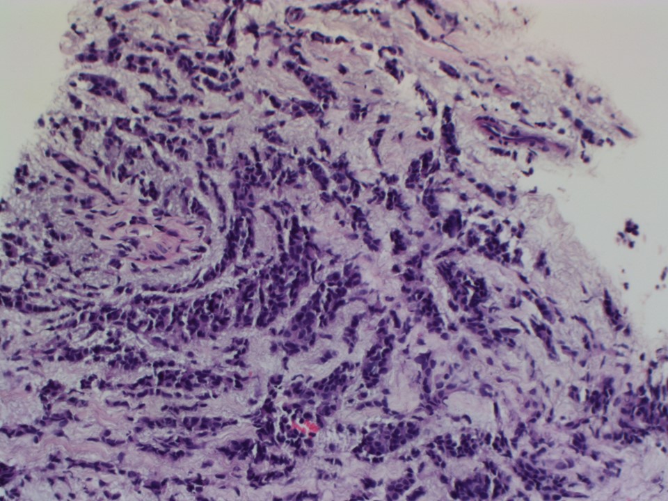 |
| Histopathology features: | |
| ‣ Specimen type: | Core needle biopsy |
| ‣ Laterality: | Left |
| ‣ Macroscopy: | Multiple whitish fragmented cores. Two long cores were 13 mm in length and 10 mm in length; the remaining multiple fragmented cores ranged in length from 2 to 3 mm |
| ‣ Histological type: | Invasive breast carcinoma of no special type |
| ‣ Histological grade: | Grade 2 (3 + 2 + 2 = 7) |
| ‣ Mitosis: | 12 |
| ‣ Maximum invasive tumour size: | |
| ‣ Lymph node status: | |
| ‣ Peritumoural lymphovascular invasion: | |
| ‣ DCIS/EIC: | |
| ‣ Margins: | |
| ‣ Pathological stage: | |
| ‣ Biomarkers: | |
| ‣ Comments: |
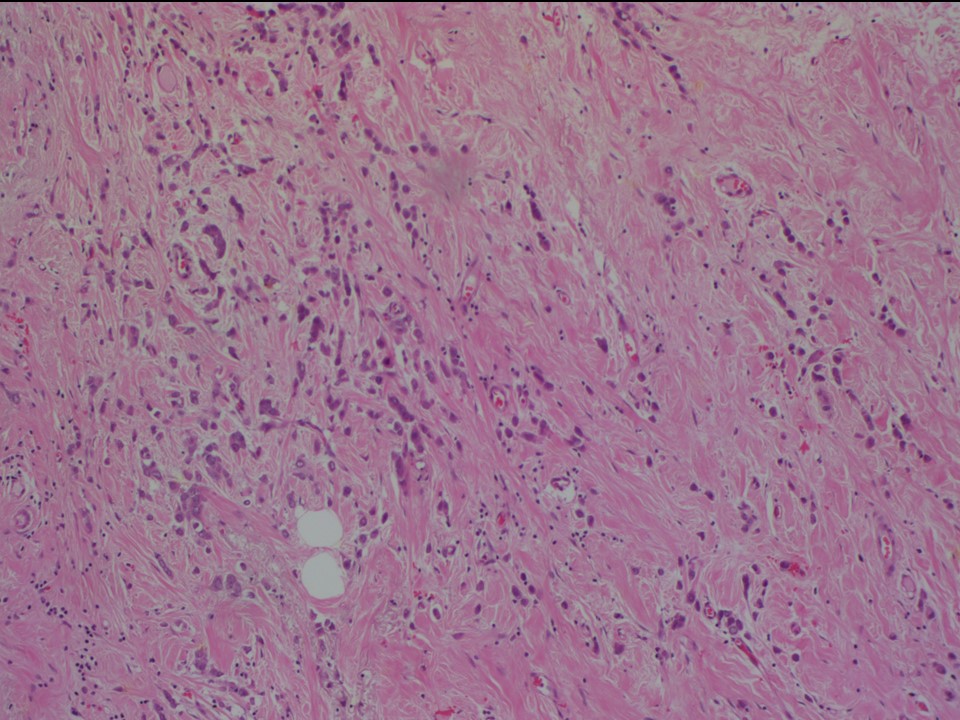
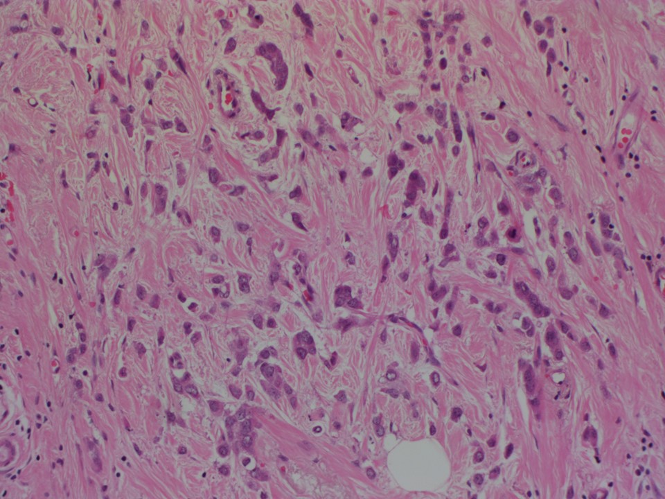
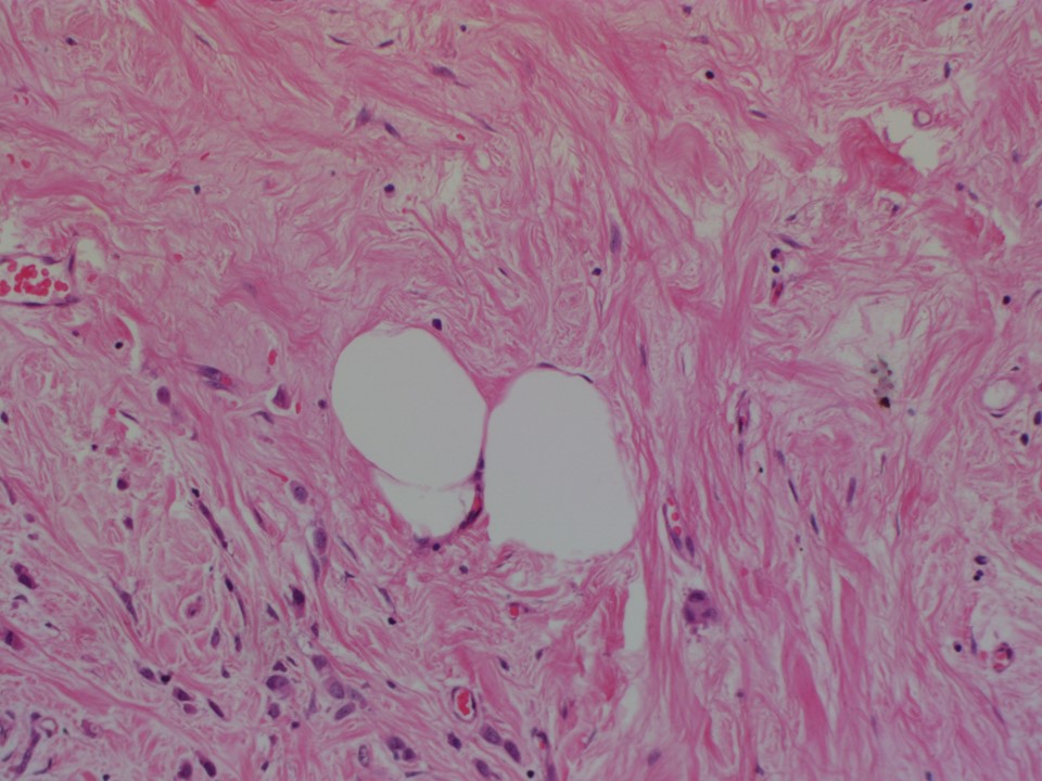
MRM left (after chemotherapy)
 |  |
 |
| Histopathology features: | |
| ‣ Specimen type: | MRM left (after chemotherapy) |
| ‣ Laterality: | Left |
| ‣ Macroscopy: | Left MRM specimen (25.0 × 25.0 × 7.0 cm) with overlying skin flap (20.0 × 7.0 cm). The nipple and areola are unremarkable. On serial sectioning a firm, greyish white area (4.5 × 4.0 × 3.0 cm) is identified, located in the upper inner quadrant. It is located 3.5 cm from the skin and 6.0 cm from the base. The remaining breast tissue is unremarkable |
| ‣ Histological type: | Invasive breast carcinoma of no special type |
| ‣ Histological grade: | Grade 2 (3 + 3 + 1 = 7) |
| ‣ Mitosis: | 5 |
| ‣ Maximum invasive tumour size: | 4.5 cm |
| ‣ Lymph node status: | 1/16 without extranodal extension |
| ‣ Peritumoural lymphovascular invasion: | |
| ‣ DCIS/EIC: | Very few DCIS. Solid, low nuclear grade without necrosis. EIC absent |
| ‣ Margins: | Free of tumour |
| ‣ Pathological stage: | yT2yN1 |
| ‣ Biomarkers: | |
| ‣ Comments: | Extensive fibrosis seen in the tumour bed and surrounding breast tissue |
Case summary:
| Postmenopausal woman presented with left breast lump. Diagnosed as architectural distortion with radiating linear shadows in the retroareolar region of the left breast with left axillary lymphadenopathy, BI-RADS 5 on imaging, as invasive breast carcinoma of no special type on needle core biopsy, and post neo-adjuvant therapy, MRM specimen showed residual invasive breast carcinoma of no special type, and yT2yN1 on histopathology. |
Learning points:
|