


Home / Training / Manuals / Atlas of breast cancer early detection / Cases
Atlas of breast cancer early detection
Filter by language: English / Русский
Go back to the list of case studies
.png) Click on the pictures to magnify and display the legends
Click on the pictures to magnify and display the legends
| Case number: | 021 |
| Age: | 48 |
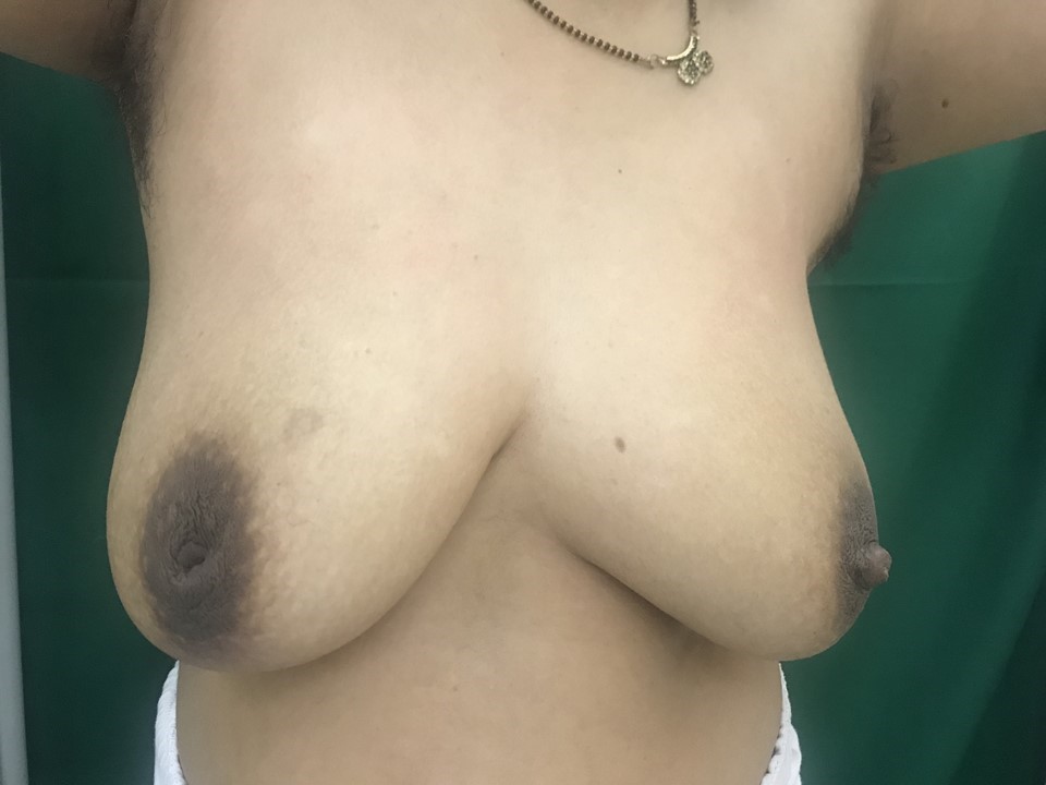
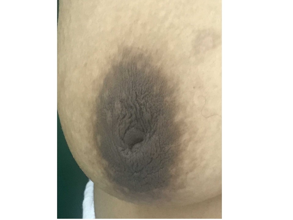
| Clinical presentation: | Premenopausal woman presented with right nipple inversion that she had had for many years. Patient had not breastfed her child from the right breast. She had an increased risk of developing breast cancer and a family history of ovarian cancer. Examination revealed nodularity in the outer quadrant of the right breast. |
 |  |
Mammography:
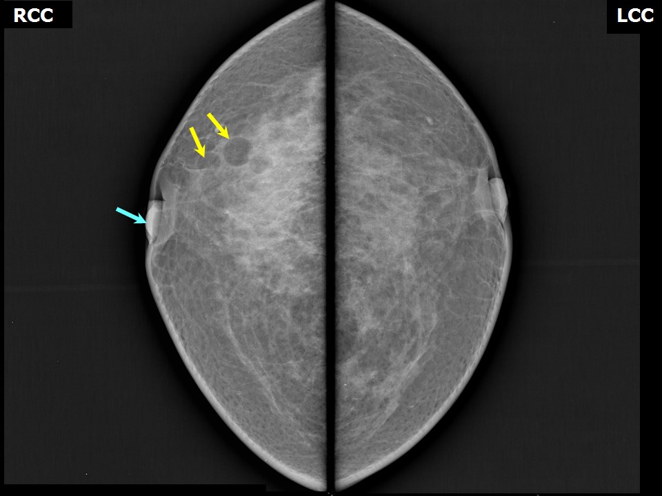 | 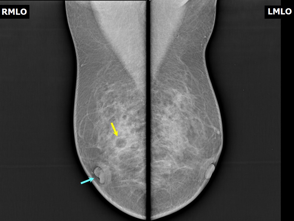 |
| Breast composition: | ACR category b (there are scattered areas of fibroglandular density) | Mammography features: |
| ‣ Location of the lesion: | Right breast, upper outer quadrant at 11 oclock, middle third |
| ‣ Mass: | |
| • Number: | 3 |
| • Size: | 1.1 × 0.7 × 0.6 cm |
| • Shape: | Oval |
| • Margins: | Circumscribed |
| • Density: | Fat-containing |
| ‣ Calcifications: | |
| • Typically benign: | None |
| • Suspicious: | None |
| • Distribution: | None |
| ‣ Architectural distortion: | None |
| ‣ Asymmetry: | None |
| ‣ Intramammary node: | None |
| ‣ Skin lesion: | None |
| ‣ Solitary dilated duct: | None |
| ‣ Associated features: | Inverted nipple |
Ultrasound:
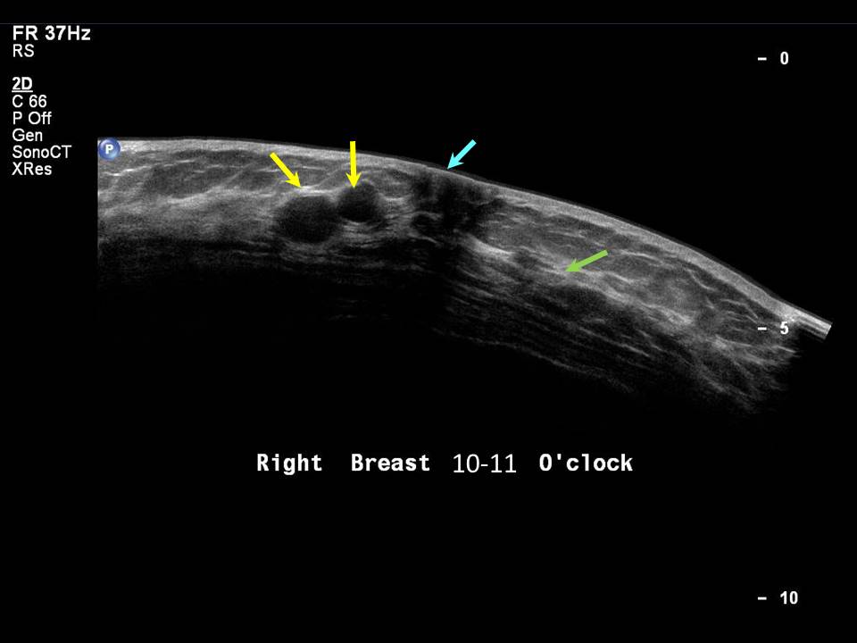 |
| Ultrasound features: Right breast, upper outer quadrant at 11 o'clock | |
| ‣ Mass | |
| • Location: | Right breast, upper outer quadrant at 11 o'clock |
| • Number: | 3 |
| • Size: | 1.1 x 0.7 x 0.6 cm in greatest dimension |
| • Shape: | Oval |
| • Orientation: | Parallel |
| • Margins: | Circumscribed |
| • Echo pattern: | Hypoechoic |
| • Posterior features: | No posterior features |
| ‣ Calcifications: | None |
| ‣ Associated features: | None |
| ‣ Special cases: | None |
BI-RADS:
BI-RADS Category: 2 (benign)Case summary:
| Premenopausal woman presented with inverted right nipple for many years and right breast nodularity. Diagnosed as oil cysts, BI-RADS 2 on imaging. |
Learning points:
|