


Home / Training / Manuals / Atlas of breast cancer early detection / Cases
Atlas of breast cancer early detection
Filter by language: English / Русский
Go back to the list of case studies
.png) Click on the pictures to magnify and display the legends
Click on the pictures to magnify and display the legends
| Case number: | 016 |
| Age: | 44 |
| Clinical presentation: | Premenopausal woman with average risk of developing breast cancer presented with a left breast lump. She had had the lump for 4 years and recently noticed a gradual increase in its size on self-examination. CBE revealed a firm lump in the upper outer quadrant of the left breast, with clinical increase in the size of the left breast lesion. |
Mammography:
 |  |
| Breast composition: | ACR category c (the breasts are heterogeneously dense, which may obscure small masses) | Mammography features: |
| ‣ Location of the lesion: | Left breast, upper outer quadrant at 12 oclock, middle and posterior thirds |
| ‣ Mass: | |
| • Number: | 1 |
| • Size: | 3.4 × 2.2 cm |
| • Shape: | Oval |
| • Margins: | Partly circumscribed and partly obscured |
| • Density: | Equal |
| ‣ Calcifications: | |
| • Typically benign: | None |
| • Suspicious: | None |
| • Distribution: | None |
| ‣ Architectural distortion: | None |
| ‣ Asymmetry: | None |
| ‣ Intramammary node: | None |
| ‣ Skin lesion: | None |
| ‣ Solitary dilated duct: | None |
| ‣ Associated features: | None |
Ultrasound:
 |
| Ultrasound features: Left breast, upper outer quadrant at 2 oclock | |
| ‣ Mass | |
| • Location: | Left breast, upper outer quadrant at 2 oclock |
| • Number: | 1 |
| • Size: | 3.4 × 2.0 cm |
| • Shape: | Oval |
| • Orientation: | Parallel |
| • Margins: | Circumscribed |
| • Echo pattern: | Hypoechoic |
| • Posterior features: | No posterior features |
| ‣ Calcifications: | None |
| ‣ Associated features: | None |
| ‣ Special cases: | None |
BI-RADS:
BI-RADS Category: 2 (benign)Further assessment:
Further assessment advised: Referral for cytologyCytology:
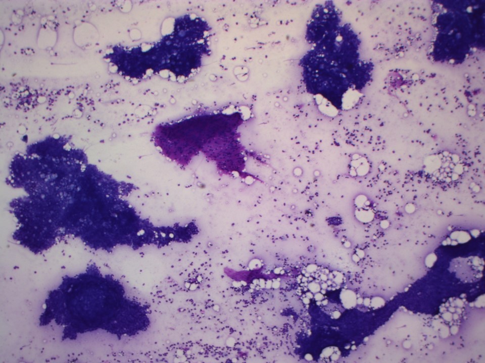 | 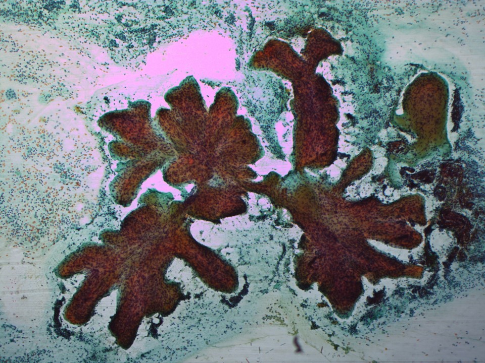 |
| Cytology features: | |
| ‣ Type of sample: | FNAC |
| ‣ Site of biopsy: | |
| • Laterality: | Left |
| • Quadrant: | Lower outer |
| • Localization technique: | Palpation |
| • Nature of aspirate: | Whitish |
| ‣ Cytological description: | Cellular smears show cohesive sheets of ductal cells with finger-like shapes. Stromal fragments, some with a sharp, club-shaped or clover-leaf appearance, are present. Many bare nuclei are seen in the background |
| ‣ Reporting category: | Benign |
| ‣ Diagnosis: | Fibroadenoma |
| ‣ Comments: | None |
Histopathology:
Lumpectomy specimen
 |
| Histopathology features: | |
| ‣ Specimen type: | Lumpectomy specimen |
| ‣ Laterality: | Left |
| ‣ Macroscopy: | Specimen (7.0 × 6.0 × 6.0 cm) with a skin flap (4.0 × 1.0 cm). Cut surface shows a firm, bulging, whitish mass 3.2 cm in greatest dimension |
| ‣ Histological type: | Fibroadenoma |
| ‣ Histological grade: | |
| ‣ Mitosis: | |
| ‣ Maximum invasive tumour size: | |
| ‣ Lymph node status: | |
| ‣ Peritumoural lymphovascular invasion: | |
| ‣ DCIS/EIC: | |
| ‣ Margins: | |
| ‣ Pathological stage: | |
| ‣ Biomarkers: | |
| ‣ Comments: |
Case summary:
| Premenopausal woman presented with left breast lump. Diagnosed as fibroadenoma, BI-RADS 2 on imaging and as fibroadenoma on cytology. |
Learning points:
|