


Home / Training / Manuals / Atlas of breast cancer early detection / Cases
Atlas of breast cancer early detection
Filter by language: English / Русский
Go back to the list of case studies
.png) Click on the pictures to magnify and display the legends
Click on the pictures to magnify and display the legends
| Case number: | 012 |
| Age: | 65 |
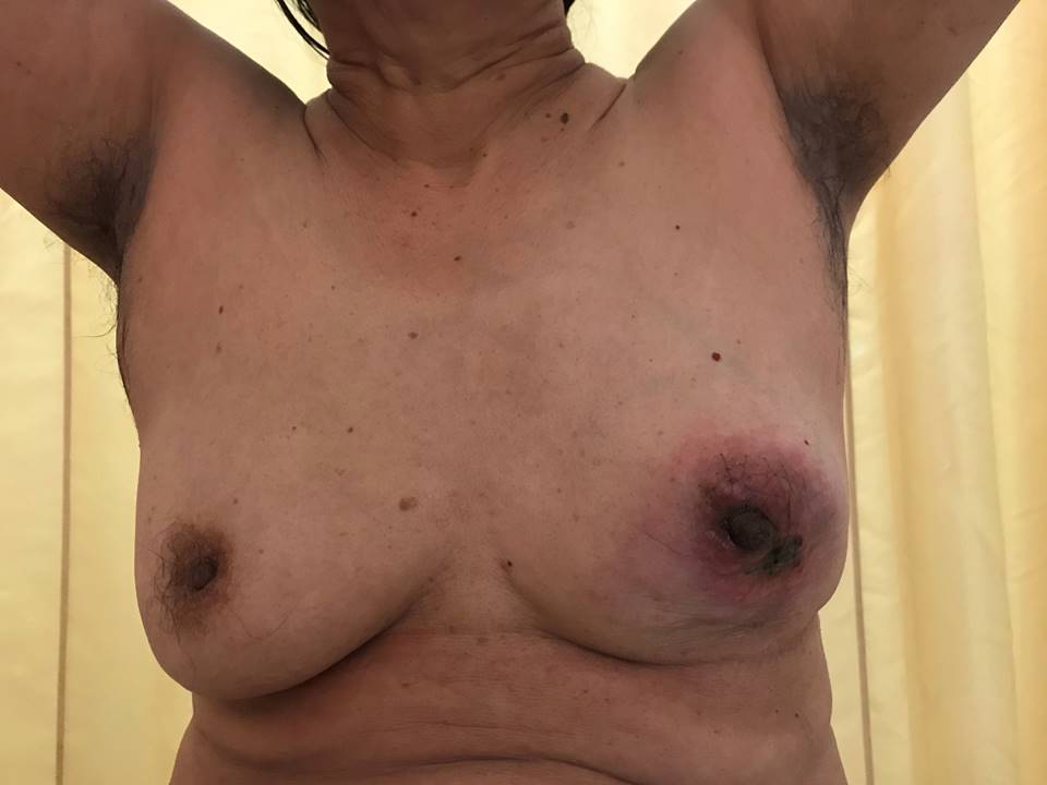
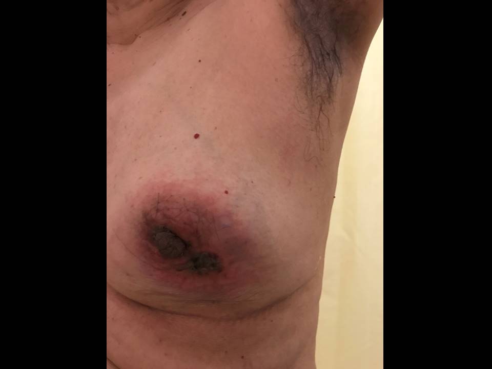
| Clinical presentation: | Postmenopausal woman with average risk of developing breast cancer presented with a left breast lump that she first noticed more than 4 months ago. Now presented with overlying skin changes and pain. |
 |  |
Mammography:
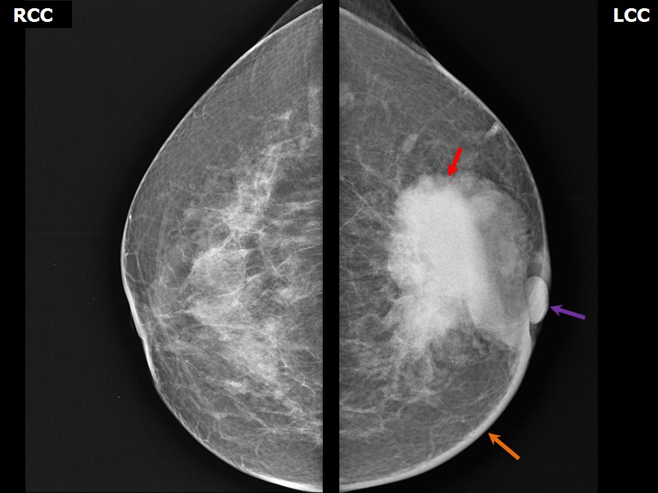 | 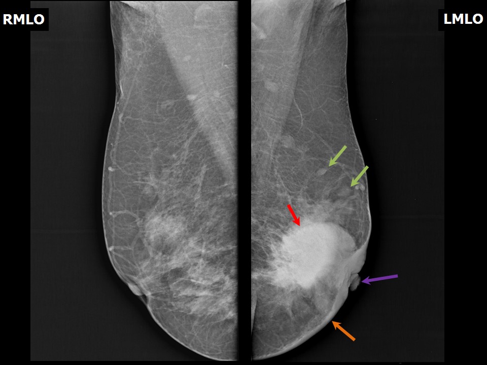 |
| Breast composition: | ACR category b (there are scattered areas of fibroglandular density) | Mammography features: |
| ‣ Location of the lesion: | Left breast, central portion of the breast, central zone, anterior and middle thirds |
| ‣ Mass: | |
| • Number: | Multiple |
| • Size: | Left retroareolar: Largest 5.5 × 3.0 cm, multiple satellite lesions (largest 2.5 × 1.0 cm) in outer quadrant at 4 oclock |
| • Shape: | Irregular |
| • Margins: | Spiculated |
| • Density: | High |
| ‣ Calcifications: | |
| • Typically benign: | None |
| • Suspicious: | None |
| • Distribution: | None |
| ‣ Architectural distortion: | None |
| ‣ Asymmetry: | None |
| ‣ Intramammary node: | None |
| ‣ Skin lesion: | None |
| ‣ Solitary dilated duct: | None |
| ‣ Associated features: | Skin thickening, nipple retraction |
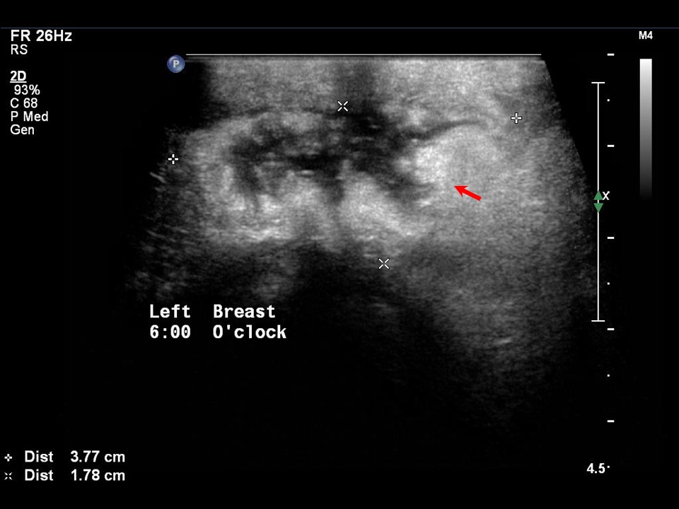
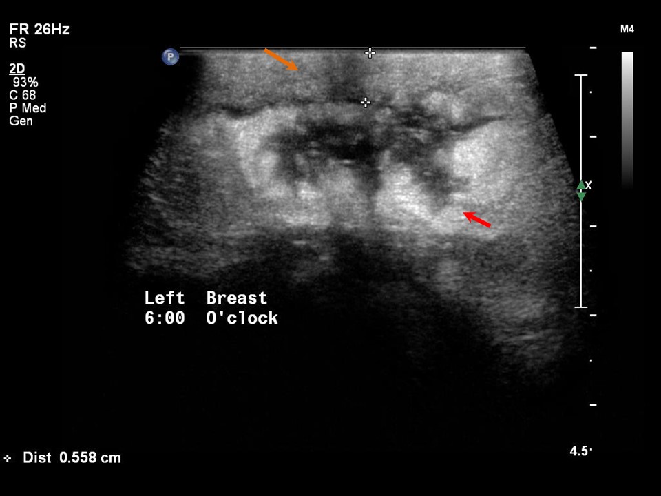
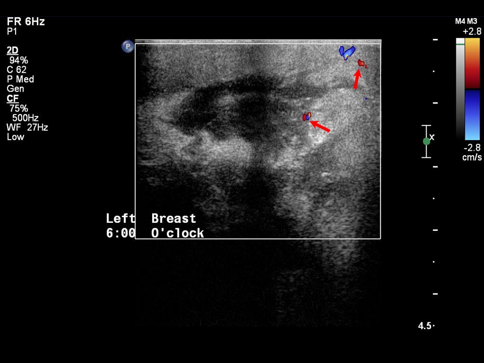
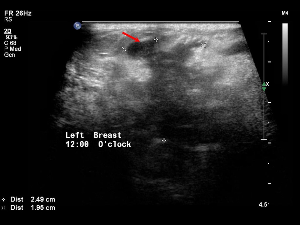
Ultrasound:
 |  |
 |  |
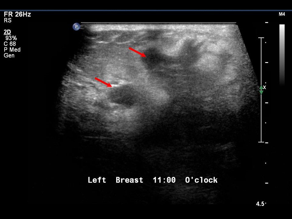 |  |
| Ultrasound features: Left breast, lower quadrants at 6 o'clock | |
| ‣ Mass | |
| • Location: | Left breast, lower quadrants at 6 o'clock |
| • Number: | Multiple |
| • Size: | Largest 3.8 × 1.8 cm |
| • Shape: | Irregular |
| • Orientation: | Not parallel |
| • Margins: | Spiculated |
| • Echo pattern: | Heteroechoic |
| • Posterior features: | No posterior features |
| ‣ Calcifications: | None |
| ‣ Associated features: | Skin thickening, vascularity in mass, axillary lymphadenopathy |
| ‣ Special cases: | None |
BI-RADS:
BI-RADS Category: 5 (highly suggestive of malignancy)Further assessment:
Further assessment advised: Referral for core biopsyHistopathology:
Core needle biopsy
 |
| Histopathology features: | |
| ‣ Specimen type: | Core needle biopsy |
| ‣ Laterality: | Left |
| ‣ Macroscopy: | Seven linear cores: longest is 20 mm in length and smallest is 7 mm in length |
| ‣ Histological type: | Invasive breast carcinoma of no special type |
| ‣ Histological grade: | Grade 2 (3 + 2 + 1 = 6) |
| ‣ Mitosis: | 4 |
| ‣ Maximum invasive tumour size: | |
| ‣ Lymph node status: | |
| ‣ Peritumoural lymphovascular invasion: | |
| ‣ DCIS/EIC: | |
| ‣ Margins: | |
| ‣ Pathological stage: | |
| ‣ Biomarkers: | |
| ‣ Comments: |
Case summary:
| Postmenopausal woman presented with palpable left breast lump with skin changes. Diagnosed as large irregular high-density mass with spiculated margins in left breast with associated features of left breast areolar skin thickening, left nipple retraction, and left axillary lymphadenopathy, BI-RADS 5 on imaging and as invasive breast carcinoma of no special type on needle core biopsy histopathology. |
Learning points:
|