


Home / Training / Manuals / Atlas of breast cancer early detection / Cases
Atlas of breast cancer early detection
Filter by language: English / Русский
Go back to the list of case studies
.png) Click on the pictures to magnify and display the legends
Click on the pictures to magnify and display the legends
| Case number: | 042 |
| Age: | 67 |
| Clinical presentation: | Postmenopausal woman with average risk of developing breast cancer presented with a left breast lump that she noticed many months ago. Now has left nipple and areolar ulceration and erosion. |
 |
Mammography:
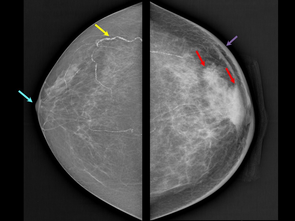 | 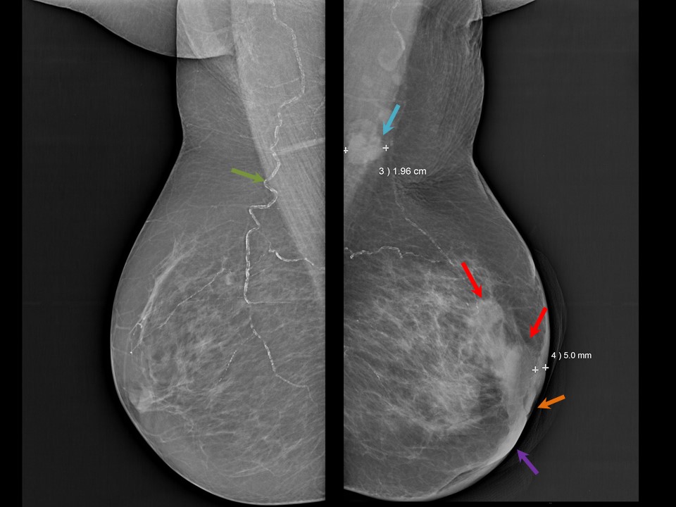 |
| Breast composition: | ACR category b (there are scattered areas of fibroglandular density) | Mammography features: |
| ‣ Location of the lesion: | Left breast, central portion of the breast, central zone, anterior third |
| ‣ Mass: | |
| • Number: | 1 |
| • Size: | 3.0 × 2.0 cm |
| • Shape: | Irregular |
| • Margins: | Indistinct |
| • Density: | High |
| ‣ Calcifications: | |
| • Typically benign: | None |
| • Suspicious: | None |
| • Distribution: | None |
| ‣ Architectural distortion: | None |
| ‣ Asymmetry: | None |
| ‣ Intramammary node: | None |
| ‣ Skin lesion: | None |
| ‣ Solitary dilated duct: | None |
| ‣ Associated features: | Skin thickening, trabecular thickening, areolar skin ulceration, nipple deformation, and axillary lymphadenopathy |
| Breast composition: | ACR category b (there are scattered areas of fibroglandular density) | Mammography features: |
| ‣ Location of the lesion: | Left breast, upper outer quadrant at 2 oclock, anterior and middle thirds |
| ‣ Mass: | |
| • Number: | 1 |
| • Size: | 1.8 × 1.5 cm |
| • Shape: | Irregular |
| • Margins: | Spiculated |
| • Density: | High |
| ‣ Calcifications: | |
| • Typically benign: | None |
| • Suspicious: | None |
| • Distribution: | None |
| ‣ Architectural distortion: | None |
| ‣ Asymmetry: | None |
| ‣ Intramammary node: | None |
| ‣ Skin lesion: | None |
| ‣ Solitary dilated duct: | None |
| ‣ Associated features: | Skin thickening, trabecular thickening, areolar skin ulceration, nipple deformation, and axillary lymphadenopathy |
Ultrasound:
 |  |
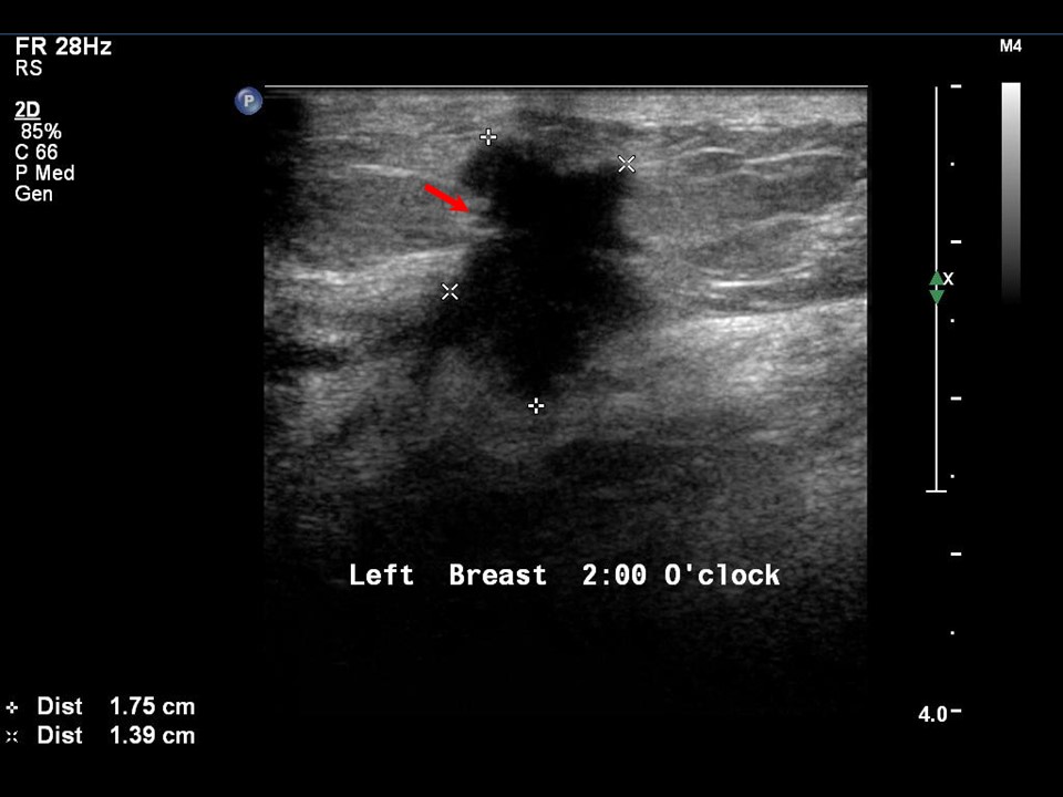 | 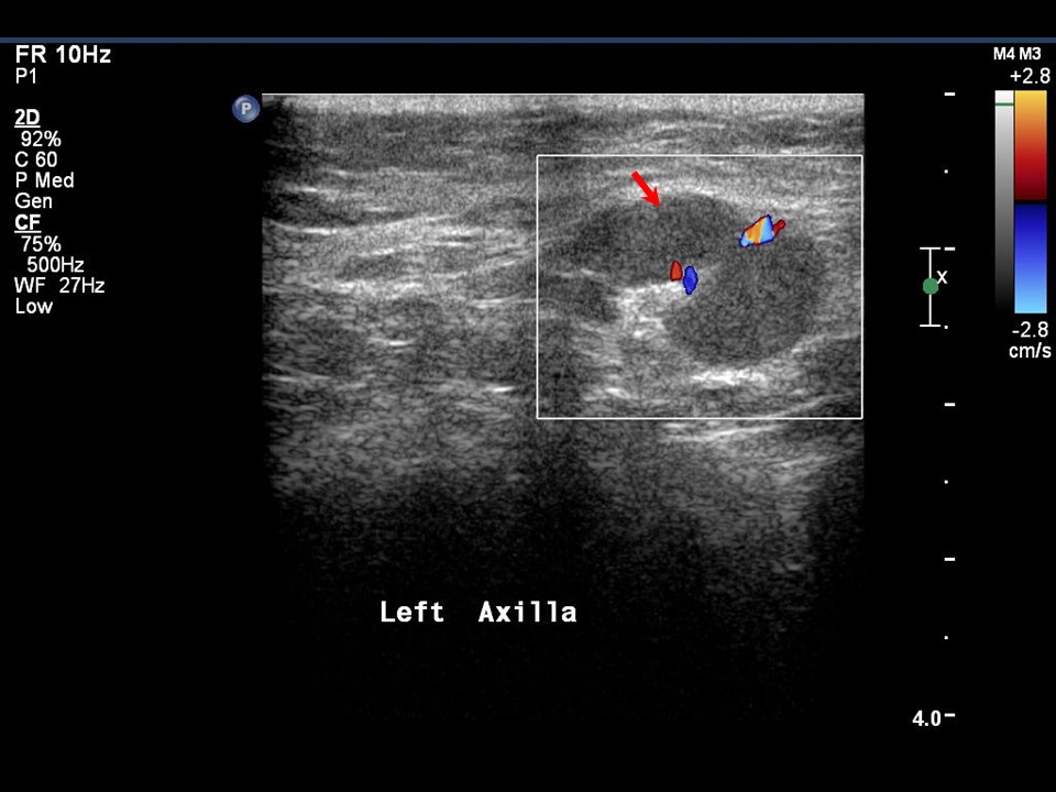 |
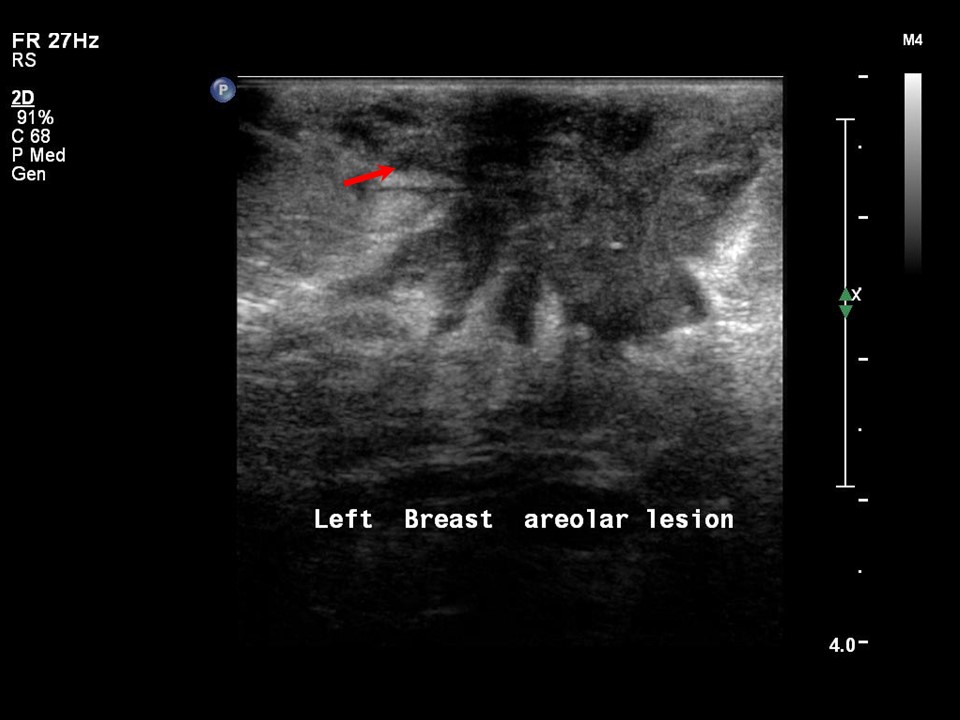
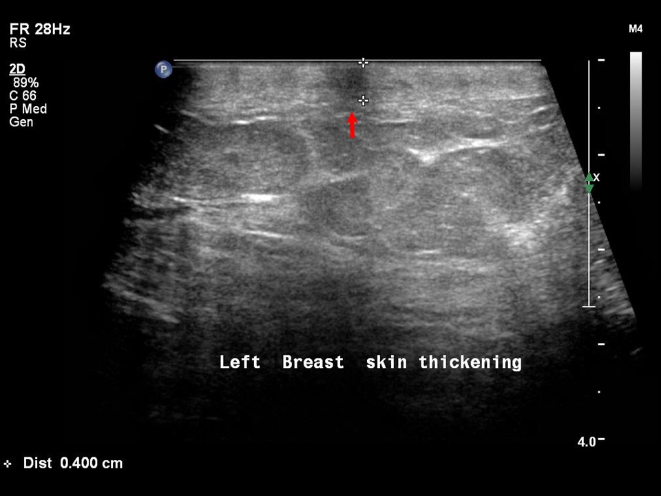
| Ultrasound features: Left breast, upper outer quadrant at 12 oclock and in subareolar region | |
| ‣ Mass | |
| • Location: | Left breast, upper outer quadrant at 12 oclock and in subareolar region |
| • Number: | 2 |
| • Size: | 3.0 × 2.0 cm and 2.0 × 1.4 cm |
| • Shape: | Irregular |
| • Orientation: | Not parallel |
| • Margins: | Spiculated |
| • Echo pattern: | Hypoechoic |
| • Posterior features: | No posterior features |
| ‣ Calcifications: | None |
| ‣ Associated features: | Skin thickening, vascularity in mass, nipple erosion, axillary lymphadenopathy, and areolar ulceration and erosion |
| ‣ Special cases: | None |
BI-RADS:
BI-RADS Category: 5 (highly suggestive of malignancy)Histopathology:
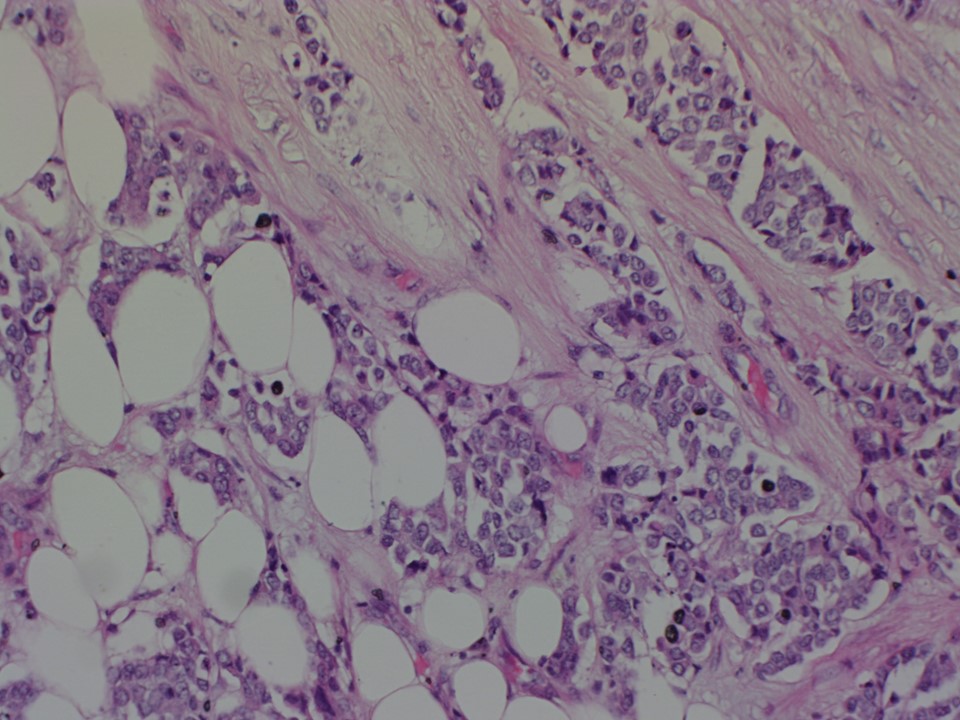
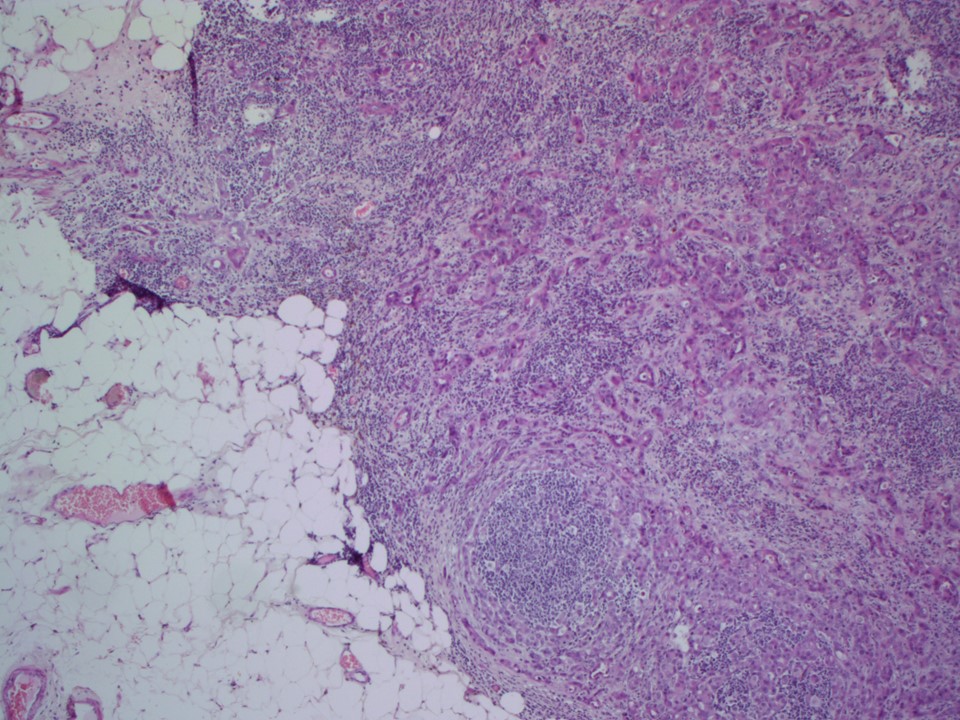
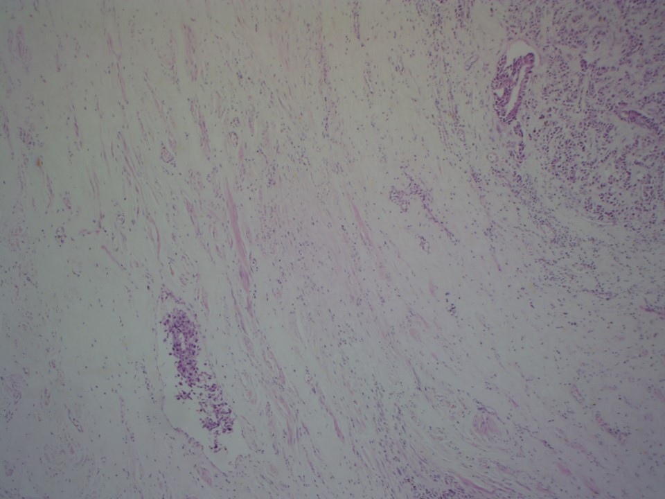
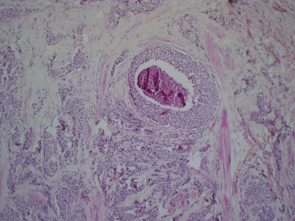
MRM
 |  |
 |  |
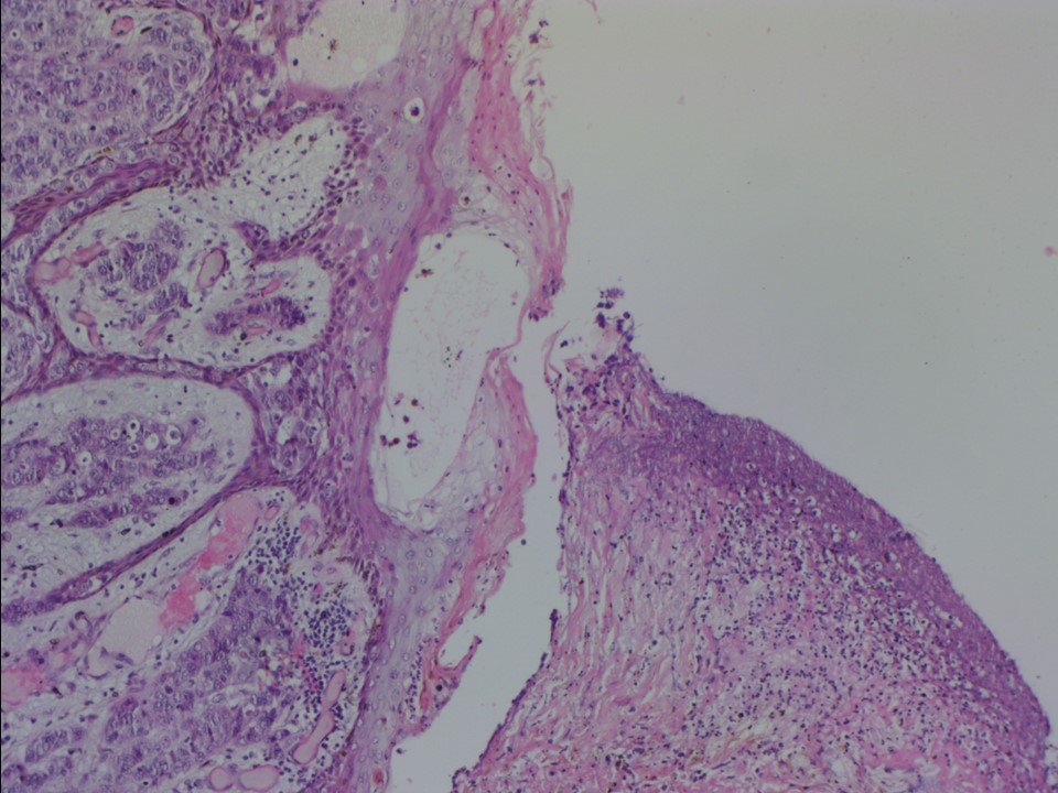 |
| Histopathology features: | |
| ‣ Specimen type: | MRM |
| ‣ Laterality: | Left |
| ‣ Macroscopy: | MRM specimen 30.0 × 12.0 × 4.5 cm, with overlying skin flap 15.0 × 5.0 cm. The nipple and areola (3.5 × 3.4 cm) are ulcerated and the ulcer is 2.0 × 2.5 cm. On serial sectioning, firm grey white tumour is identified (4.0 × 3.6 × 3.2 cm) in the central quadrant. It involves the skin and is located 4.5 cm from the base. The remaining breast tissue reveals fibrotic white areas. Representative sections are submitted. Lymph nodes: On dissection of the axillary tail, 11 lymph nodes are identified. The largest node is 2.0 × 1.5 cm and the cut surface shows grey white areas |
| ‣ Histological type: | Invasive breast carcinoma of no special type |
| ‣ Histological grade: | Grade 3 (3 + 3 + 2 = 8) |
| ‣ Mitosis: | 12 |
| ‣ Maximum invasive tumour size: | 4.0 cm in greatest dimension |
| ‣ Lymph node status: | 3/11 showing metastasis with perinodal extension |
| ‣ Peritumoural lymphovascular invasion: | Present |
| ‣ DCIS/EIC: | Comedo type DCIS of high nuclear grade; EIC absent |
| ‣ Margins: | Overlying skin is involved by tumour. The nipple is involved by carcinoma. The base is free of tumour |
| ‣ Pathological stage: | pT4bN1 |
| ‣ Biomarkers: | |
| ‣ Comments: |
Case summary:
| Postmenopausal woman presented with left breast lump with left nippleareolar ulceration and erosion. Diagnosed as left breast carcinoma with skin thickening, nippleareolar ulceration with excoriation and left axillary metastatic node, BI-RADS 5 on imaging and as invasive breast carcinoma of no special type, pT4bN1 on histopathology. |
Learning points:
|