


Home / Training / Manuals / Atlas of breast cancer early detection / Cases
Atlas of breast cancer early detection
Filter by language: English / Русский
Go back to the list of case studies
.png) Click on the pictures to magnify and display the legends
Click on the pictures to magnify and display the legends
| Case number: | 139 |
| Age: | 62 |
| Clinical presentation: | Postmenopausal woman with average risk of breast cancer presented for the evaluation of a chest CT scan, which had detected a lump in the left breast. Patient first noticed a lump many years ago and reports that it has not increased in size. |
Mammography:
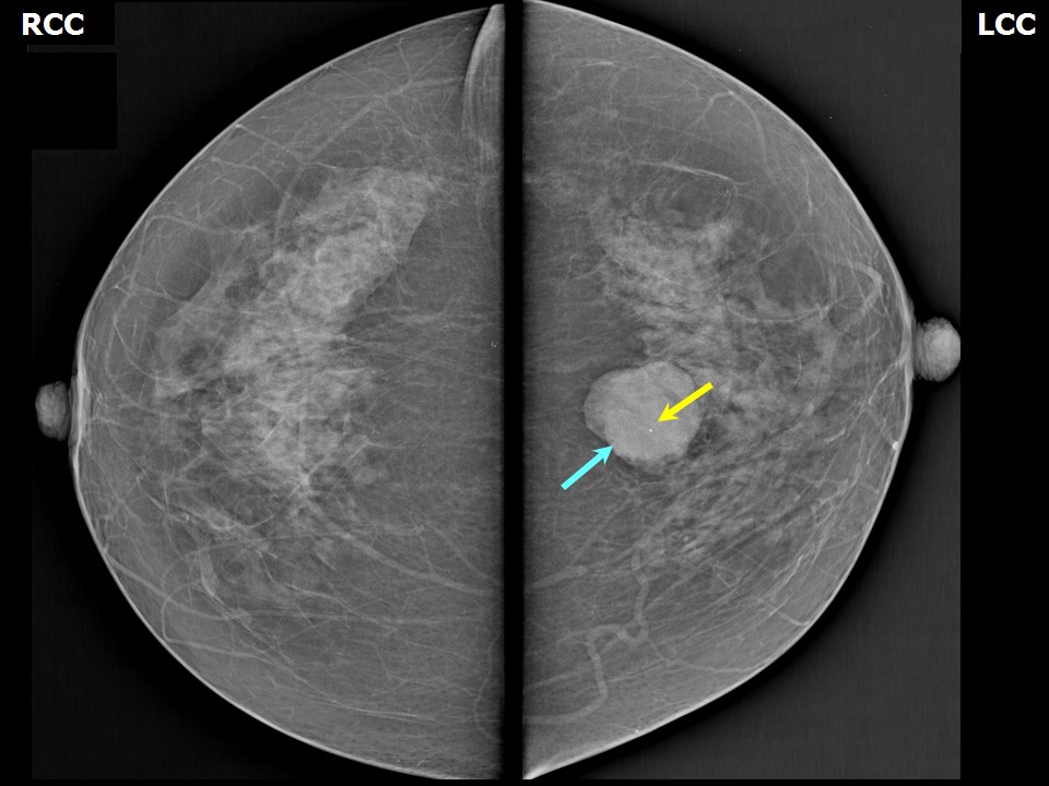 | 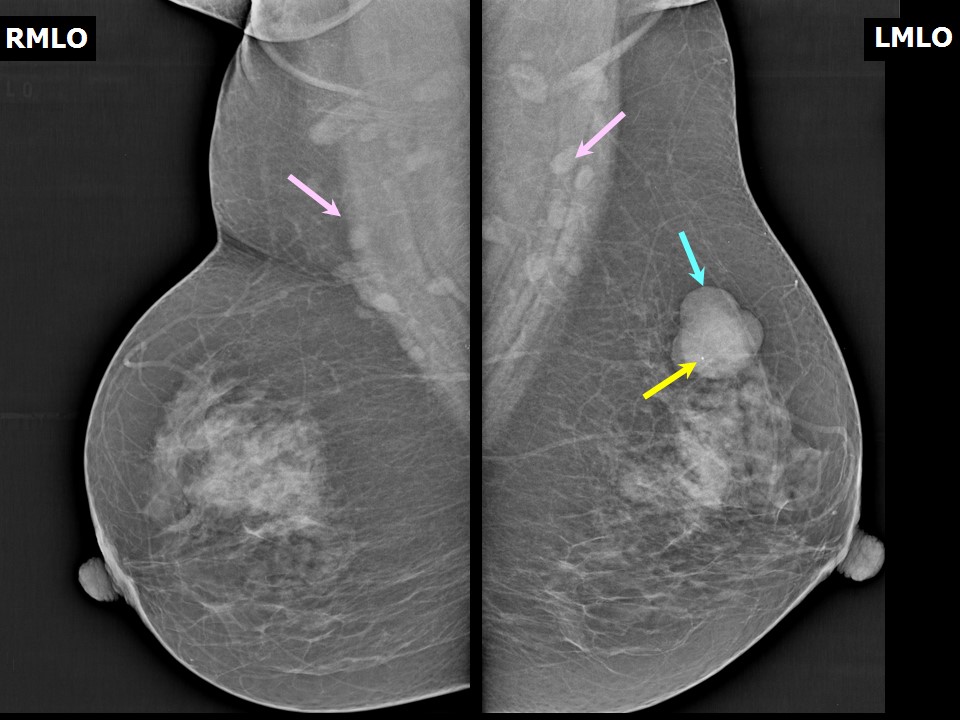 |
| Breast composition: | ACR category b (there are scattered areas of fibroglandular density) | Mammography features: |
| ‣ Location of the lesion: | Left breast, upper inner quadrant at 11 oclock, middle and posterior thirds |
| ‣ Mass: | |
| • Number: | 1 |
| • Size: | 3.3 × 3.2 × 3.0 cm |
| • Shape: | Oval |
| • Margins: | Circumscribed |
| • Density: | Equal |
| ‣ Calcifications: | |
| • Typically benign: | Macrocalcifications |
| • Suspicious: | None |
| • Distribution: | None |
| ‣ Architectural distortion: | None |
| ‣ Asymmetry: | None |
| ‣ Intramammary node: | None |
| ‣ Skin lesion: | None |
| ‣ Solitary dilated duct: | None |
| ‣ Associated features: | None |
Ultrasound:
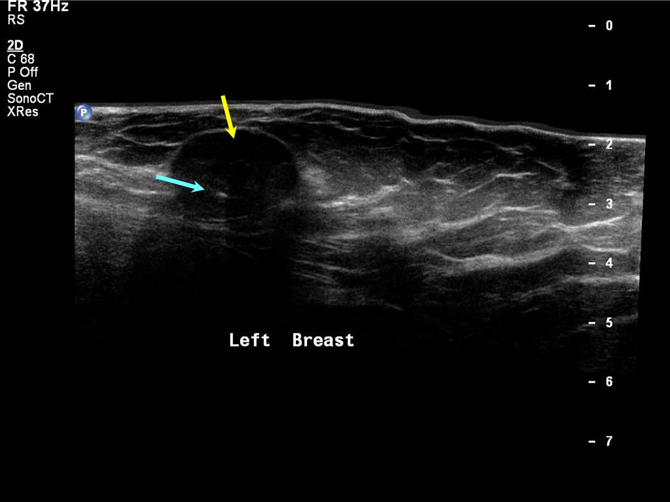 | 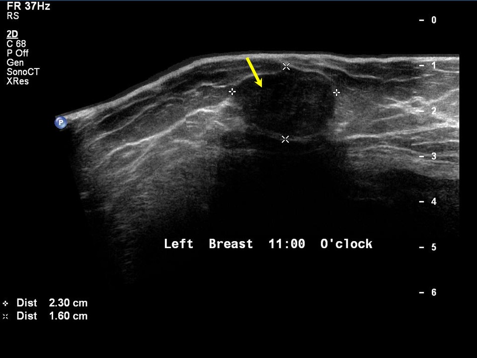 |
| Ultrasound features: Left breast, upper inner quadrant at 11 oclock | |
| ‣ Mass | |
| • Location: | Left breast, upper inner quadrant at 11 oclock |
| • Number: | 1 |
| • Size: | 3.0 × 2.0 cm |
| • Shape: | Oval |
| • Orientation: | Parallel |
| • Margins: | Circumscribed |
| • Echo pattern: | Hypoechoic |
| • Posterior features: | No posterior features |
| ‣ Calcifications: | Typically benign in mass |
| ‣ Associated features: | Calcification in mass and poor vascularity |
| ‣ Special cases: | None |
BI-RADS:
BI-RADS Category: 2 (benign)Further assessment:
Further assessment advised: Referral for cytologyCytology:
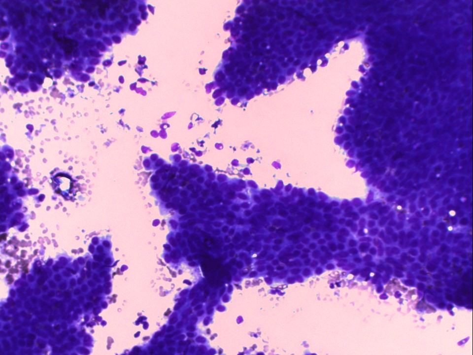 |
| Cytology features: | |
| ‣ Type of sample: | FNAC |
| ‣ Site of biopsy: | |
| • Laterality: | Left |
| • Quadrant: | Upper inner |
| • Localization technique: | Palpation |
| • Nature of aspirate: | Whitish |
| ‣ Cytological description: | Very cellular smears with tightly cohesive ductal epithelial cells. A few stromal fragments and bare nuclei are seen in the background |
| ‣ Reporting category: | Benign |
| ‣ Diagnosis: | Fibroadenoma |
| ‣ Comments: | None |
Case summary:
| Postmenopausal woman presented with left breast lump. Diagnosed as fibroadenoma, BI-RADS 2 on imaging and as fibroadenoma on cytology. |
Learning points:
|