


Home / Training / Manuals / Atlas of breast cancer early detection / Cases
Atlas of breast cancer early detection
Filter by language: English / Русский
Go back to the list of case studies
.png) Click on the pictures to magnify and display the legends
Click on the pictures to magnify and display the legends
| Case number: | 078 |
| Age: | 65 |
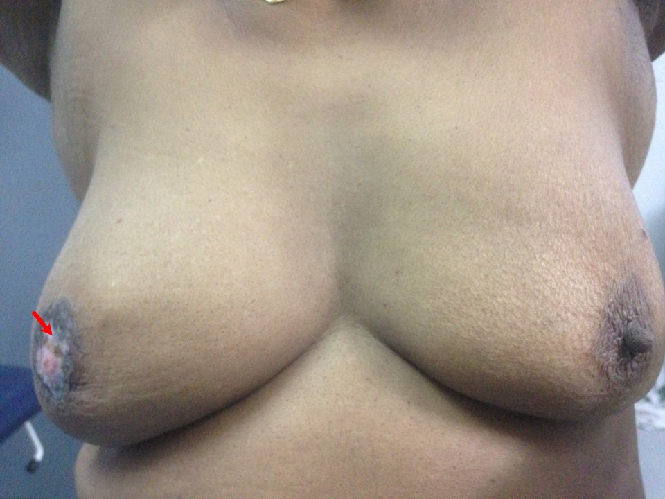
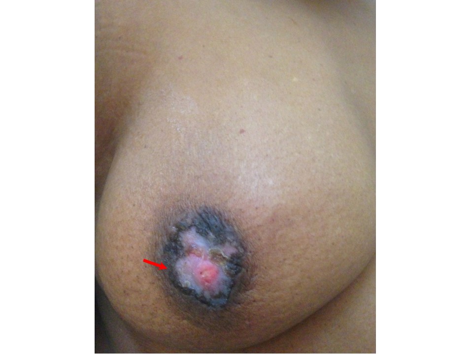
| Clinical presentation: | Postmenopausal woman with average risk of developing breast cancer presented with right nippleareolar ulceration and itching. On clinical examination, a hard lump was felt in the right breast in the retroareolar region. |
 |  |
Mammography:
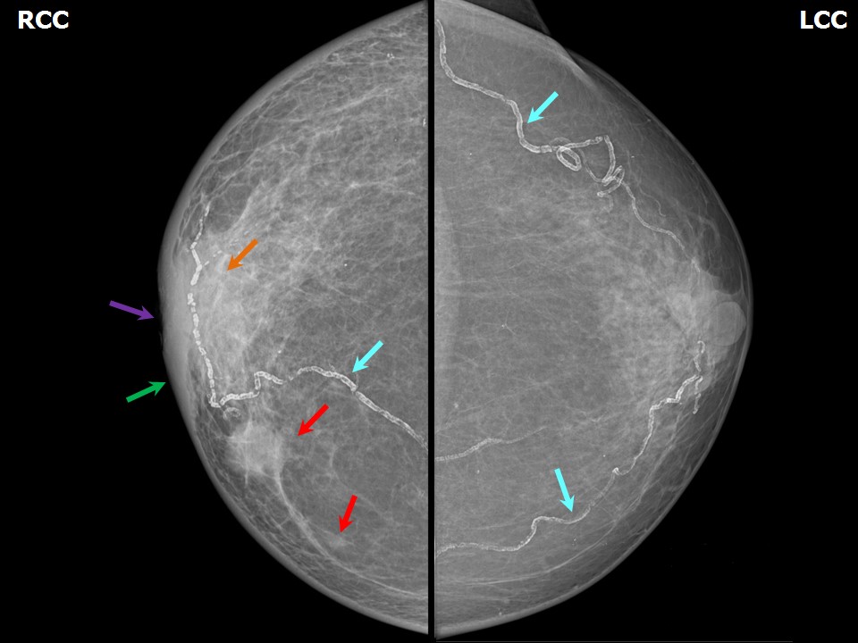 | 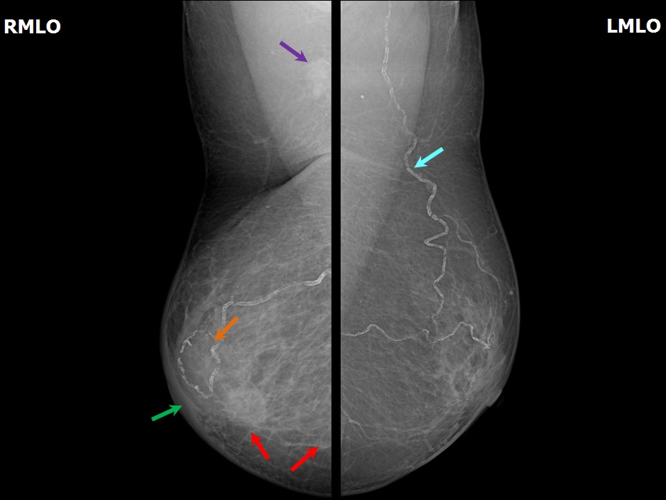 |
| Breast composition: | ACR category b (there are scattered areas of fibroglandular density) | Mammography features: |
| ‣ Location of the lesion: | Right breast, lower inner quadrant at 5 oclock, anterior third |
| ‣ Mass: | |
| • Number: | 1 |
| • Size: | 1.9 × 1.8 cm |
| • Shape: | Round |
| • Margins: | Indistinct |
| • Density: | High |
| ‣ Calcifications: | |
| • Typically benign: | None |
| • Suspicious: | None |
| • Distribution: | None |
| ‣ Architectural distortion: | Present |
| ‣ Asymmetry: | Focal, central quadrant |
| ‣ Intramammary node: | None |
| ‣ Skin lesion: | None |
| ‣ Solitary dilated duct: | None |
| ‣ Associated features: | Skin thickening, axillary adenopathy, and architectural distortion |
| Breast composition: | ACR category b (there are scattered areas of fibroglandular density) | Mammography features: |
| ‣ Location of the lesion: | Right breast, lower inner quadrant at 4 oclock, middle third |
| ‣ Mass: | |
| • Number: | 2 |
| • Size: | 0.5 cm in greatest dimension |
| • Shape: | Irregular |
| • Margins: | Indistinct |
| • Density: | Equal |
| ‣ Calcifications: | |
| • Typically benign: | None |
| • Suspicious: | None |
| • Distribution: | None |
| ‣ Architectural distortion: | None |
| ‣ Asymmetry: | None |
| ‣ Intramammary node: | None |
| ‣ Skin lesion: | None |
| ‣ Solitary dilated duct: | None |
| ‣ Associated features: | None |
Ultrasound:
 |  |
 |  |
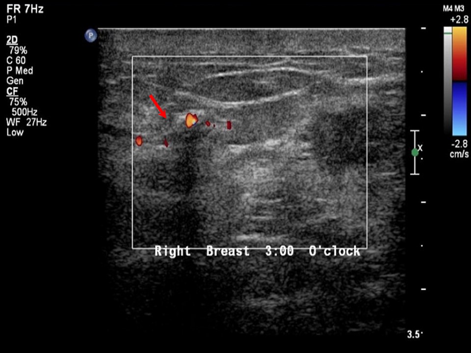 |
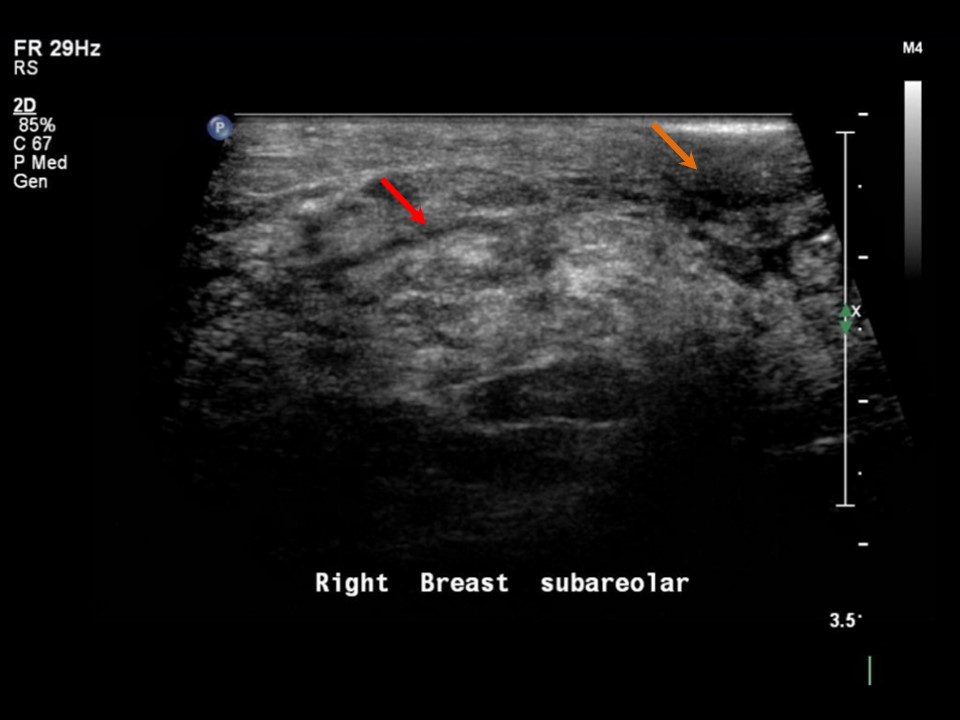
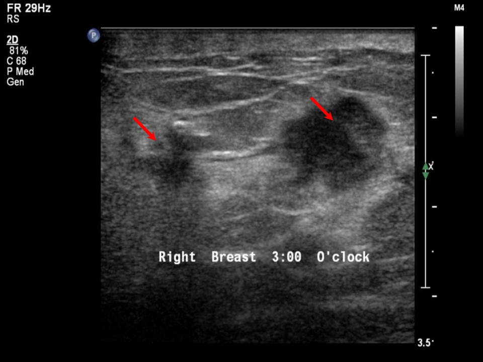
| Ultrasound features: Right breast, lower inner quadrant at 5 oclock | |
| ‣ Mass | |
| • Location: | Right breast, lower inner quadrant at 5 oclock |
| • Number: | 1 |
| • Size: | 1.7 × 1.3 cm |
| • Shape: | Irregular |
| • Orientation: | Not parallel |
| • Margins: | Spiculated |
| • Echo pattern: | Hypoechoic |
| • Posterior features: | No posterior features |
| ‣ Calcifications: | None |
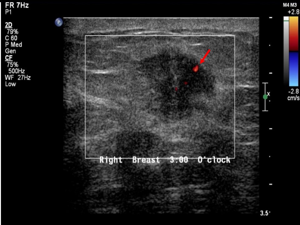
| ‣ Associated features: | Skin thickening, architectural distortion, and internal vascularity |
| ‣ Special cases: | None |
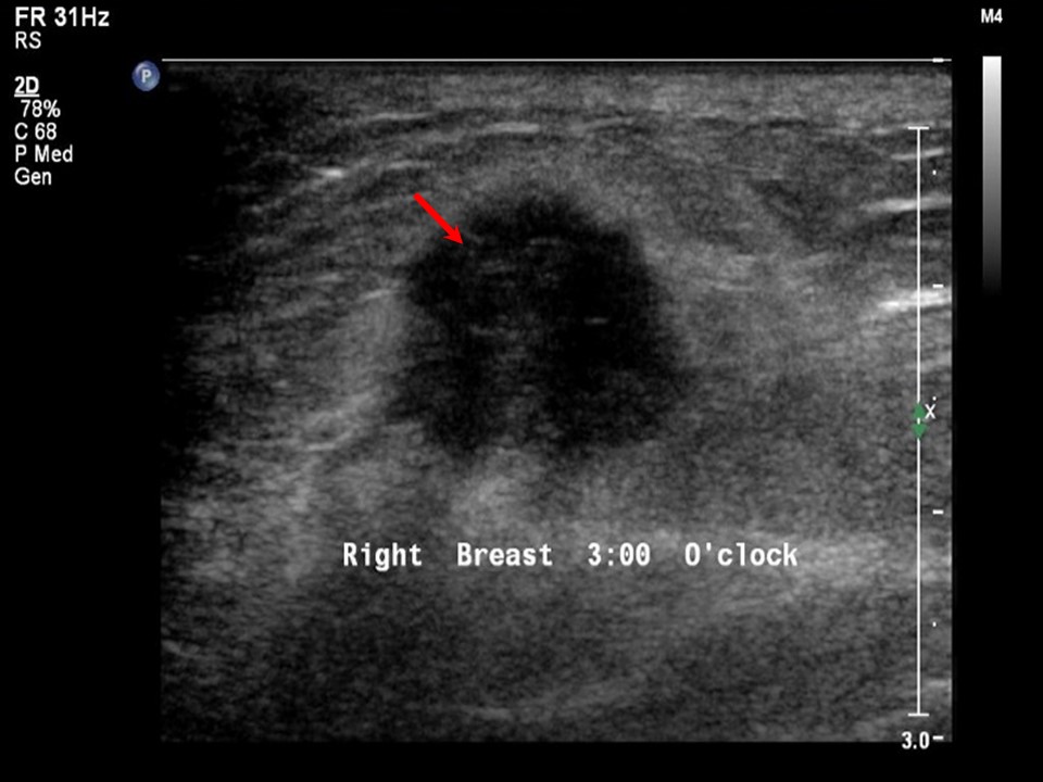
| Ultrasound features: Right breast, lower inner quadrant at 4 oclock | |
| ‣ Mass | |
| • Location: | Right breast, lower inner quadrant at 4 oclock |
| • Number: | 1 |
| • Size: | 0.5 cm in greatest dimension |
| • Shape: | Irregular |
| • Orientation: | Not parallel |
| • Margins: | Spiculated |
| • Echo pattern: | Hypoechoic |
| • Posterior features: | No posterior features |
| ‣ Calcifications: | None |
| ‣ Associated features: | Internal vascularity |
| ‣ Special cases: | None |
BI-RADS:
BI-RADS Category: 5 (highly suggestive of malignancy)Further assessment:
Further assessment advised: Referral for cytologyCytology:
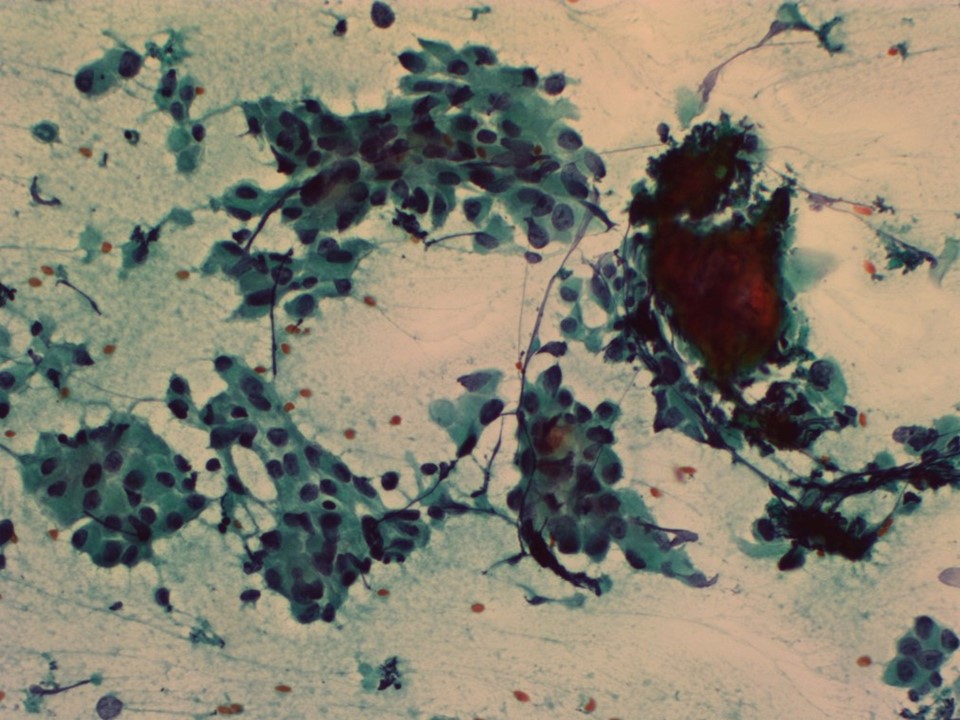 |
| Cytology features: | |
| ‣ Type of sample: | FNAC (solid lesion) |
| ‣ Site of biopsy: | |
| • Laterality: | Right |
| • Quadrant: | |
| • Localization technique: | Palpation |
| • Nature of aspirate: | |
| ‣ Cytological description: | Cellular smears with many single isolated malignant cells and dyscohesive malignant cell clusters |
| ‣ Reporting category: | Malignant |
| ‣ Diagnosis: | Carcinoma |
| ‣ Comments: | None |
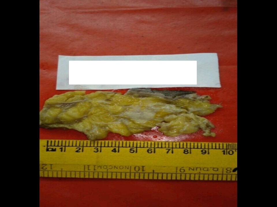
Histopathology:
MRM
 |  |
 |  |
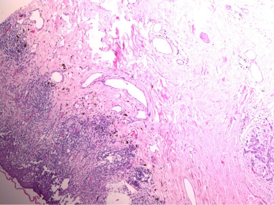 |  |
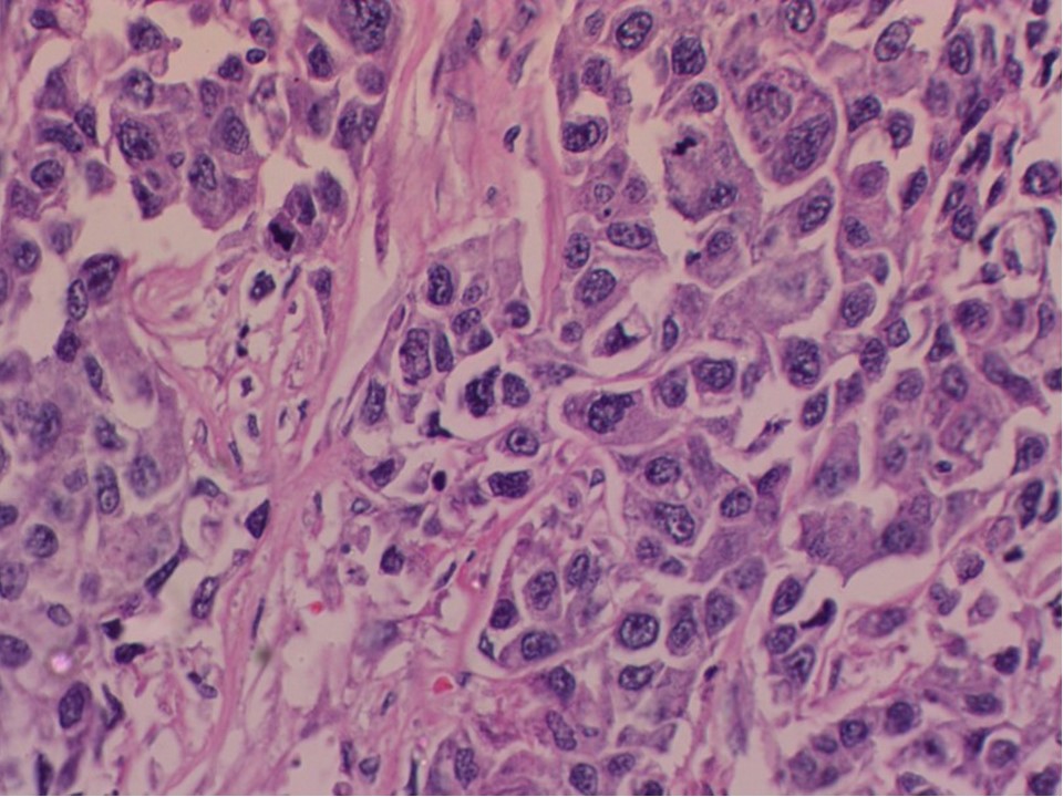
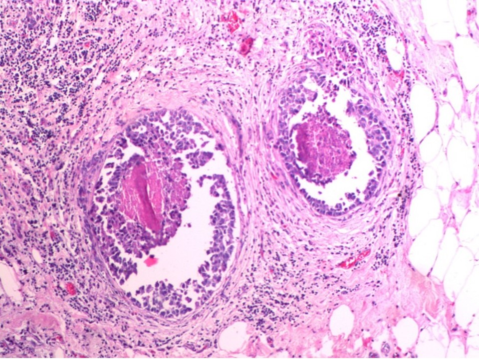
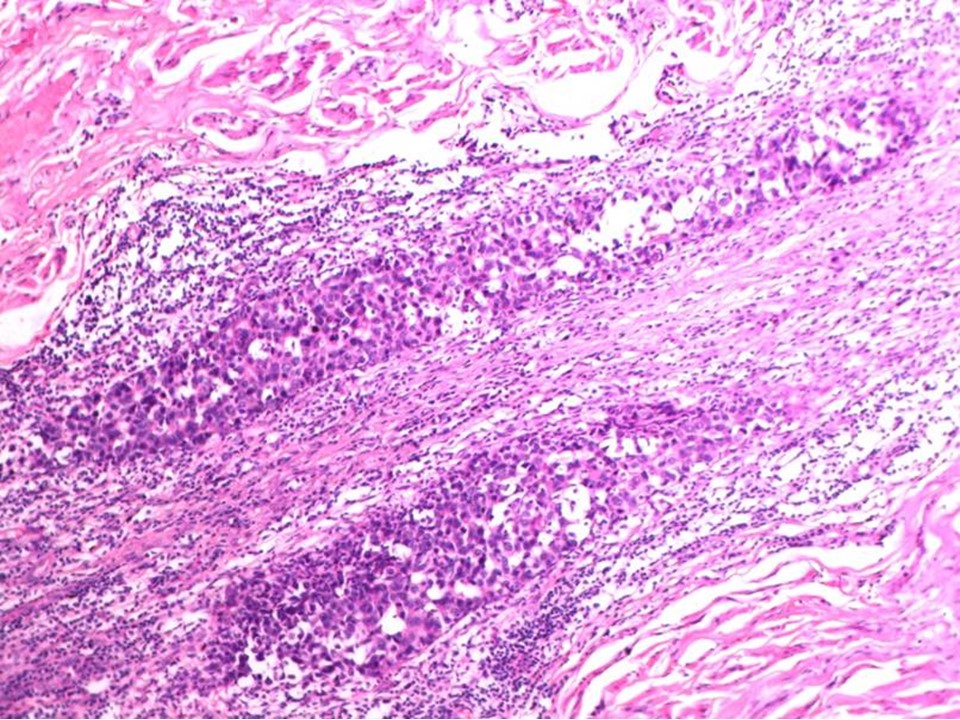
| Histopathology features: | |
| ‣ Specimen type: | MRM |
| ‣ Laterality: | Right |
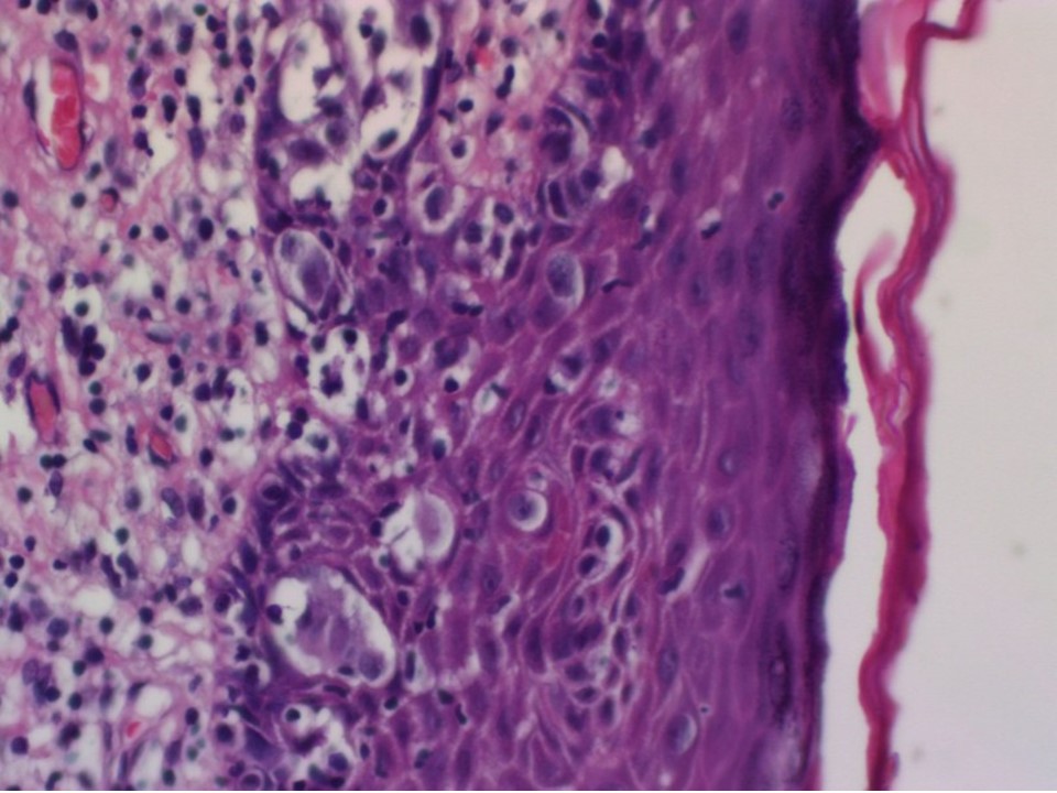
| ‣ Macroscopy: | The nipple and areola are flattened and this area is seen as an indurated firm whitish area (3.0 × 2.5 cm). On serial sectioning, a greyish white tumour (2.3 × 2.0 × 2.0 cm) is identified, located below the areola. It is 1.0 cm from the skin and 2.5 cm from the base |
| ‣ Histological type: | Invasive carcinoma of no special type |
| ‣ Histological grade: | Grade 3 (3 + 3 + 3 = 9) |
| ‣ Mitosis: | 22 |
| ‣ Maximum invasive tumour size: | 2.3 cm in greatest dimension |
| ‣ Lymph node status: | 24/24 |
| ‣ Peritumoural lymphovascular invasion: | Present |
| ‣ DCIS/EIC: | Comedo high grade |
| ‣ Margins: | Posterior margin is free of tumour and areolar skin is ulcerated |
| ‣ Pathological stage: | pT4bN3 |
| ‣ Biomarkers: | |
| ‣ Comments: |
Case summary:
| Postmenopausal woman presented with right breast lump, nippleareolar ulceration, and itching. Diagnosed as right breast carcinoma with right areolar skin thickening and right nipple excoriation, BI-RADS 5 on imaging, as right breast carcinoma on cytology, and as invasive breast carcinoma of no special type, pT4bN3 on histopathology. |
Learning points:
|