Home / Training / Manuals / Atlas of breast cancer early detection / Cases
Atlas of breast cancer early detection
Filter by language: English / Русский
Go back to the list of case studies
.png) Click on the pictures to magnify and display the legends
Click on the pictures to magnify and display the legends
| Case number: | 118 |
| Age: | 75 |
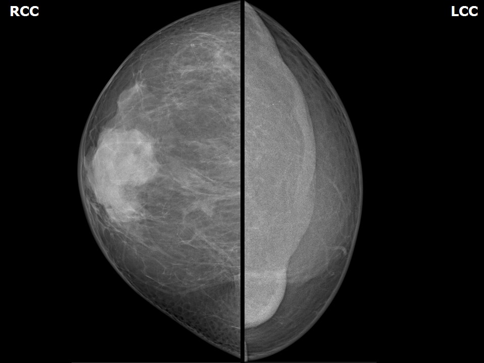
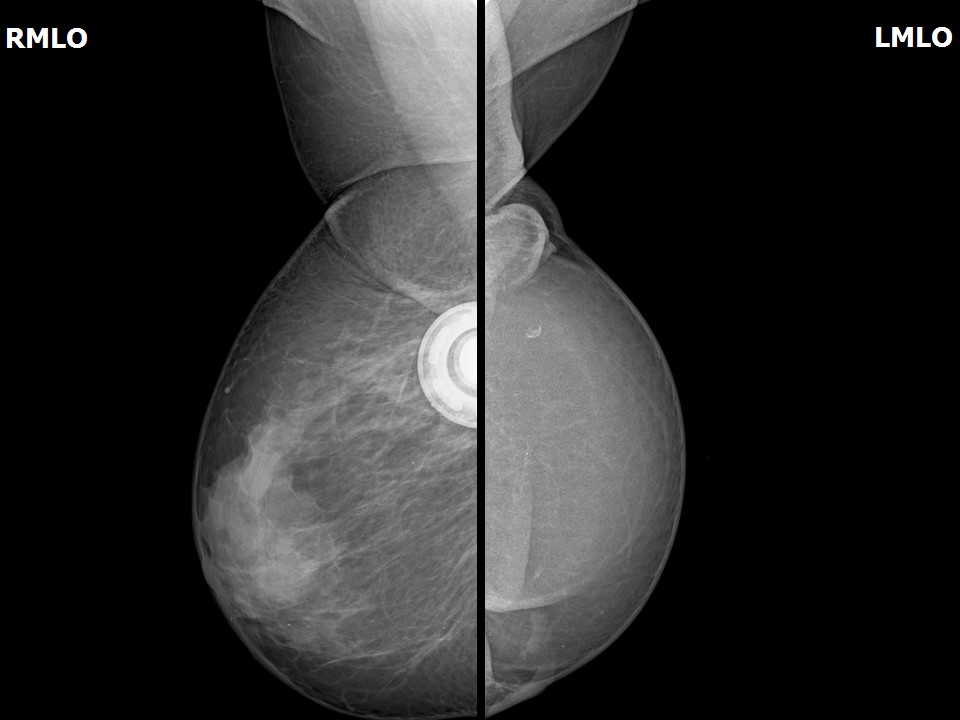
| Clinical presentation: | Postmenopausal woman who was a known case of bilateral breast cancer and had undergone right breast-conserving surgery, left MRM, and a reconstruction with rectus abdominis muscle presented for a follow-up mammography. |
Mammography:
| Breast composition: | ACR category b (there are scattered areas of fibroglandular density) | Mammography features: |
| ‣ Location of the lesion: | Right breast, upper quadrant breast-conserving surgery scar tissue with chemoport in situ |
| ‣ Mass: | |
| • Number: | 0 |
| • Size: | No |
| • Shape: | None |
| • Margins: | None |
| • Density: | None |
| ‣ Calcifications: | |
| • Typically benign: | None |
| • Suspicious: | None |
| • Distribution: | None |
| ‣ Architectural distortion: | None |
| ‣ Asymmetry: | None |
| ‣ Intramammary node: | None |
| ‣ Skin lesion: | None |
| ‣ Solitary dilated duct: | None |
| ‣ Associated features: | None |
| Breast composition: | ACR category b (there are scattered areas of fibroglandular density) | Mammography features: |
| ‣ Location of the lesion: | Left breast, MRM status, reconstructed TRAM flap seen |
| ‣ Mass: | |
| • Number: | 0 |
| • Size: | No |
| • Shape: | None |
| • Margins: | None |
| • Density: | None |
| ‣ Calcifications: | |
| • Typically benign: | Solitary round calcification focus in upper quadrant of the flap |
| • Suspicious: | None |
| • Distribution: | None |
| ‣ Architectural distortion: | None |
| ‣ Asymmetry: | None |
| ‣ Intramammary node: | None |
| ‣ Skin lesion: | None |
| ‣ Solitary dilated duct: | None |
| ‣ Associated features: | None |
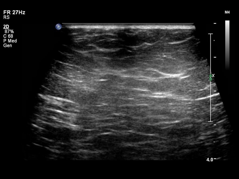
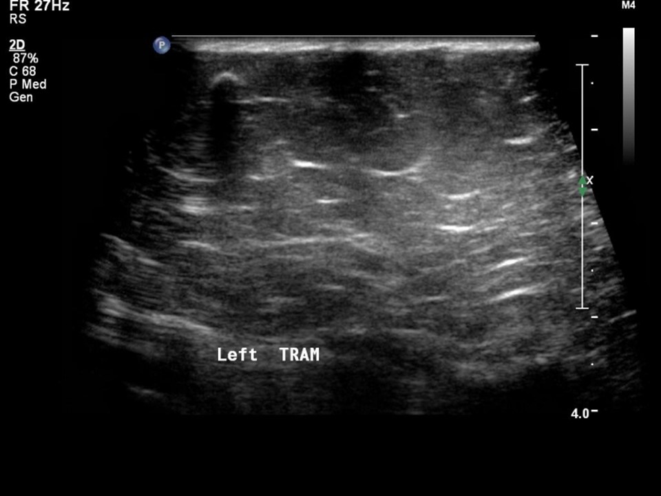
Ultrasound:
BI-RADS:
BI-RADS Category: 2 (benign)Case summary:
| Postmenopausal woman, a known case of bilateral breast cancer who had undergone right breast-conserving surgery and left MRM with rectus abdominis muscle reconstruction, is on regular follow-up mammography. On imaging, heterogeneously dense breast parenchyma in the right breast and TRAM flap reconstruction of the left breast are seen. A chemoport is seen in situ in the right breast. |
Learning points:
| <
25 avenue Tony Garnier CS 90627 69366, LYON CEDEX 07 France - Tel: +33 (0)4 72 73 84 85
© IARC 2026 - Terms of use - Privacy Policy.
© IARC 2026 - Terms of use - Privacy Policy.


