


Home / Training / Manuals / Atlas of breast cancer early detection / Cases
Atlas of breast cancer early detection
Filter by language: English / Русский
Go back to the list of case studies
.png) Click on the pictures to magnify and display the legends
Click on the pictures to magnify and display the legends
| Case number: | 116 |
| Age: | 44 |
| Clinical presentation: | Premenopausal woman with average risk of developing breast cancer presented with a breast lump. Examination revealed a mobile breast lump on the right side. |
Ultrasound:
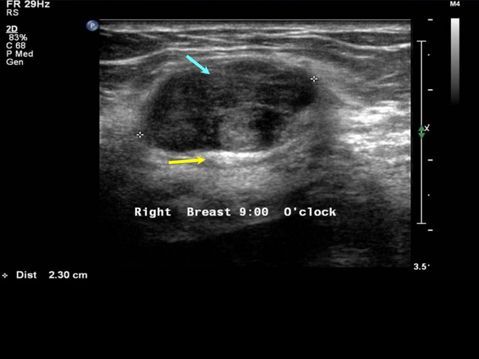 | 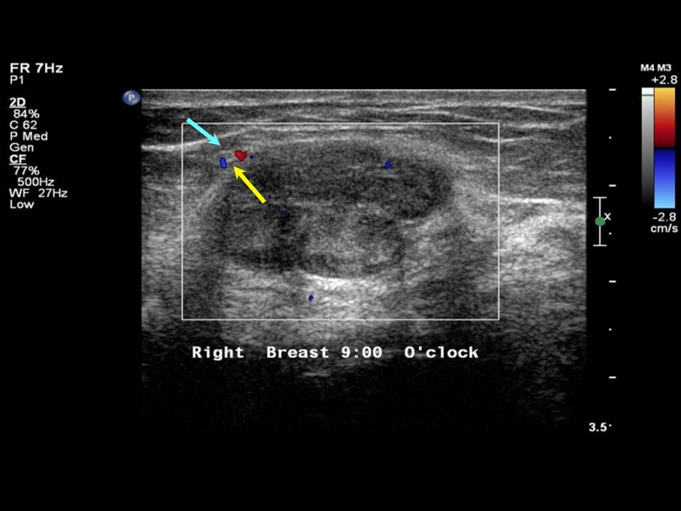 |
| Ultrasound features: Right breast, outer quadrants at 9 oclock | |
| ‣ Mass | |
| • Location: | Right breast, outer quadrants at 9 oclock |
| • Number: | 1 |
| • Size: | 2.3 × 1.4 cm |
| • Shape: | Oval |
| • Orientation: | Parallel |
| • Margins: | Circumscribed, smooth lobulations |
| • Echo pattern: | Hypoechoic |
| • Posterior features: | Posterior Enhancement |
| ‣ Calcifications: | None |
| ‣ Associated features: | None |
| ‣ Special cases: | None |
BI-RADS:
BI-RADS Category: 2 (benign)Further assessment:
Further assessment advised: Referral for cytologyCytology:
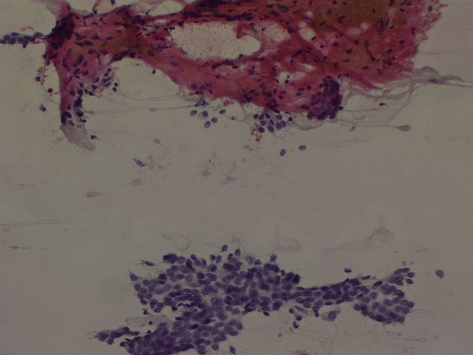 |
| Cytology features: | |
| ‣ Type of sample: | FNAC |
| ‣ Site of biopsy: | |
| • Laterality: | Right |
| • Quadrant: | Outer central |
| • Localization technique: | Palpation |
| • Nature of aspirate: | Whitish |
| ‣ Cytological description: | Smears reveal many clusters of ductal epithelial cells with myoepithelial cells. A few papillary clusters with mild atypia are seen. There are many stromal and adipose tissue fragments. Background shows a few bare nuclei |
| ‣ Reporting category: | Benign |
| ‣ Diagnosis: | Benign proliferative breast lesion |
| ‣ Comments: | None |
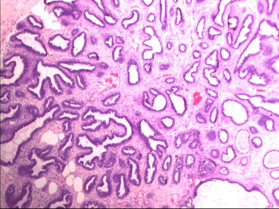
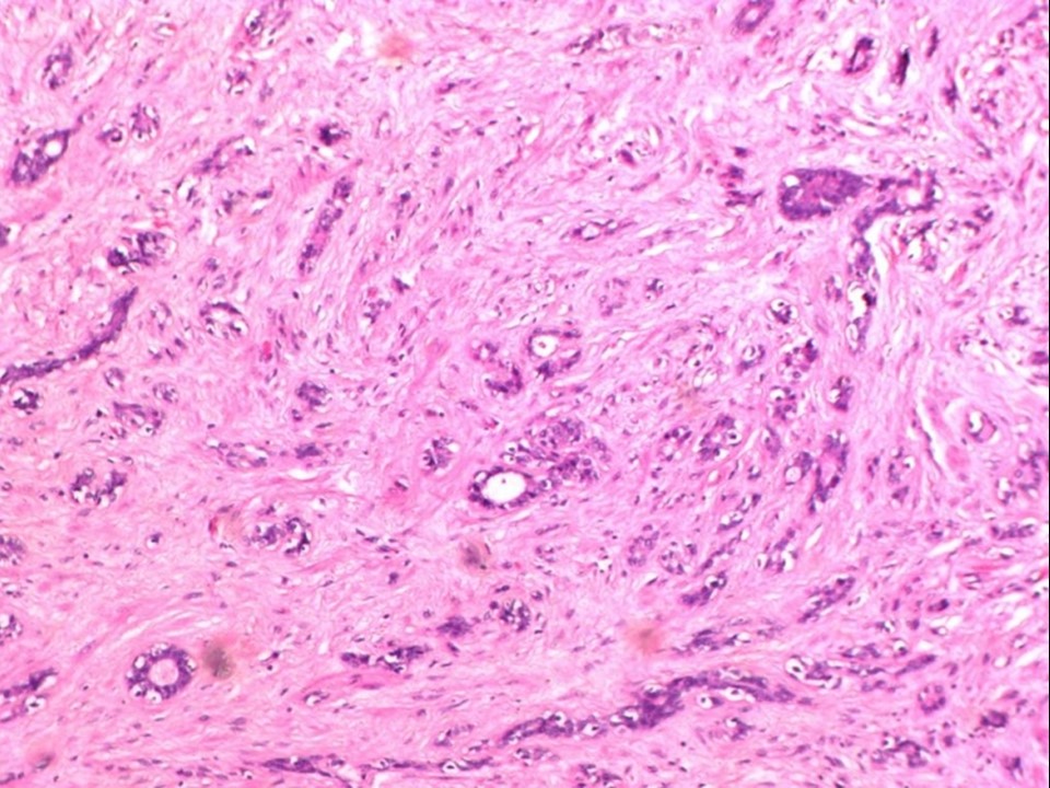
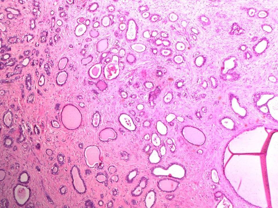
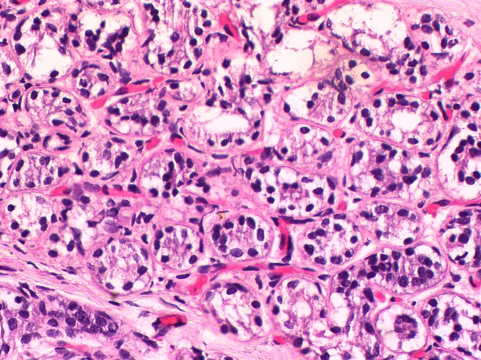
Histopathology:
Lumpectomy specimen
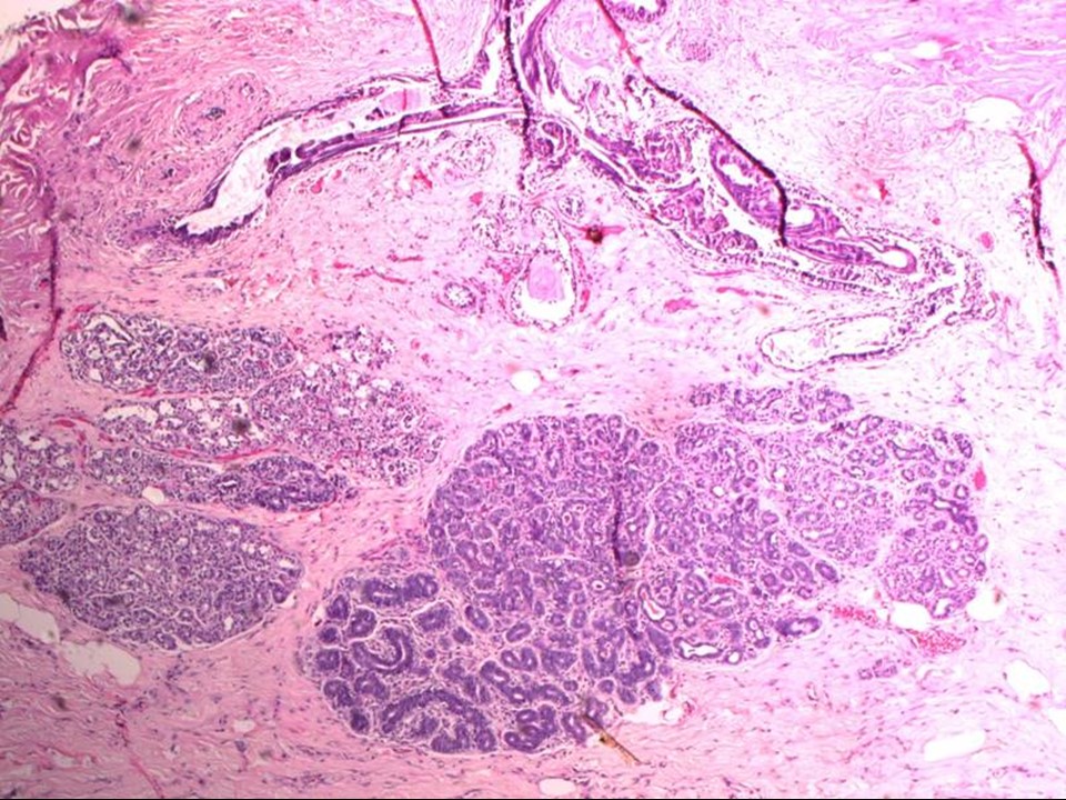 |  |
 |  |
 | 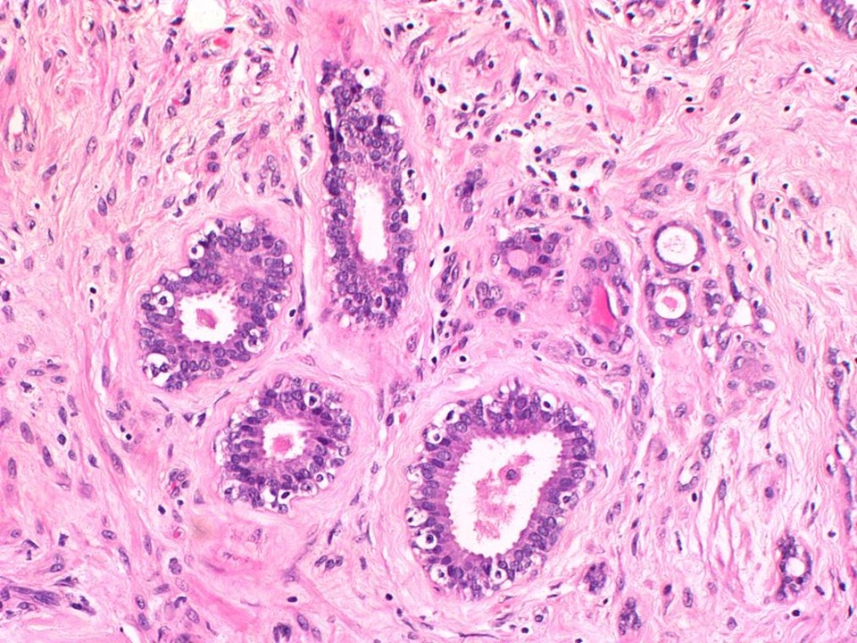 |
| Histopathology features: | |
| ‣ Specimen type: | Lumpectomy specimen |
| ‣ Laterality: | Right |
| ‣ Macroscopy: | Lumpectomy specimen (8.5 × 7.5 × 3.0 cm) with skin flap (3.0 × 1.2 cm). On serial sectioning, three well-circumscribed greyish white areas are identified (2.5 × 2.0 × 2.0 cm, 1.7 × 1.5 × 1.0 cm, and 1.4 × 1.0 × 1.0 cm) |
| ‣ Histological type: | Multiple sections studied show circumscribed centrilobular areas of florid adenosis with myoepithelial proliferation and a few ducts with epithelial proliferation. The surrounding stroma is proliferative with a few lymphocytes. A few nodular foci show histology consistent with sclerosing adenosis. Microglandular adenosis and UDH is noted. A few ducts show features of ductal hyperplasia with atypia and flat epithelial atypia. There is surrounding fibrosis with dilated ducts and occasional ducts with apocrine metaplasia and columnar cell metaplasia |
| ‣ Histological grade: | |
| ‣ Mitosis: | |
| ‣ Maximum invasive tumour size: | |
| ‣ Lymph node status: | |
| ‣ Peritumoural lymphovascular invasion: | |
| ‣ DCIS/EIC: | Absent |
| ‣ Margins: | |
| ‣ Pathological stage: | |
| ‣ Biomarkers: | |
| ‣ Comments: | Proliferative breast disease with florid adenosis, sclerosing adenosis, UDH, microglandular adenosis, and foci of ADH. There is no evidence of in situ or invasive malignancy |
Case summary:
| Premenopausal woman presented with a right breast lump. Diagnosed as BI-RADS 2 on imaging and as benign proliferative breast disease on cytology and histopathology. |
Learning points:
|