


Home / Training / Manuals / Atlas of breast cancer early detection / Cases
Atlas of breast cancer early detection
Filter by language: English / Русский
Go back to the list of case studies
.png) Click on the pictures to magnify and display the legends
Click on the pictures to magnify and display the legends
| Case number: | 108 |
| Age: | 52 |
| Clinical presentation: | Premenopausal woman presented for mammography screening. She had an increased risk of breast cancer because of family history, and was scheduled for mammographic surveillance. |
Mammography:
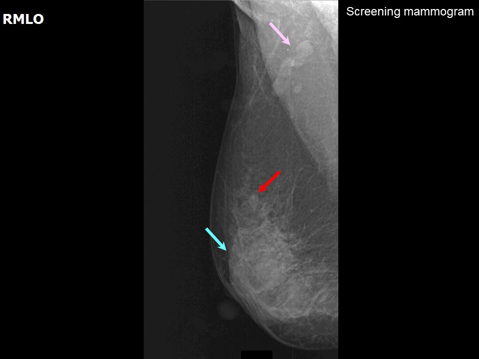 |
| Breast composition: | ACR category b (there are scattered areas of fibroglandular density) | Mammography features: |
| ‣ Location of the lesion: | Right breast, upper outer quadrant at 10 oclock, middle third |
| ‣ Mass: | |
| • Number: | 1 |
| • Size: | 0.7 cm in greatest dimension |
| • Shape: | Irregular |
| • Margins: | Indistinct |
| • Density: | Equal |
| ‣ Calcifications: | |
| • Typically benign: | None |
| • Suspicious: | None |
| • Distribution: | None |
| ‣ Architectural distortion: | None |
| ‣ Asymmetry: | None |
| ‣ Intramammary node: | None |
| ‣ Skin lesion: | None |
| ‣ Solitary dilated duct: | None |
| ‣ Associated features: | None |
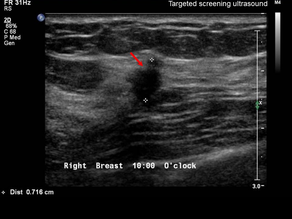
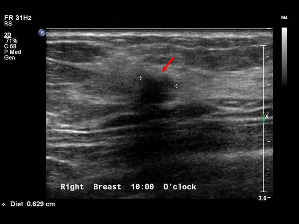
Ultrasound:
 |  |
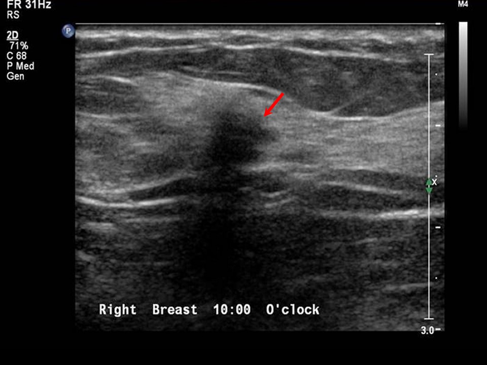 | 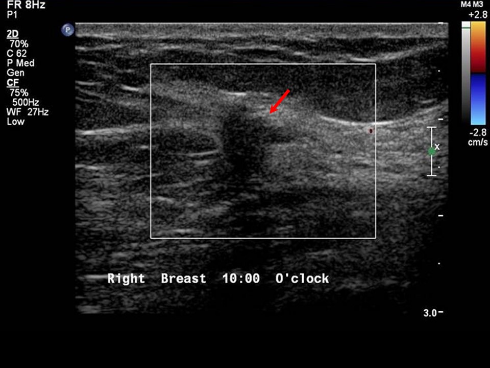 |
| Ultrasound features: Right breast, upper outer quadrant at 10 oclock | |
| ‣ Mass | |
| • Location: | Right breast, upper outer quadrant at 10 oclock |
| • Number: | 1 |
| • Size: | 0.7 cm in greatest dimension |
| • Shape: | Irregular |
| • Orientation: | Not parallel |
| • Margins: | Spiculated |
| • Echo pattern: | Hypoechoic |
| • Posterior features: | Posterior shadowing |
| ‣ Calcifications: | None |
| ‣ Associated features: | None |
| ‣ Special cases: | None |
BI-RADS:
BI-RADS Category: 5 (highly suggestive of malignancy)Further assessment:
Further assessment advised: Further imaging with breast MRIMRI:
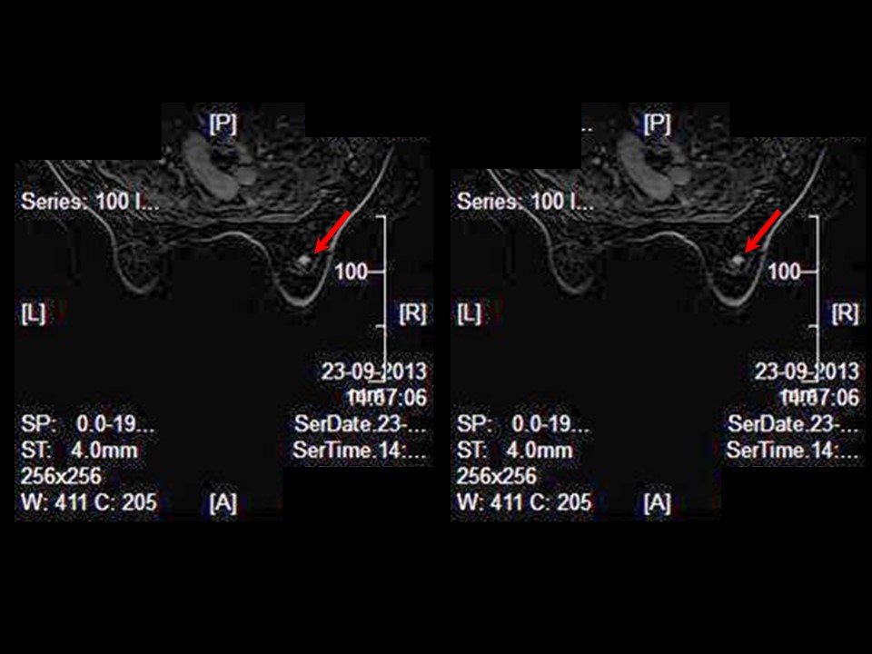 | 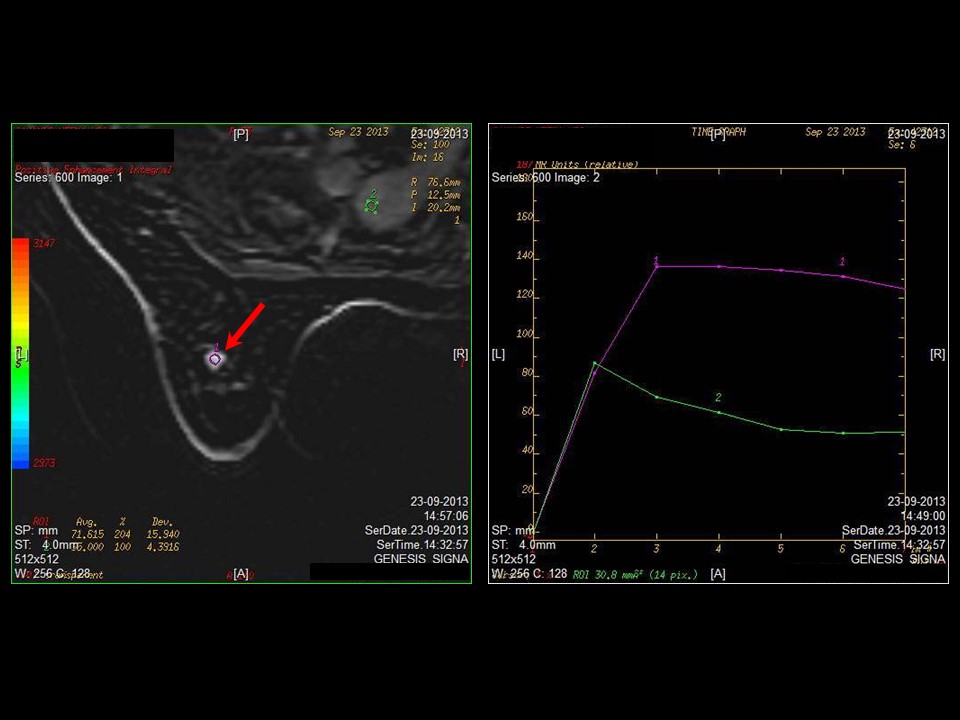 |
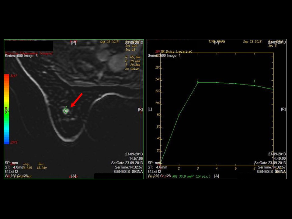 |  |
| MRI features: | ||
| ‣ MRI features: | Amount of fibroglandular tissue: category b (there are scattered areas of fibroglandular density). Background parenchymal enhancement: Mild (2550%), symmetrical | |
| ‣ Location: | Right breast, upper outer quadrant | |
| ‣ Focus: | No | |
| ‣ Mass: | ||
| • Shape: | Round | |
| • Margin: | Spiculated | |
| • Internal enhancement: | Intense homogeneous | |
| • Kinetic curve: | Type 3 | |
| ‣ Non-mass enhancement: | ||
| • Distribution: | No | |
| • Internal enhancement: | No | |
| ‣ Non-enhancing findings: | No | |
| ‣ Associated features: | No | |
| ‣ Axillary nodes: | No | |
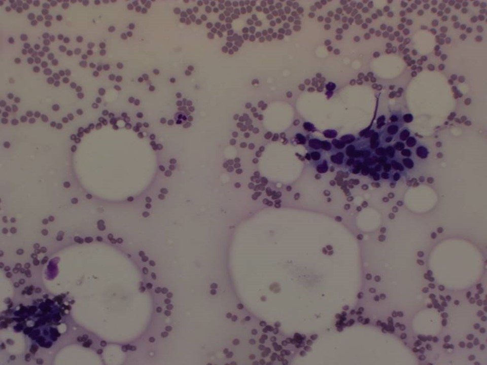
Cytology:
 |
| Cytology features: | |
| ‣ Type of sample: | FNAC |
| ‣ Site of biopsy: | |
| • Laterality: | Right |
| • Quadrant: | Upper outer |
| • Localization technique: | Palpation |
| • Nature of aspirate: | Whitish |
| ‣ Cytological description: | Smears reveal many sheets of tightly cohesive, benign ductal epithelial cells and bare nuclei. A few sheets of ductal epithelial cells show anisonucleosis, enlarged, mildly hyperchromatic nuclei and moderate amounts of cytoplasm |
| ‣ Reporting category: | Suspicious, probably in situ or invasive carcinoma |
| ‣ Diagnosis: | Predominantly benign cells with a few cells suspicious for malignancy |
| ‣ Comments: | None |
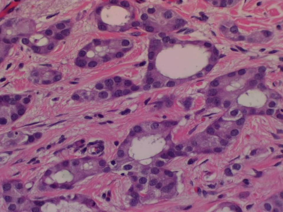
Histopathology:
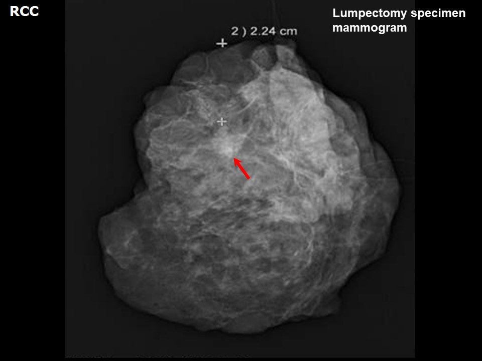
Breast-conserving surgery
 |
| Histopathology features: | |
| ‣ Specimen type: | Breast-conserving surgery |
| ‣ Laterality: | Right |
| ‣ Macroscopy: | On serial sectioning, a small firm greyish white area (0.6 × 0.5 × 0.5 cm) is seen. The closest cut margin is the superior margin, which is 1.5 cm from the tumour |
| ‣ Histological type: | Invasive carcinoma of no special type, a few with tubular carcinoma morphology |
| ‣ Histological grade: | Grade 1 (2 + 1 + 1 = 4) |
| ‣ Mitosis: | 3 |
| ‣ Maximum invasive tumour size: | 0.6 cm in greatest dimension |
| ‣ Lymph node status: | 0/14 |
| ‣ Peritumoural lymphovascular invasion: | Not identified |
| ‣ DCIS/EIC: | Small foci of cribriform type low grade |
| ‣ Margins: | Free of tumour |
| ‣ Pathological stage: | pT1N0 |
| ‣ Biomarkers: | |
| ‣ Comments: | Amount of fibroglandular tissue: category b (scattered fibroglandular tissue). Background: parenchymal enhancement: Mild (2550%), symmetrical. |
Case summary:
| Premenopausal woman with increased risk of developing breast cancer presented for screening. Mammography, breast ultrasound, and breast MRI reveal right breast irregular subcentimeter-size lesion of suspicious morphology, diagnosed as BI-RADS category 4C on imaging, suspicious for malignancy on cytology, and invasive breast carcinoma of no special type, pT1N0 on histopathology. |
Learning points:
|