


Home / Training / Manuals / Atlas of breast cancer early detection / Cases
Atlas of breast cancer early detection
Filter by language: English / Русский
Go back to the list of case studies
.png) Click on the pictures to magnify and display the legends
Click on the pictures to magnify and display the legends
BI-RADS Category (2012): 4C (high suspicion for malignancy)
| Case number: | 097 |
| Age: | 55 |
| Clinical presentation: | Postmenopausal woman presented for follow-up screening with increased risk of developing breast cancer. She has a family history of breast carcinoma. Examination did not reveal any lumps in either of the breasts or in the axillae. |
Mammography:
 |  |
| Breast composition: | ACR category c (the breasts are heterogeneously dense, which may obscure small masses) | Mammography features: |
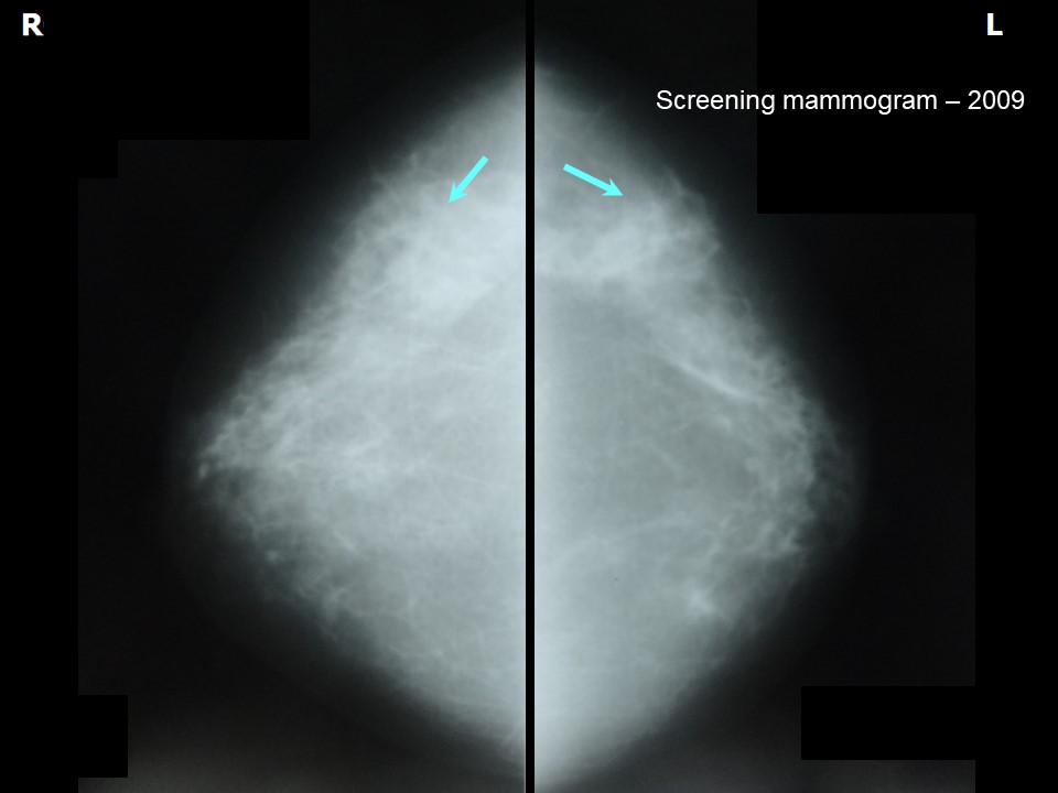
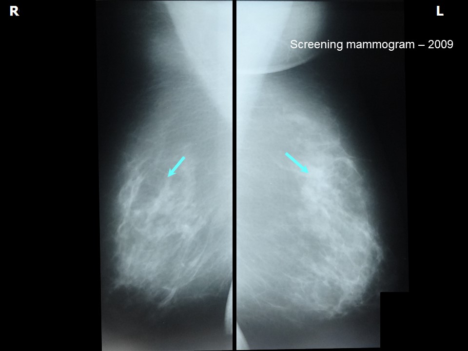
| ‣ Location of the lesion: | Screening mammogram dated 2009: No mass |
| ‣ Mass: | |
| • Number: | 0 |
| • Size: | No |
| • Shape: | None |
| • Margins: | None |
| • Density: | None |
| ‣ Calcifications: | |
| • Typically benign: | None |
| • Suspicious: | None |
| • Distribution: | None |
| ‣ Architectural distortion: | None |
| ‣ Asymmetry: | None |
| ‣ Intramammary node: | None |
| ‣ Skin lesion: | None |
| ‣ Solitary dilated duct: | None |
| ‣ Associated features: | None |
 |  |
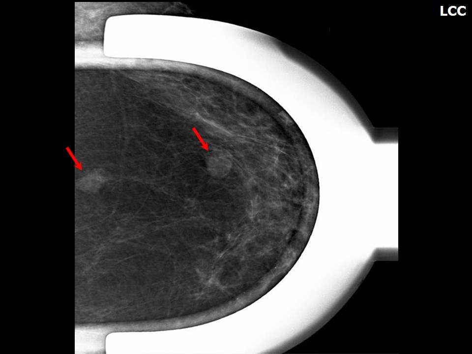 |
| Breast composition: | ACR category b (there are scattered areas of fibroglandular density) | Mammography features: |
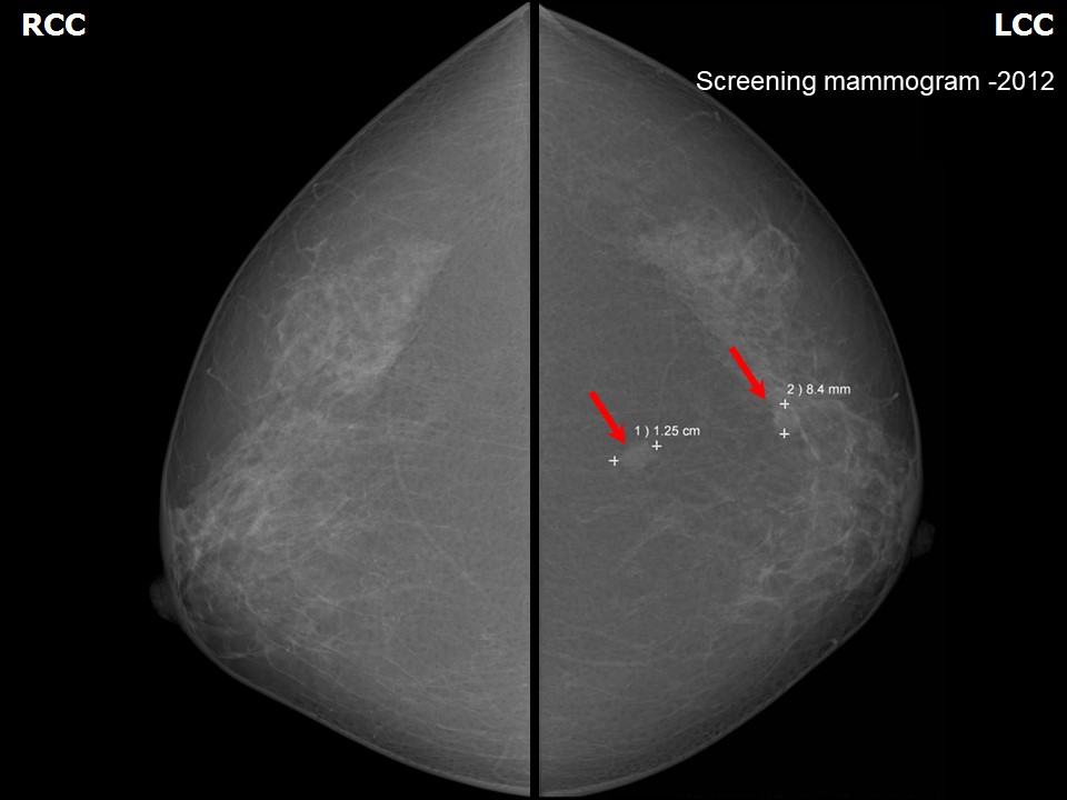
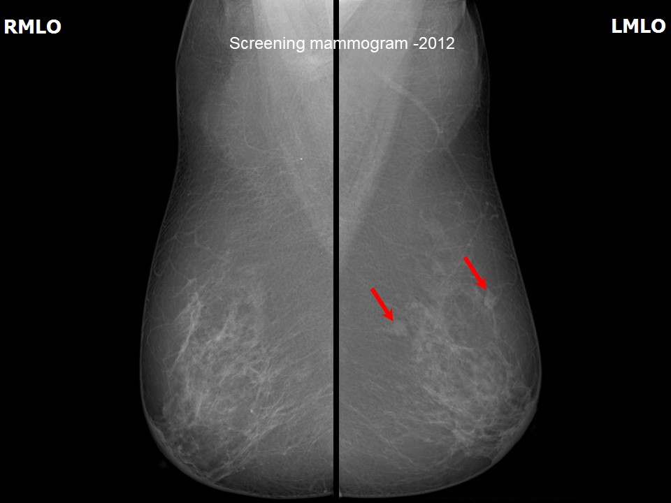
| ‣ Location of the lesion: | Screening mammogram follow-up dated 2012: Left breast, upper outer quadrant at 1 oclock, middle and posterior thirds |
| ‣ Mass: | |
| • Number: | 2 (this is a new finding, developing asymmetry) |
| • Size: | 1.2 cm and 0.8 cm in greatest dimension |
| • Shape: | Irregular |
| • Margins: | Indistinct |
| • Density: | Equal |
| ‣ Calcifications: | |
| • Typically benign: | None |
| • Suspicious: | None |
| • Distribution: | None |
| ‣ Architectural distortion: | None |
| ‣ Asymmetry: | None |
| ‣ Intramammary node: | None |
| ‣ Skin lesion: | None |
| ‣ Solitary dilated duct: | None |
| ‣ Associated features: | None |
Ultrasound:
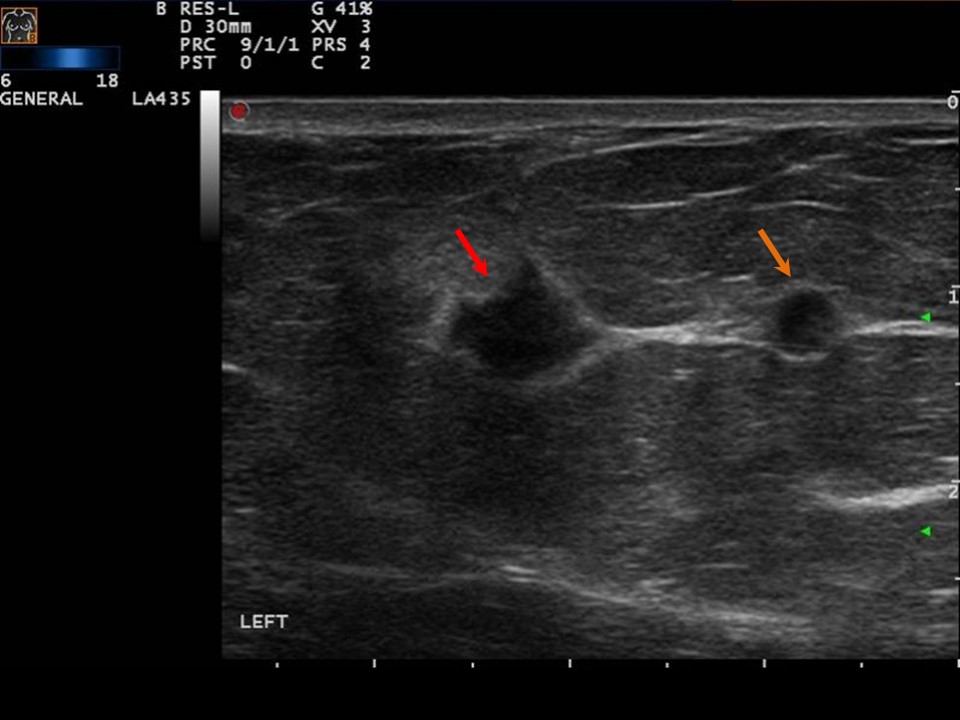 |  |
| Ultrasound features: Left breast, upper outer quadrant at 1 oclock | |
| ‣ Mass | |
| • Location: | Left breast, upper outer quadrant at 1 oclock |
| • Number: | 2 (this is a new finding, developing asymmetry) |
| • Size: | 0.8 cm and 0.4 cm in greatest dimension |
| • Shape: | Irregular |
| • Orientation: | Parallel |
| • Margins: | Spiculated |
| • Echo pattern: | Hypoechoic |
| • Posterior features: | No posterior features |
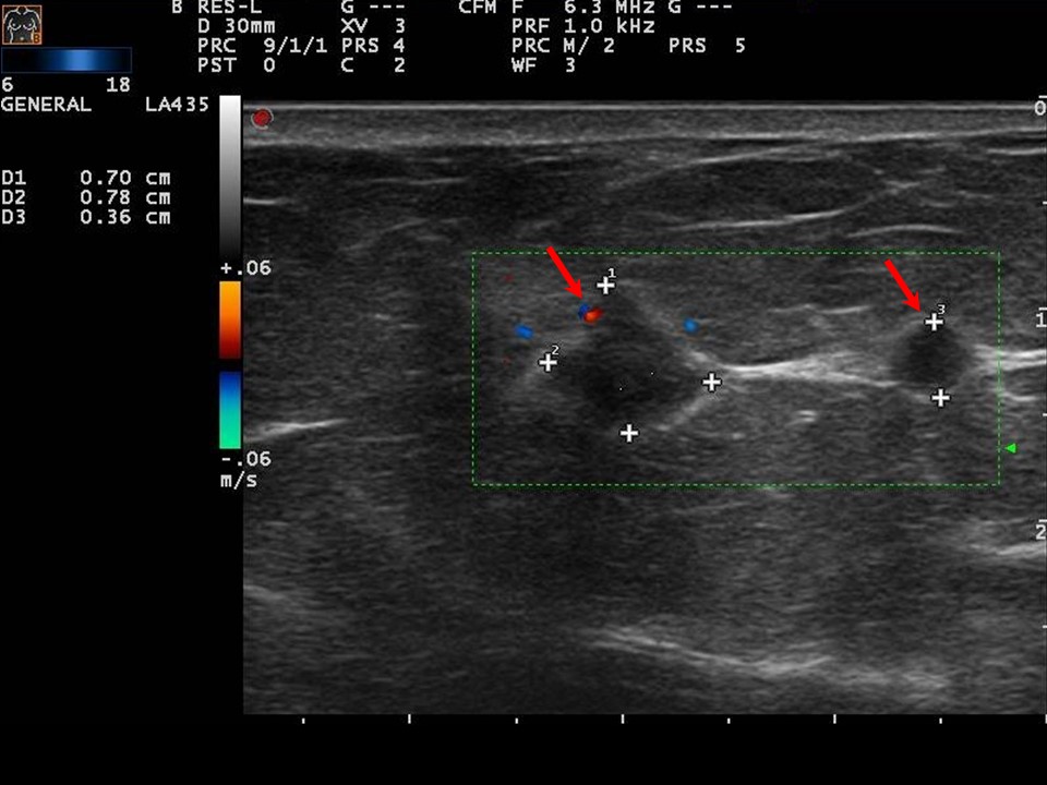
| ‣ Calcifications: | None |
| ‣ Associated features: | Internal vascularity |
| ‣ Special cases: | None |
BI-RADS:
BI-RADS Category (2009): 1 (negative)BI-RADS Category (2012): 4C (high suspicion for malignancy)
Further assessment:
Further assessment advised: Referral for cytologyCytology:
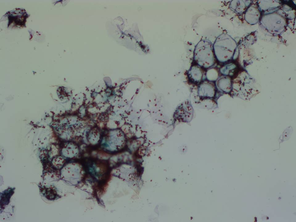 |
| Cytology features: | |
| ‣ Type of sample: | FNAC |
| ‣ Site of biopsy: | |
| • Laterality: | Left |
| • Quadrant: | 1 oclock on ultrasound, non-palpable lesion |
| • Localization technique: | Ultrasound-guided |
| • Nature of aspirate: | Haemorrhagic |
| ‣ Cytological description: | Smears show only RBCs |
| ‣ Reporting category: | Insufficient material |
| ‣ Diagnosis: | Inadequate |
| ‣ Comments: | None |
Histopathology:
Breast-conserving surgery
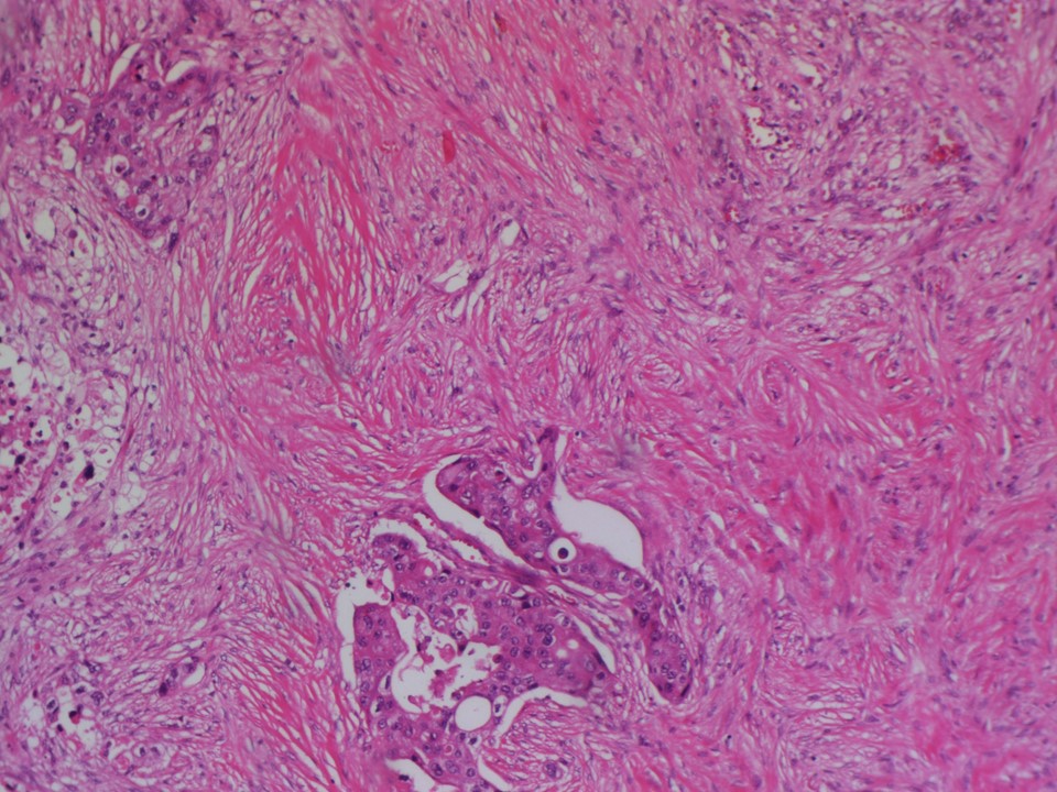 | 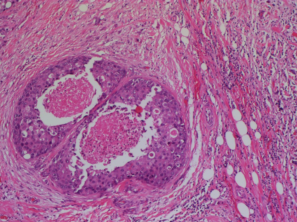 |
| Histopathology features: | |
| ‣ Specimen type: | Breast-conserving surgery |
| ‣ Laterality: | Left |
| ‣ Macroscopy: | On serial sectioning of the lumpectomy specimen, a firm greyish white tumour (0.9 × 0.7 × 0.7 cm) is identified |
| ‣ Histological type: | Invasive breast carcinoma of no special type |
| ‣ Histological grade: | Grade 3 (3 + 3 + 3 = 9) |
| ‣ Mitosis: | 30 |
| ‣ Maximum invasive tumour size: | 0.9 cm |
| ‣ Lymph node status: | 0/19 |
| ‣ Peritumoural lymphovascular invasion: | Present |
| ‣ DCIS/EIC: | DCIS of comedo type, high grade with necrosis |
| ‣ Margins: | Free of tumour |
| ‣ Pathological stage: | pT1bN0 |
| ‣ Biomarkers: | ER positive, PR positive, and HER2/neu negative |
| ‣ Comments: |
Case summary:
| Postmenopausal woman with increased risk of developing breast cancer presented for mammography follow-up screening. Examination did not reveal lumps in either of the breasts or in the axillae. Mammography and breast ultrasound dated 2012 revealed new suspicious irregular lesions with indistinct margins in the left breast. Compared with previous mammogram dated 2009, follow-up screening revealed developing asymmetries in the left breast. Interval change is seen. Diagnosed as suspicious for malignancy, BI-RADS 4C on imaging and as invasive breast carcinoma of no special type, pT1bN0 on histopathology. |
Learning points:
|