


Home / Training / Manuals / Atlas of breast cancer early detection / Cases
Atlas of breast cancer early detection
Filter by language: English / Русский
Go back to the list of case studies
.png) Click on the pictures to magnify and display the legends
Click on the pictures to magnify and display the legends
| Case number: | 096 |
| Age: | 60 |
| Clinical presentation: | Postmenopausal woman with average risk of developing breast cancer presented with nipple discharge from the right nipple of duration 10 days. Examination revealed a lump 4 cm in diameter in the right retroareolar region. |
Mammography:
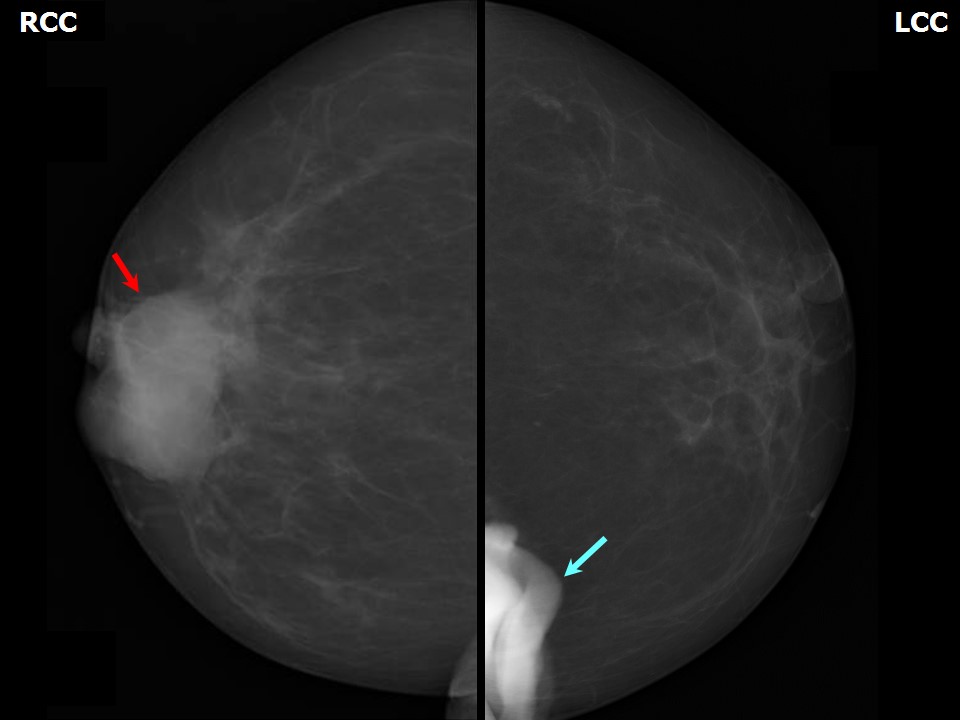 | 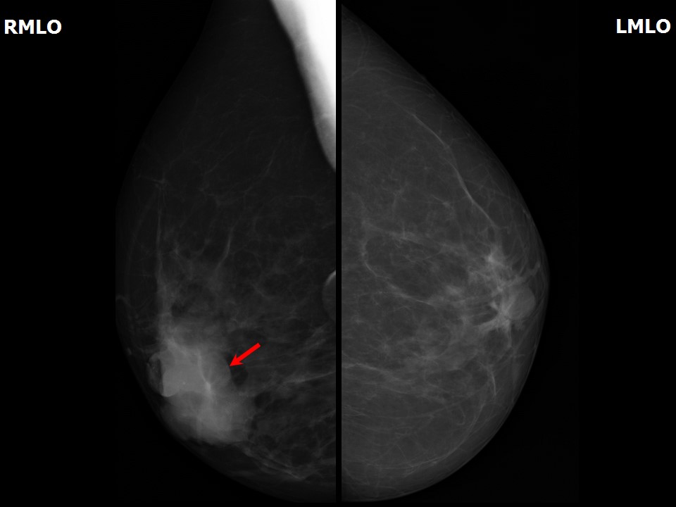 |
| Breast composition: | ACR category a (the breasts are almost entirely fatty) | Mammography features: |
| ‣ Location of the lesion: | Right breast, central portion of the breast, central zone, anterior third |
| ‣ Mass: | |
| • Number: | 1 |
| • Size: | 3.0 × 2.5 cm |
| • Shape: | Irregular |
| • Margins: | Circumscribed |
| • Density: | High |
| ‣ Calcifications: | |
| • Typically benign: | None |
| • Suspicious: | None |
| • Distribution: | None |
| ‣ Architectural distortion: | None |
| ‣ Asymmetry: | None |
| ‣ Intramammary node: | None |
| ‣ Skin lesion: | None |
| ‣ Solitary dilated duct: | None |
| ‣ Associated features: | Trabecular thickening |
Ultrasound:
 | 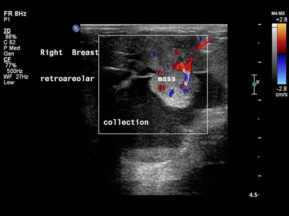 |
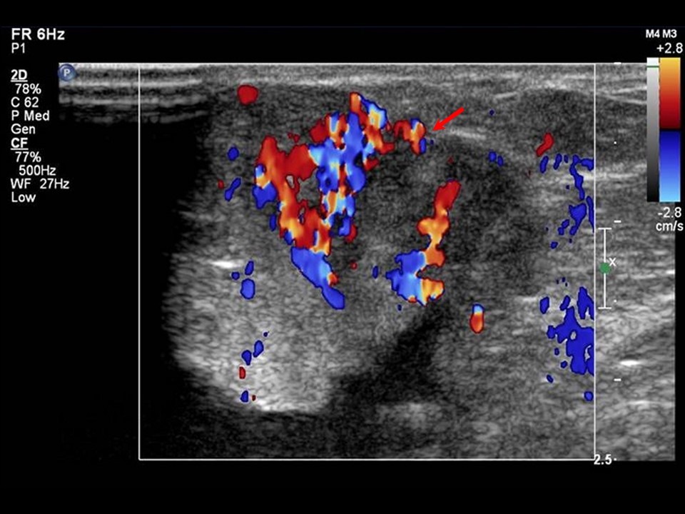 |
| Ultrasound features: Right breast, central portion of the breast | |
| ‣ Mass | |
| • Location: | Right breast, central portion of the breast |
| • Number: | 1 |
| • Size: | 2.1 × 2.0 cm |
| • Shape: | Irregular |
| • Orientation: | Parallel |
| • Margins: | Indistinct |
| • Echo pattern: | Complex cystic and solid |
| • Posterior features: | Posterior shadowing |
| ‣ Calcifications: | None |
| ‣ Associated features: | Internal vascularity |
| ‣ Special cases: | None |
BI-RADS:
BI-RADS Category: 4C (high suspicion for malignancy)Further assessment:
Further assessment advised: Referral for cytologyCytology:
 |
| Cytology features: | |
| ‣ Type of sample: | FNAC (cystic lesion) |
| ‣ Site of biopsy: | |
| • Laterality: | Right |
| • Quadrant: | Inner, retroareolar region |
| • Localization technique: | Palpation |
| • Nature of aspirate: | 0.5 mL of brownish yellow fluid |
| ‣ Cytological description: | Smear shows thick proteinaceous debris in the background with scattered small clusters of benign ductal epithelial cells |
| ‣ Reporting category: | Benign |
| ‣ Diagnosis: | Fibrocystic change, benign |
| ‣ Comments: | None |
Histopathology:
Breast lumpectomy specimen
 | 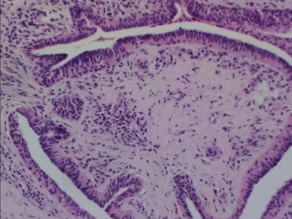 |
| Histopathology features: | |
| ‣ Specimen type: | Breast lumpectomy specimen |
| ‣ Laterality: | Right |
| ‣ Macroscopy: | Specimen 4.0 × 3.0 × 1.5 cm. Cut surface shows two cystic areas (2.0 × 1.0 × 0.5 cm and 0.5 × 0.6 × 0.3 cm) |
| ‣ Histological type: | Multiple sections taken from the entire lumpectomy specimen show changes of epithelial hyperplasia, sclerosing adenosis, and intraductal papillomas. Some areas show densely fibrotic stroma with entrapped small tubules and varying degrees of cyst formation and hyperplasia. A few of the epithelial cells show apocrine metaplasia |
| ‣ Histological grade: | |
| ‣ Mitosis: | |
| ‣ Maximum invasive tumour size: | |
| ‣ Lymph node status: | |
| ‣ Peritumoural lymphovascular invasion: | |
| ‣ DCIS/EIC: | Not identified |
| ‣ Margins: | |
| ‣ Pathological stage: | |
| ‣ Biomarkers: | |
| ‣ Comments: | Negative for malignancy |
Case summary:
| Postmenopausal woman presented with right breast lump. Diagnosed as a complex cystic and solid mass in the subareolar region of the right breast, BI-RADS 4C on imaging, as benign fibrocystic change on cytology, and as epithelial hyperplasia, sclerosing adenosis, and intraductal papillomas, negative for malignancy on histopathology. |
Learning points:
|