


Home / Training / Manuals / Atlas of breast cancer early detection / Cases
Atlas of breast cancer early detection
Filter by language: English / Русский
Go back to the list of case studies
.png) Click on the pictures to magnify and display the legends
Click on the pictures to magnify and display the legends
| Case number: | 090 |
| Age: | 27 |
| Clinical presentation: | Premenopausal woman with average risk of developing breast cancer presented with pain in the right breast. Examination revealed a lump measuring 1.5 cm in diameter in the right upper outer quadrant. |
Ultrasound:
 |  |
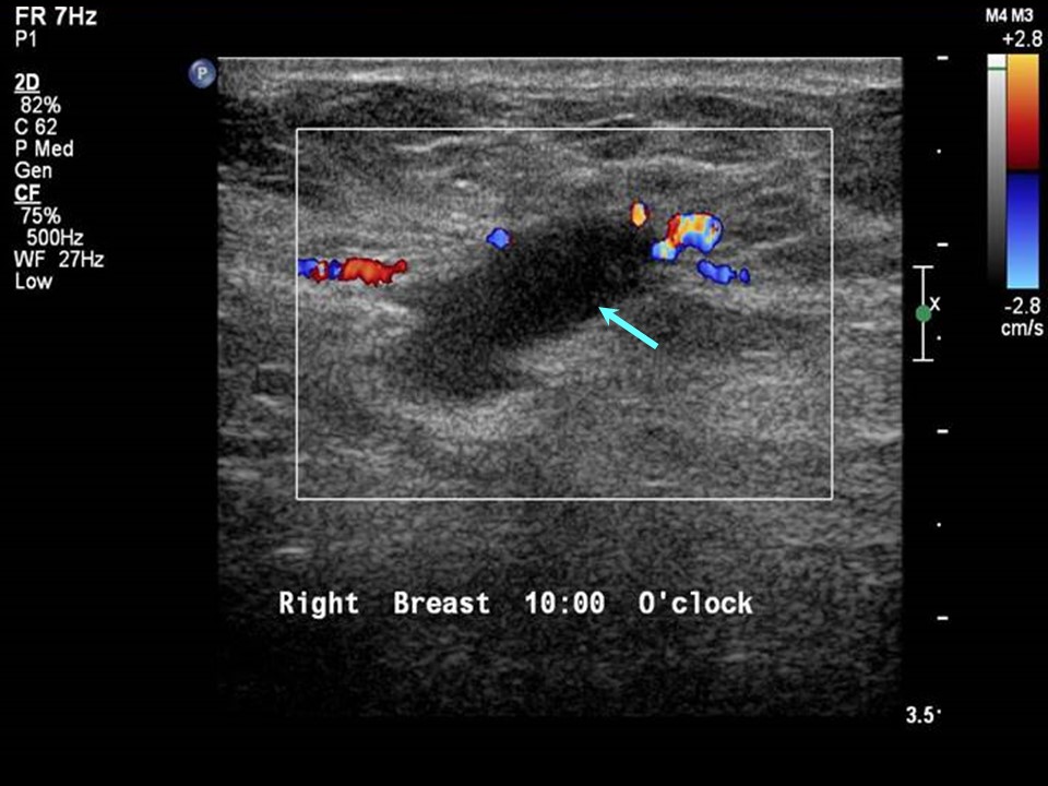 |
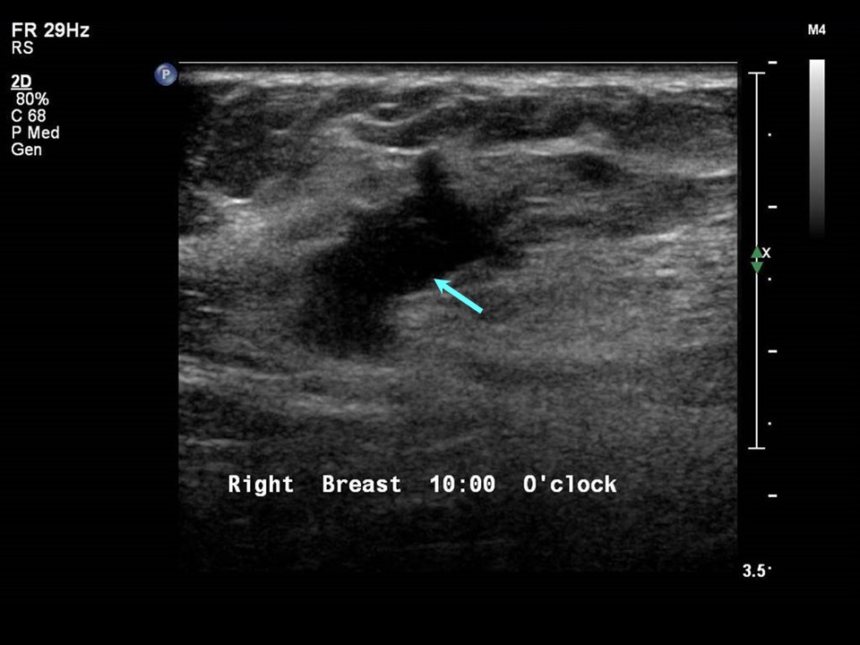
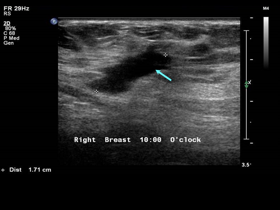
| Ultrasound features: Right breast, upper outer quadrant, 10 oclock in axillary tail | |
| ‣ Mass | |
| • Location: | Right breast, upper outer quadrant, 10 oclock in axillary tail |
| • Number: | 1 |
| • Size: | 1.7 × 1.0 cm |
| • Shape: | Irregular |
| • Orientation: | Not parallel |
| • Margins: | Angular |
| • Echo pattern: | Hypoechoic |
| • Posterior features: | No posterior features |
| ‣ Calcifications: | None |
| ‣ Associated features: | Peripheral vascularity and vessels in rim |
| ‣ Special cases: | None |
BI-RADS:
BI-RADS Category: 2 (benign)Further assessment:
Further assessment advised: Referral for cytologyCytology:
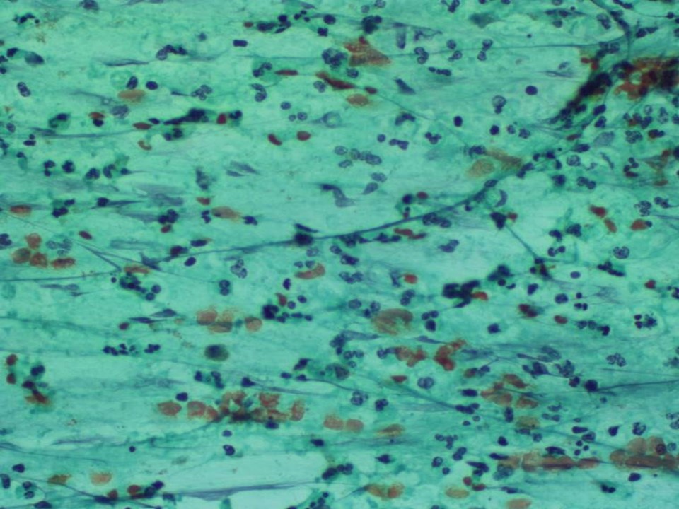 |
| Cytology features: | |
| ‣ Type of sample: | FNAC (solid lesion) |
| ‣ Site of biopsy: | |
| • Laterality: | Right |
| • Quadrant: | Upper outer |
| • Localization technique: | Palpation |
| • Nature of aspirate: | 2 mL of purulent yellowish white material |
| ‣ Cytological description: | Smears show plenty of neutrophils admixed with a few lymphocytes on a necrotic background |
| ‣ Reporting category: | Benign |
| ‣ Diagnosis: | Acute inflammation and breast abscess |
| ‣ Comments: | None |
Case summary:
| Premenopausal woman presented with right breast pain. Diagnosed as inflammatory lesion in right breast, BI-RADS 2 on imaging and as acute inflammation, breast abscess on cytology. |
Learning points:
|