


Home / Training / Manuals / Atlas of breast cancer early detection / Cases
Atlas of breast cancer early detection
Filter by language: English / Русский
Go back to the list of case studies
.png) Click on the pictures to magnify and display the legends
Click on the pictures to magnify and display the legends
| Case number: | 084 |
| Age: | 71 |
| Clinical presentation: | Postmenopausal woman after surgery for right breast carcinoma. On postoperative surveillance, examination revealed a palpable right breast lump at the operated site. |
Mammography:
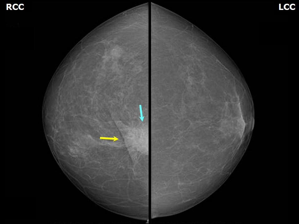 | 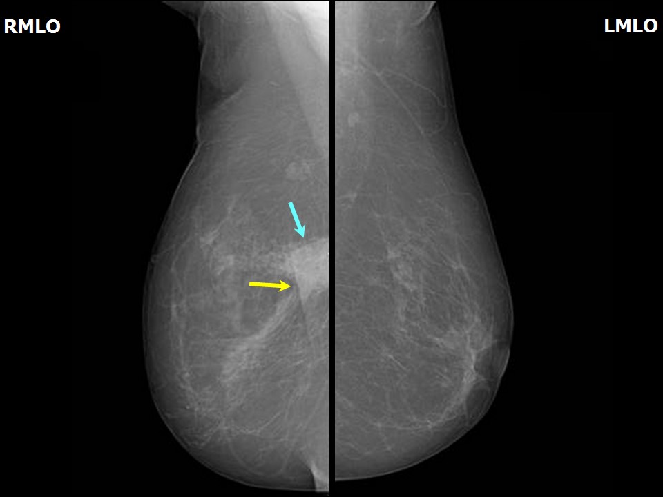 |
| Breast composition: | ACR category a (the breasts are almost entirely fatty) | Mammography features: |
| ‣ Location of the lesion: | Right breast, upper inner quadrant at 1 oclock, posterior third, beneath surgical scar |
| ‣ Mass: | |
| • Number: | 1 |
| • Size: | 3.0 × 1.5 cm |
| • Shape: | Oval |
| • Margins: | Indistinct |
| • Density: | High |
| ‣ Calcifications: | |
| • Typically benign: | None |
| • Suspicious: | None |
| • Distribution: | None |
| ‣ Architectural distortion: | Present |
| ‣ Asymmetry: | Focal |
| ‣ Intramammary node: | None |
| ‣ Skin lesion: | None |
| ‣ Solitary dilated duct: | None |
| ‣ Associated features: | Skin retraction, architectural distortion, and trabecular thickening in postoperative breast |
Ultrasound:
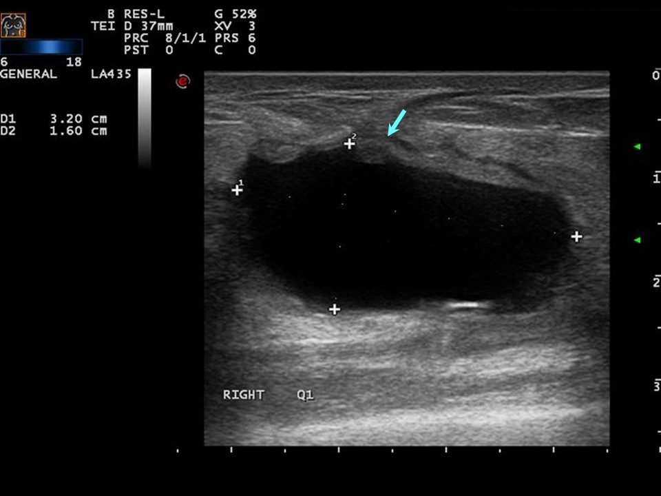 |
| Ultrasound features: Right breast, upper inner quadrant, 1 oclock position | |
| ‣ Mass | |
| • Location: | Right breast, upper inner quadrant, 1 oclock position |
| • Number: | 1 |
| • Size: | 3.2 × 1.6 cm |
| • Shape: | Oval |
| • Orientation: | Not parallel |
| • Margins: | Indistinct |
| • Echo pattern: | Anechoic |
| • Posterior features: | Posterior shadowing |
| ‣ Calcifications: | None |
| ‣ Associated features: | Postoperative fluid collection, architectural distortion, and absent vascularity |
| ‣ Special cases: | None |
BI-RADS:
BI-RADS Category (Right BCS, post operative breast): 2 (benign)Further assessment:
Further assessment advised: Referral for cytologyCytology:
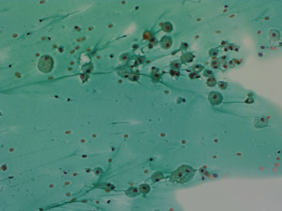 |
| Cytology features: | |
| ‣ Type of sample: | FNAC (cystic lesion) |
| ‣ Site of biopsy: | |
| • Laterality: | Right |
| • Quadrant: | Upper inner |
| • Localization technique: | Palpation |
| • Nature of aspirate: | 10 mL of yellowish fluid |
| ‣ Cytological description: | Smears do not show any ductal epithelial cells. Many histiocytes with abundant foamy cytoplasm are seen on a proteinaceous background. Inflammatory cells are not seen |
| ‣ Reporting category: | Benign |
| ‣ Diagnosis: | Consistent with seroma |
| ‣ Comments: | None |
Case summary:
| Postmenopausal woman after right breast-conserving surgery on postoperative surveillance presented with right breast lump at site of surgery. Diagnosed as organized seroma in right breast with surgical scar, BI-RADS 2 on imaging and as right breast seroma collection on cytology. |
Learning points:
|