


Home / Training / Manuals / Atlas of breast cancer early detection / Cases
Atlas of breast cancer early detection
Filter by language: English / Русский
Go back to the list of case studies
.png) Click on the pictures to magnify and display the legends
Click on the pictures to magnify and display the legends
| Case number: | 065 |
| Age: | 60 |
| Clinical presentation: | Postmenopausal woman with average risk of developing breast cancer presented with painless right breast lump. On examination, a hard non-tender lump was found in the right breast. |
Mammography:
 | 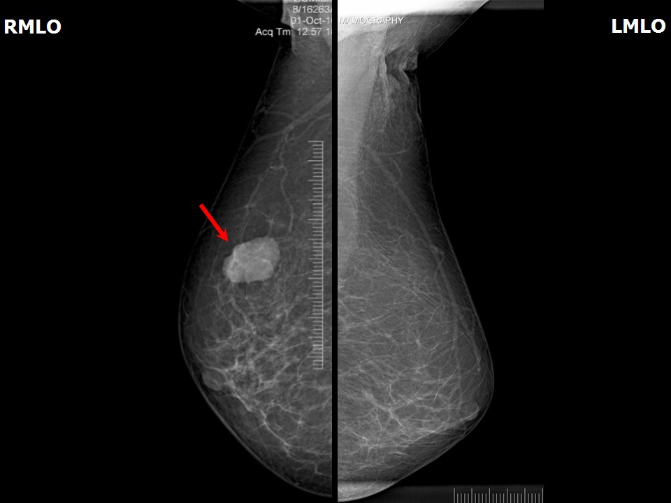 |
| Breast composition: | ACR category a (the breasts are almost entirely fatty) | Mammography features: |
| ‣ Location of the lesion: | Right breast, upper outer quadrant at 10 oclock, middle third |
| ‣ Mass: | |
| • Number: | 1 |
| • Size: | 3.1 × 2.2 cm |
| • Shape: | Oval |
| • Margins: | Partly circumscribed and partly indistinct |
| • Density: | High |
| ‣ Calcifications: | |
| • Typically benign: | None |
| • Suspicious: | None |
| • Distribution: | None |
| ‣ Architectural distortion: | None |
| ‣ Asymmetry: | None |
| ‣ Intramammary node: | None |
| ‣ Skin lesion: | None |
| ‣ Solitary dilated duct: | None |
| ‣ Associated features: | None |
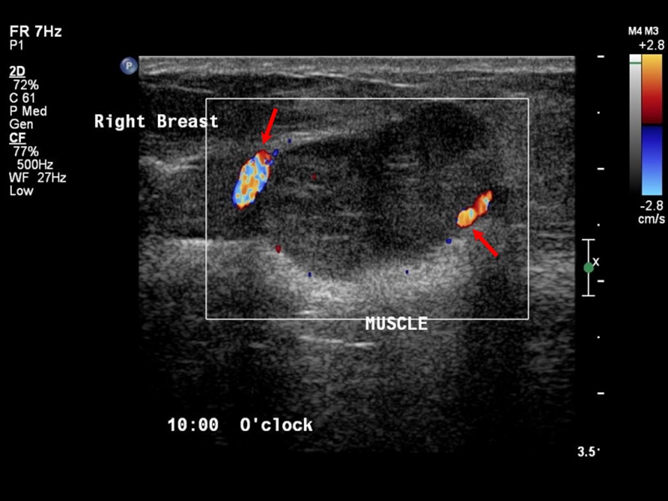
Ultrasound:
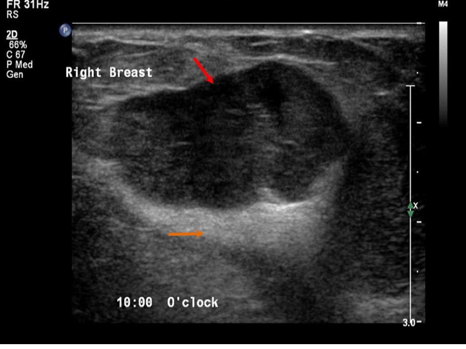 |  |
| Ultrasound features: Right breast, upper outer quadrant at 10 oclock | |
| ‣ Mass | |
| • Location: | Right breast, upper outer quadrant at 10 oclock |
| • Number: | 1 |
| • Size: | 3.0 × 1.4 cm |
| • Shape: | Irregular |
| • Orientation: | Parallel |
| • Margins: | Partly circumscribed and partly indistinct margins |
| • Echo pattern: | Hypoechoic |
| • Posterior features: | Posterior shadowing |
| ‣ Calcifications: | None |
| ‣ Associated features: | Internal vascularity |
| ‣ Special cases: | None |
BI-RADS:
BI-RADS Category: 4B (moderate suspicion of malignancy)Further assessment:
Further assessment advised: Referral for cytologyCytology:
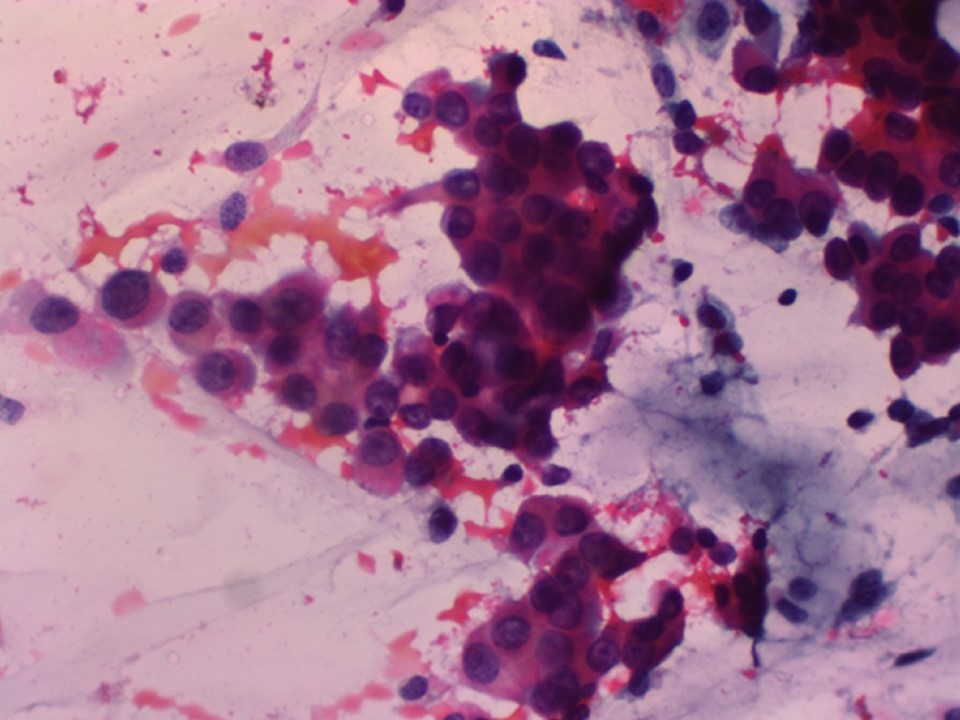 |
| Cytology features: | |
| ‣ Type of sample: | FNAC (solid lesion) |
| ‣ Site of biopsy: | |
| • Laterality: | Right |
| • Quadrant: | |
| • Localization technique: | Palpation |
| • Nature of aspirate: | Mucoid |
| ‣ Cytological description: | Smears show malignant cells with a uniform regular nucleus; many cells are plasmacytoid with eccentric nuclei. Background shows mucinous material |
| ‣ Reporting category: | Malignant |
| ‣ Diagnosis: | Carcinoma |
| ‣ Comments: | None |
Histopathology:
MRM
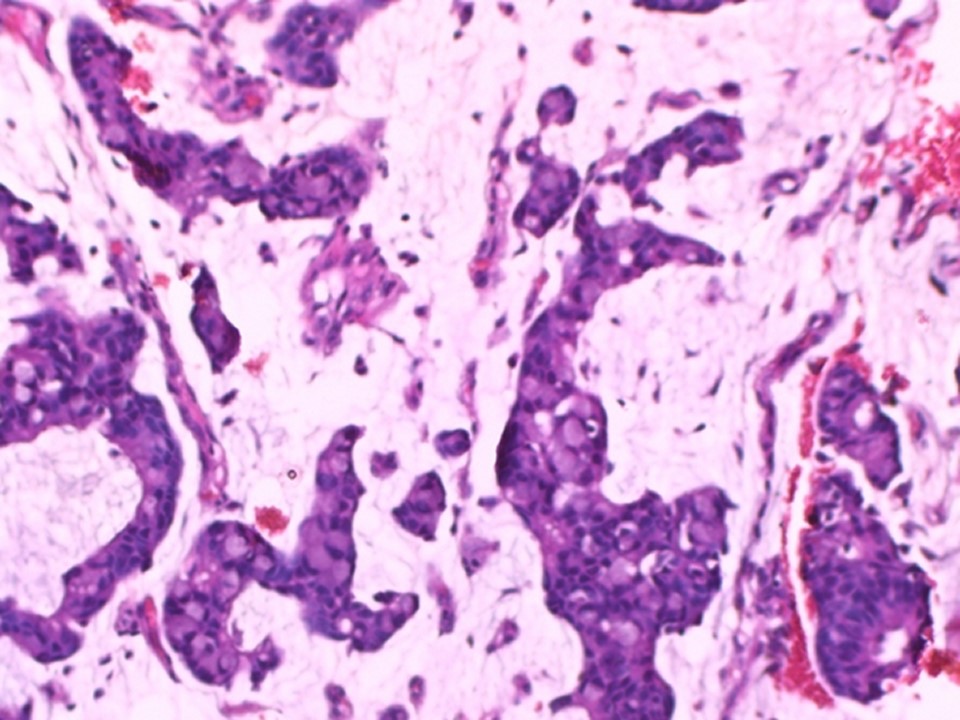 |
| Histopathology features: | |
| ‣ Specimen type: | MRM |
| ‣ Laterality: | Right |
| ‣ Macroscopy: | Cut section shows a well-circumscribed greyish white tumour (2.2 × 2.0 × 1.5 cm), with mucinous areas of soft consistency |
| ‣ Histological type: | Mucinous |
| ‣ Histological grade: | Grade 1 (3 + 1 + 1 = 5) |
| ‣ Mitosis: | Not identified |
| ‣ Maximum invasive tumour size: | 2.2 cm in greatest dimension |
| ‣ Lymph node status: | 0/22 |
| ‣ Peritumoural lymphovascular invasion: | Not identified |
| ‣ DCIS/EIC: | Not identified |
| ‣ Margins: | Free of tumour |
| ‣ Pathological stage: | pT2N0 |
| ‣ Biomarkers: | |
| ‣ Comments: |
Case summary:
| Postmenopausal woman presented with lump in the right breast. Diagnosed as right breast mass of suspicious morphology, BI-RADS 4B on imaging, as mucinous carcinoma on cytology, and as mucinous carcinoma, pT2N0 on histopathology. |
Learning points:
|