


Home / Training / Manuals / Atlas of breast cancer early detection / Cases
Atlas of breast cancer early detection
Filter by language: English / Русский
Go back to the list of case studies
.png) Click on the pictures to magnify and display the legends
Click on the pictures to magnify and display the legends
| Case number: | 057 |
| Age: | 70 |
| Clinical presentation: | Postmenopausal woman with average risk of developing breast cancer presented with lump of duration 34 months in the left breast. Subsequently she also developed discharge from the left nipple. Examination revealed a 1 × 2 cm lump in the inner lower quadrant of the left breast. Nipple discharge was serous in nature. |
Mammography:
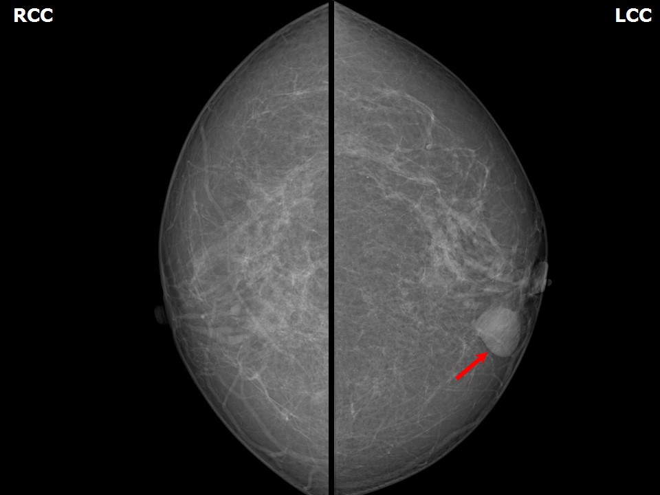 | 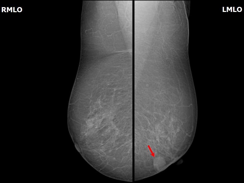 |
| Breast composition: | ACR category b (there are scattered areas of fibroglandular density) | Mammography features: |
| ‣ Location of the lesion: | Left breast, lower inner quadrant at 78 oclock, anterior third |
| ‣ Mass: | |
| • Number: | 1 |
| • Size: | 2.0 × 1.1 cm |
| • Shape: | Oval |
| • Margins: | Circumscribed |
| • Density: | Equal |
| ‣ Calcifications: | |
| • Typically benign: | None |
| • Suspicious: | None |
| • Distribution: | None |
| ‣ Architectural distortion: | None |
| ‣ Asymmetry: | None |
| ‣ Intramammary node: | None |
| ‣ Skin lesion: | None |
| ‣ Solitary dilated duct: | Present |
| ‣ Associated features: | None |
Ultrasound:
 | 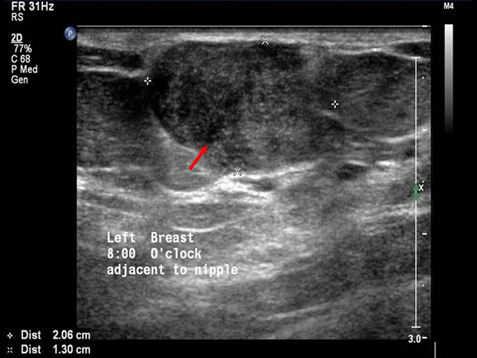 |
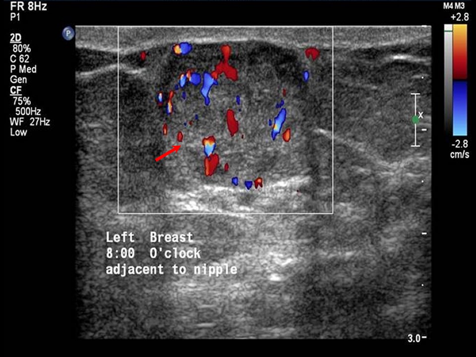 |
| Ultrasound features: Left breast, lower inner quadrant at 8 oclock | |
| ‣ Mass | |
| • Location: | Left breast, lower inner quadrant at 8 oclock |
| • Number: | 1 |
| • Size: | 2.0 × 1.3 cm |
| • Shape: | Irregular |
| • Orientation: | Parallel |
| • Margins: | Indistinct |
| • Echo pattern: | Heteroechoic |
| • Posterior features: | No posterior features |
| ‣ Calcifications: | None |
| ‣ Associated features: | Duct changes: Ectatic duct seen adjacent to mass lesion. Internal vascularity within mass |
| ‣ Special cases: | None |
BI-RADS:
BI-RADS Category: 4B (moderate suspicion of malignancy)Further assessment:
Further assessment advised: Referral for cytologyCytology:
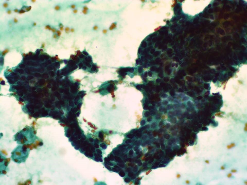 |
| Cytology features: | |
| ‣ Type of sample: | FNAC |
| ‣ Site of biopsy: | |
| • Laterality: | Left |
| • Quadrant: | |
| • Localization technique: | Palpation |
| • Nature of aspirate: | whitish |
| ‣ Cytological description: | Smears are cellular and show many clusters and sheets of ductal epithelial cells arranged as tight monolayered clusters. A few clusters show branching and papillary configuration and a few are loosely cohesive and show mild atypia. A few myoepithelial cells are seen. A few clusters show apocrine metaplastic change. Background shows proteinaceous material with many macrophages. |
| ‣ Reporting category: | Atypical, probably benign |
| ‣ Diagnosis: | Atypia, papillary lesion. Advised frozen section or excision to rule out malignancy |
| ‣ Comments: | None |
Histopathology:
Lumpectomy
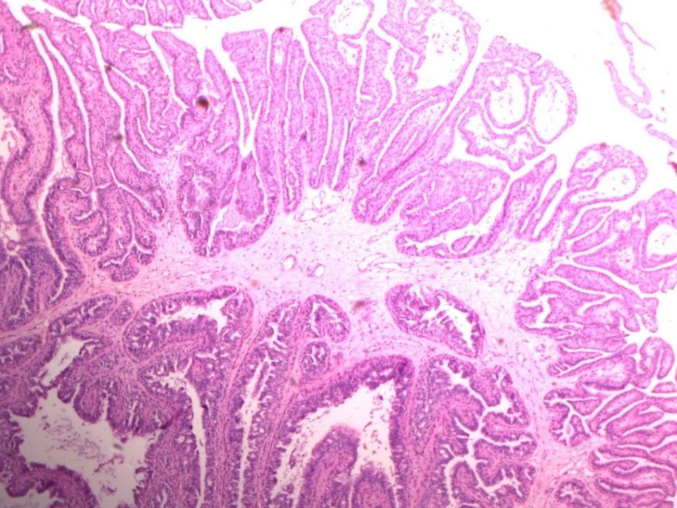 | 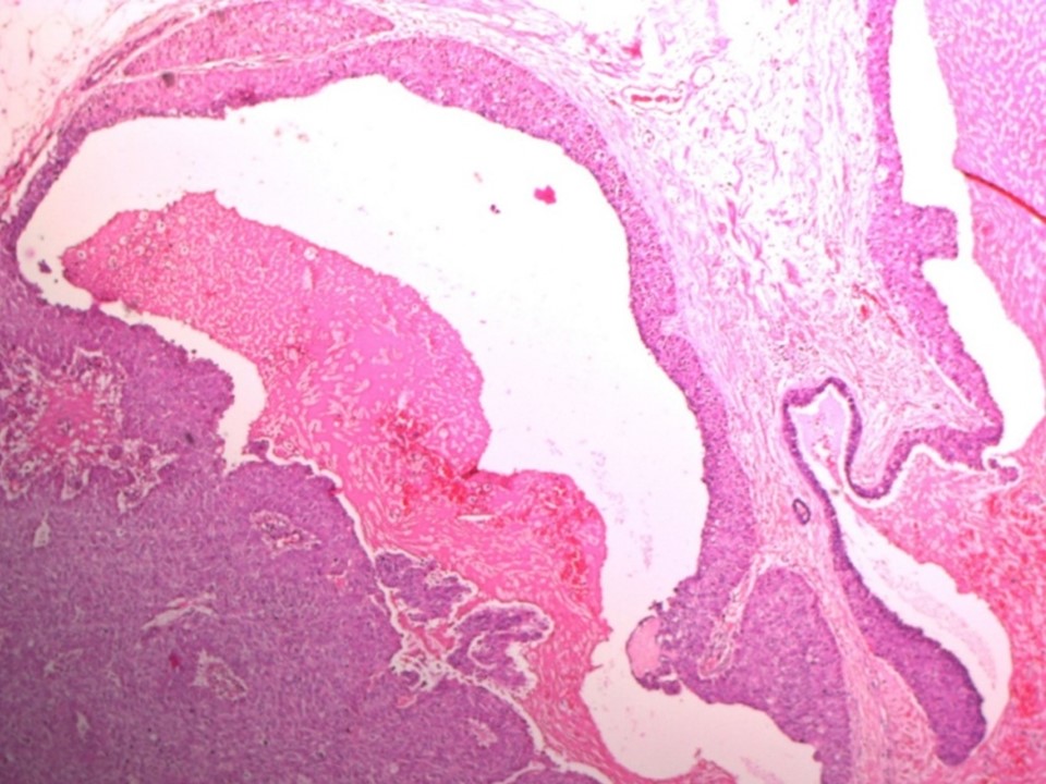 |
| Histopathology features: | |
| ‣ Specimen type: | Lumpectomy |
| ‣ Laterality: | Left |
| ‣ Macroscopy: | Specimen (8.0 × 5.0 × 5.5 cm) with a skin flap (5.0 × 1.0 cm). Skin shows a scar 3.5 cm in length. Cut surface shows central, stellate scar (2.5 × 2.0 × 2.5 cm) surrounded by fat. Scar extends to the skin. Base is 1.5 cm from scar |
| ‣ Histological type: | There is no invasive component of the lesion |
| ‣ Histological grade: | |
| ‣ Mitosis: | |
| ‣ Maximum invasive tumour size: | |
| ‣ Lymph node status: | |
| ‣ Peritumoural lymphovascular invasion: | |
| ‣ DCIS/EIC: | Multiple foci of intraductal papillary carcinoma (papillary DCIS of intermediate nuclear grade) |
| ‣ Margins: | |
| ‣ Pathological stage: | |
| ‣ Biomarkers: | |
| ‣ Comments: | Initially, surgical excision of the mass with frozen section was done followed by lumpectomy. Two fibrofatty bits (5.5 × 4.0 × 1.5 cm and 3.0 × 3.5 × 1.0 cm). Cut surface yellowish white with a few firm areas In addition to DCIS, there are numerous large ectatic ducts with intraductal papillomas. ER and PR are strongly positive in the papillomas |
Case summary:
| Postmenopausal woman presented with left breast lump and serous nipple discharge. Diagnosed as left breast subareolar dilated duct with intraductal mass at terminal end, BI-RADS 4B on imaging, as atypia, papillary lesion of the left breast on cytology, and as multiple foci of intraductal papillary carcinoma and numerous large ectatic ducts with intraductal papillomas on histopathology. |
Learning points:
|