


Home / Training / Manuals / Atlas of breast cancer early detection / Cases
Atlas of breast cancer early detection
Filter by language: English / Русский
Go back to the list of case studies
.png) Click on the pictures to magnify and display the legends
Click on the pictures to magnify and display the legends
| Case number: | 044 |
| Age: | 65 |
| Clinical presentation: | Postmenopausal woman with average risk of developing breast cancer presented with a left breast lump. Examination revealed a hard lump, 6 cm in diameter, in the upper quadrant of the left breast. |
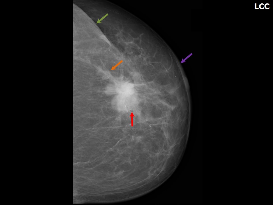
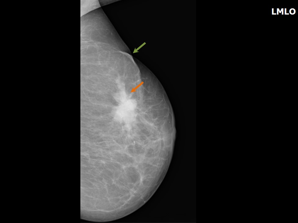
Mammography:
 |  |
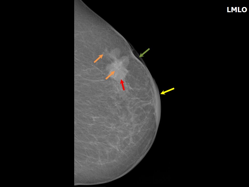 |
| Breast composition: | ACR category b (there are scattered areas of fibroglandular density) | Mammography features: |
| ‣ Location of the lesion: | Left breast, central portion of the breast at 12 oclock, anterior and middle thirds |
| ‣ Mass: | |
| • Number: | 1 |
| • Size: | 6.4 × 3.3 cm |
| • Shape: | Irregular |
| • Margins: | Spiculated |
| • Density: | High |
| ‣ Calcifications: | |
| • Typically benign: | None |
| • Suspicious: | Fine pleomorphic |
| • Distribution: | Grouped and fine-linear branching |
| ‣ Architectural distortion: | None |
| ‣ Asymmetry: | None |
| ‣ Intramammary node: | None |
| ‣ Skin lesion: | None |
| ‣ Solitary dilated duct: | None |
| ‣ Associated features: | Skin retraction and grouped and fine-linear branching calcifications |
Ultrasound:
 | 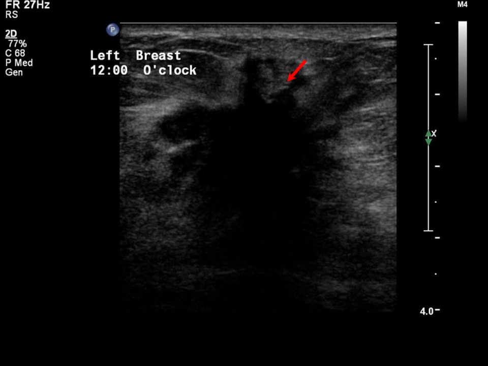 |
| Ultrasound features: Left breast, upper quadrants at 12 oclock | |
| ‣ Mass | |
| • Location: | Left breast, upper quadrants at 12 oclock |
| • Number: | 1 |
| • Size: | 5.0 × 2.0 cm |
| • Shape: | Irregular |
| • Orientation: | Not parallel |
| • Margins: | Angular and spiculated |
| • Echo pattern: | Heterogeneous |
| • Posterior features: | Posterior shadowing |
| ‣ Calcifications: | Present in mass |
| ‣ Associated features: | Internal vascularity, skin thickening, and microcalcifications |
| ‣ Special cases: | None |
BI-RADS:
BI-RADS Category: 5 (highly suggestive of malignancy)Further assessment:
Further assessment advised: Referral for cytology and for core biopsyCytology:
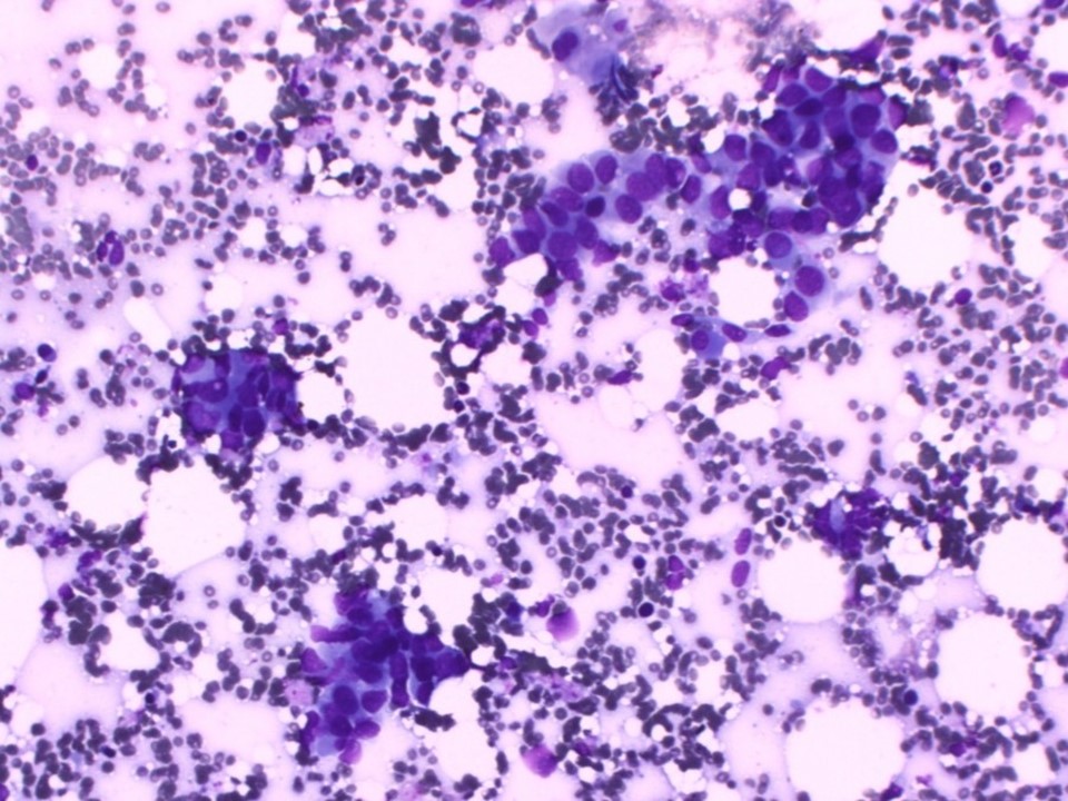 |
| Cytology features: | |
| ‣ Type of sample: | FNAC |
| ‣ Site of biopsy: | |
| • Laterality: | Left |
| • Quadrant: | |
| • Localization technique: | Palpation |
| • Nature of aspirate: | 0.5 mL of thick whitish material |
| ‣ Cytological description: | Smears show many dyscohesive clusters and a few dissociated cells with a high N:C ratio and hyperchromatic, pleomorphic nucleoli with coarse chromatin. Background shows many RBCs and proteinaceous material |
| ‣ Reporting category: | Malignant |
| ‣ Diagnosis: | Carcinoma |
| ‣ Comments: | None |
Histopathology:
Core needle biopsy
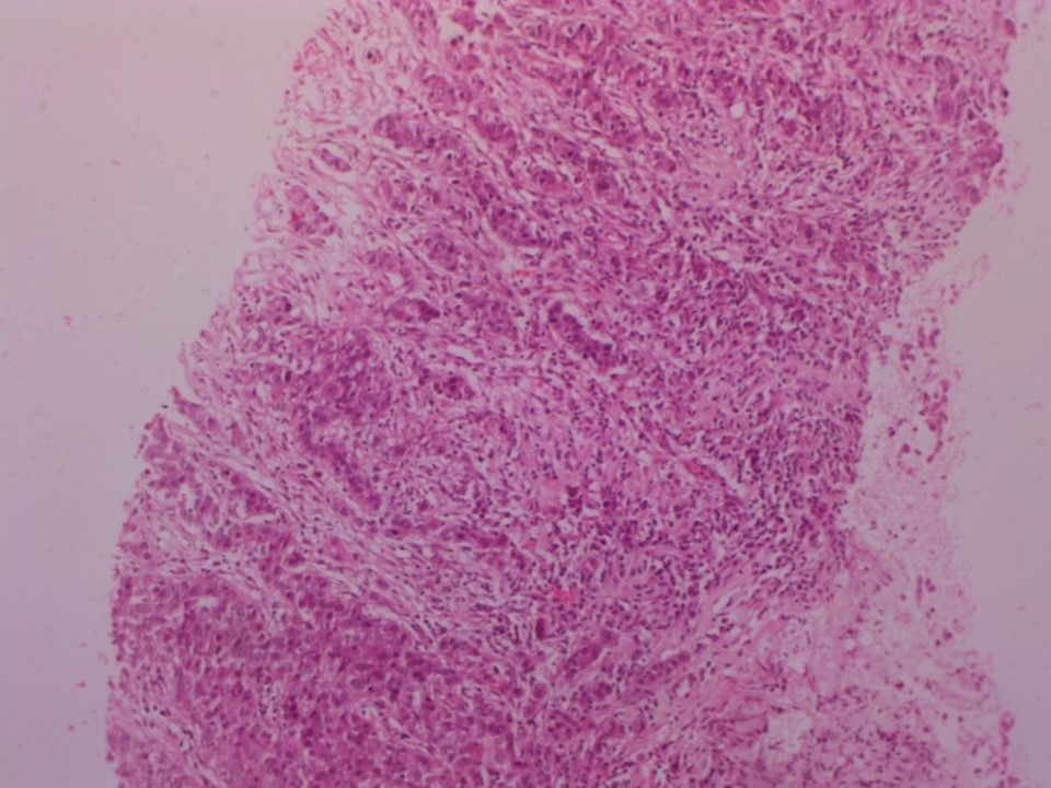 |
| Histopathology features: | |
| ‣ Specimen type: | Core needle biopsy |
| ‣ Laterality: | Left |
| ‣ Macroscopy: | Three whitish tissue cores, each 12 mm in length |
| ‣ Histological type: | Invasive breast carcinoma of no special type |
| ‣ Histological grade: | Grade 2 (3 + 2 + 2 = 7) |
| ‣ Mitosis: | 14 |
| ‣ Maximum invasive tumour size: | |
| ‣ Lymph node status: | |
| ‣ Peritumoural lymphovascular invasion: | |
| ‣ DCIS/EIC: | |
| ‣ Margins: | |
| ‣ Pathological stage: | |
| ‣ Biomarkers: | |
| ‣ Comments: |
Case summary:
| Postmenopausal woman presented with left breast lump. Diagnosed as left breast carcinoma with fine pleomorphic, grouped and fine-linear branching calcifications within, and overlying skin thickening and retraction, BI-RADS 5 on imaging, as carcinoma of the left breast on cytology, and as invasive breast carcinoma of no special type on core biopsy histopathology. |
Learning points:
|