


Home / Training / Manuals / Atlas of breast cancer early detection / Cases
Atlas of breast cancer early detection
Filter by language: English / Русский
Go back to the list of case studies
.png) Click on the pictures to magnify and display the legends
Click on the pictures to magnify and display the legends
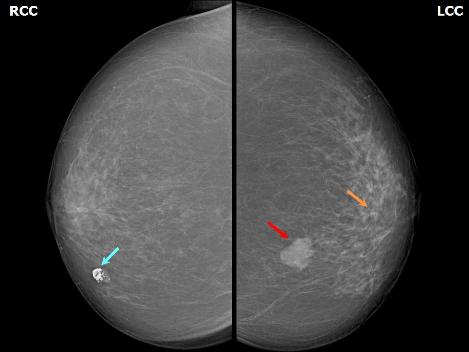
BI-RADS Category (Right breast): 2 (benign)
| Case number: | 039 |
| Age: | 77 |
| Clinical presentation: | Postmenopausal and post hysterectomy, nulliparous woman with increased risk for developing breast cancer presented with a lump in the left breast of 6 months duration. Examination revealed hard, mobile lump in the lower inner quadrant of the left breast. |
Mammography:
 |  |
 |  |
| Breast composition: | ACR category b (there are scattered areas of fibroglandular density) | Mammography features: |
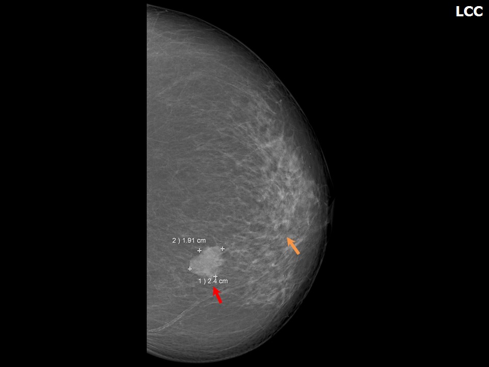
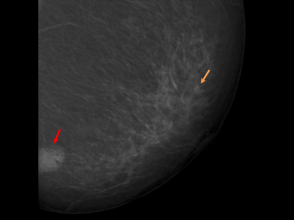
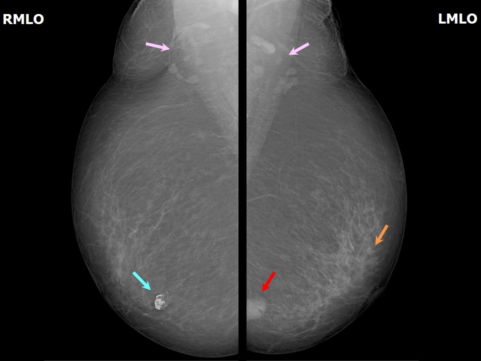
| ‣ Location of the lesion: | Lesion 1: Left breast, lower inner quadrant at 7 oclock, middle third |
| ‣ Mass: | |
| • Number: | 1 |
| • Size: | 2.0 × 1.5 cm |
| • Shape: | Irregular |
| • Margins: | Indistinct |
| • Density: | High |
| ‣ Calcifications: | |
| • Typically benign: | Vascular calcification |
| • Suspicious: | None |
| • Distribution: | None |
| ‣ Architectural distortion: | None |
| ‣ Asymmetry: | None |
| ‣ Intramammary node: | None |
| ‣ Skin lesion: | None |
| ‣ Solitary dilated duct: | None |
| ‣ Associated features: | None |
| Breast composition: | ACR category b (there are scattered areas of fibroglandular density) | Mammography features: |
| ‣ Location of the lesion: | Lesion 2: Left breast, central portion of the breast, lower outer quadrant at 5 oclock, anterior third |
| ‣ Mass: | |
| • Number: | 1 |
| • Size: | 0.7 cm in greatest dimension |
| • Shape: | Irregular |
| • Margins: | Indistinct |
| • Density: | High |
| ‣ Calcifications: | |
| • Typically benign: | None |
| • Suspicious: | None |
| • Distribution: | None |
| ‣ Architectural distortion: | None |
| ‣ Asymmetry: | None |
| ‣ Intramammary node: | None |
| ‣ Skin lesion: | None |
| ‣ Solitary dilated duct: | None |
| ‣ Associated features: | None |
| Breast composition: | ACR category b (there are scattered areas of fibroglandular density) | Mammography features: |
| ‣ Location of the lesion: | Right breast, lower inner quadrant at 5 oclock, middle third |
| ‣ Mass: | |
| • Number: | 1 |
| • Size: | 1.2 cm in greatest dimension |
| • Shape: | Oval |
| • Margins: | Circumscribed |
| • Density: | Equal |
| ‣ Calcifications: | |
| • Typically benign: | Coarse, popcorn-like |
| • Suspicious: | None |
| • Distribution: | None |
| ‣ Architectural distortion: | None |
| ‣ Asymmetry: | None |
| ‣ Intramammary node: | None |
| ‣ Skin lesion: | None |
| ‣ Solitary dilated duct: | None |
| ‣ Associated features: | Coarse, popcorn-like calcification |
Ultrasound:
 |  |
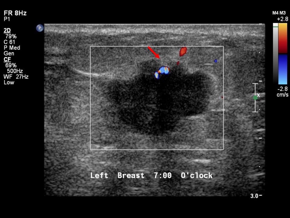 |
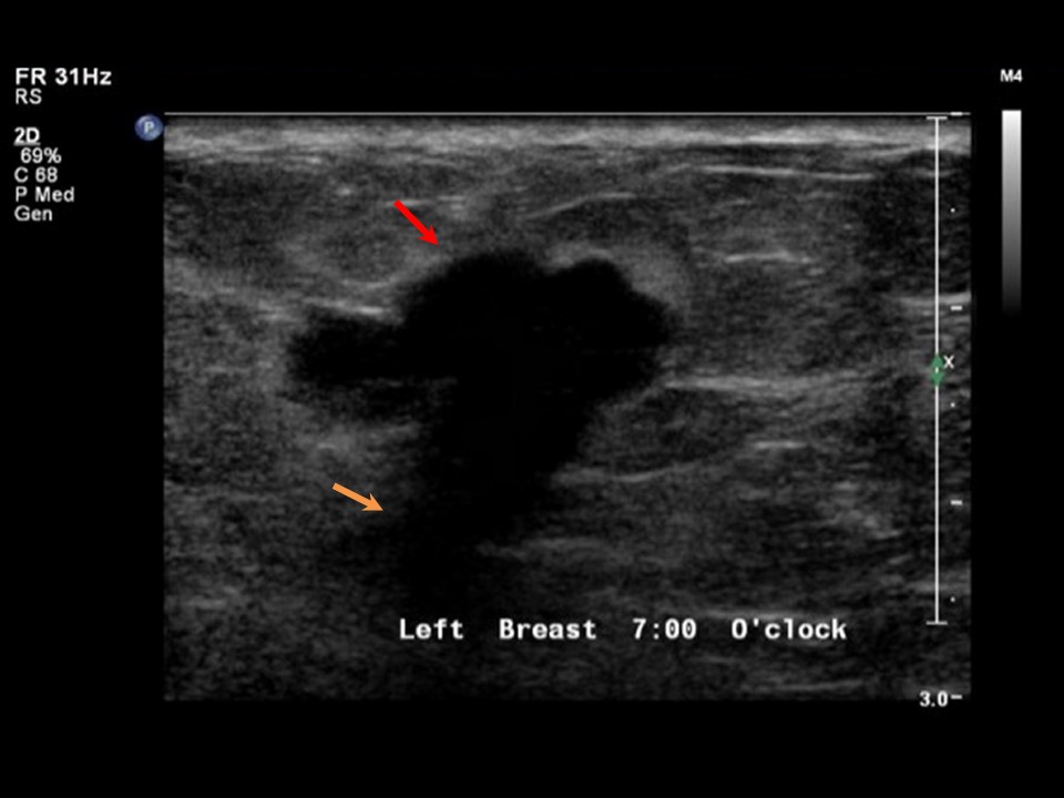
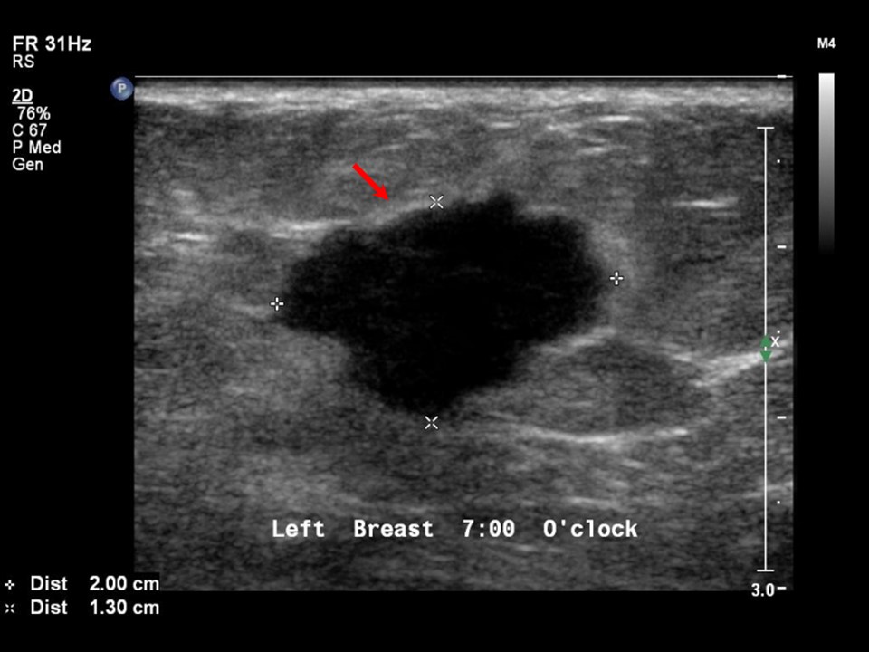
| Ultrasound features: Left breast, lower inner quadrant at 7 oclock | |
| ‣ Mass | |
| • Location: | Left breast, lower inner quadrant at 7 oclock |
| • Number: | 1 |
| • Size: | 2.0 × 1.3 cm |
| • Shape: | Irregular |
| • Orientation: | Not parallel |
| • Margins: | Angular |
| • Echo pattern: | Hypoechoic |
| • Posterior features: | Posterior shadowing |
| ‣ Calcifications: | None |
| ‣ Associated features: | Internal vascularity |
| ‣ Special cases: | None |
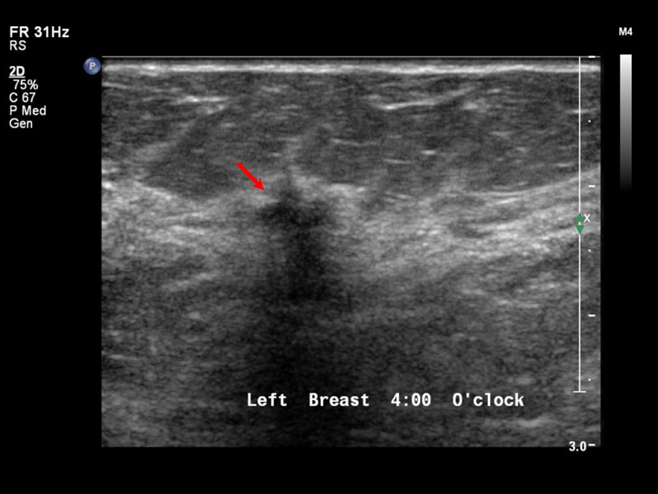 | 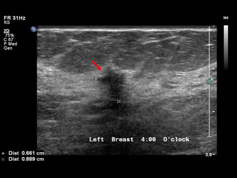 |
| Ultrasound features: Left breast, lower outer quadrant at 4 oclock | |
| ‣ Mass | |
| • Location: | Left breast, lower outer quadrant at 4 oclock |
| • Number: | 1 |
| • Size: | 0.8 × 0.6 cm |
| • Shape: | Irregular |
| • Orientation: | Not parallel |
| • Margins: | Spiculated |
| • Echo pattern: | Hypoechoic |
| • Posterior features: | No posterior features |
| ‣ Calcifications: | None |
| ‣ Associated features: | None |
| ‣ Special cases: | None |
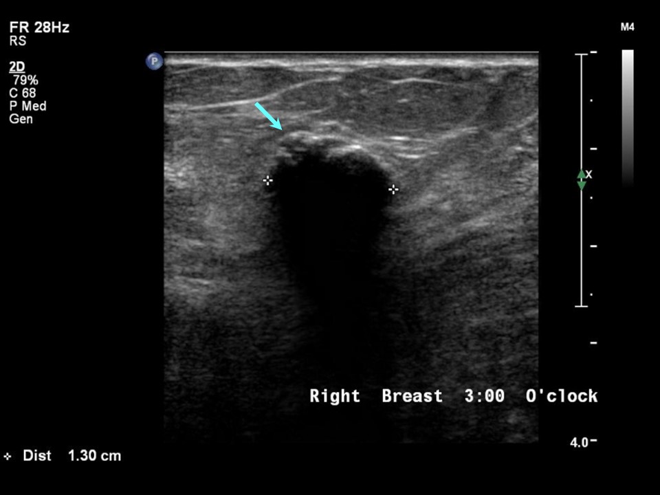 |
| Ultrasound features: Right breast, lower inner quadrant at 3 oclock | |
| ‣ Mass | |
| • Location: | Right breast, lower inner quadrant at 3 oclock |
| • Number: | 1 |
| • Size: | 1.3 × 0.8 cm |
| • Shape: | Oval |
| • Orientation: | Parallel |
| • Margins: | Circumscribed |
| • Echo pattern: | Hypoechoic |
| • Posterior features: | Posterior shadowing |
| ‣ Calcifications: | Macrocalcifications in mass |
| ‣ Associated features: | None |
| ‣ Special cases: | None |
BI-RADS:
BI-RADS Category (Left breast, 2 nodules): 5 (highly suggestive of malignancy)BI-RADS Category (Right breast): 2 (benign)
Further assessment:
Further assessment advised: Referral for cytologyCytology:
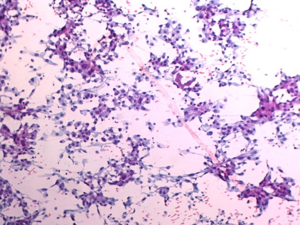 |
| Cytology features: | |
| ‣ Type of sample: | FNAC |
| ‣ Site of biopsy: | |
| • Laterality: | Left |
| • Quadrant: | Lower inner |
| • Localization technique: | Palpation |
| • Nature of aspirate: | Whitish |
| ‣ Cytological description: | Cellular smears are from the lower inner quadrant breast lump. Many clusters and sheets of atypical ductal cells are seen. Many scattered solitary cells are also present. These cells have large, pleomorphic, and hyperchromatic nuclei with prominent nucleoli. Background shows abundant lymphocytes |
| ‣ Reporting category: | Malignant |
| ‣ Diagnosis: | Carcinoma |
| ‣ Comments: | None |
| Cytology features: | |
| ‣ Type of sample: | FNAC |
| ‣ Site of biopsy: | |
| • Laterality: | Left |
| • Quadrant: | Upper outer |
| • Localization technique: | Palpation |
| • Nature of aspirate: | Fatty material |
| ‣ Cytological description: | Smears show a few fibroadipose tissue fragments against a background of RBCs |
| ‣ Reporting category: | Insufficient material |
| ‣ Diagnosis: | Insufficient |
| ‣ Comments: | None |
Histopathology:
MRM
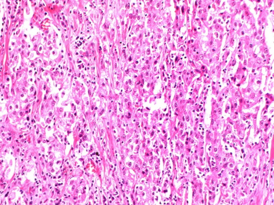 | 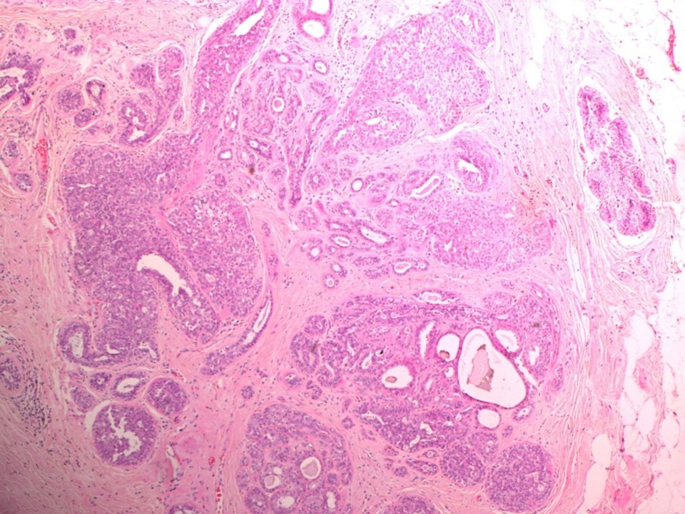 |
| Histopathology features: | |
| ‣ Specimen type: | MRM |
| ‣ Laterality: | Left |
| ‣ Macroscopy: | Specimen (25.0 × 20.0 × 60.0 cm), with overlying skin flap (20.0 × 6.0 cm). The nipple appears flattened. On serial sectioning an irregular greyish white tumour (2.2 × 1.7 × 1.7 cm) with a haemorrhagic area is identified in the lower inner quadrant. It is located 1.5 cm from the skin and 2.0 cm from the base. Closest inferior margin is 1.0 cm away. There is another small firm whitish nodule (0.5 cm) in the lateral half of the specimen, 2.5 cm from the base and 12.0 cm from the first tumour |
| ‣ Histological type: | Invasive breast carcinoma |
| ‣ Histological grade: | Grade 2 (3 + 2 + 2 = 7) |
| ‣ Mitosis: | 12 |
| ‣ Maximum invasive tumour size: | 2.2 cm |
| ‣ Lymph node status: | 0/15 |
| ‣ Peritumoural lymphovascular invasion: | Present |
| ‣ DCIS/EIC: | Absent |
| ‣ Margins: | Free of tumour |
| ‣ Pathological stage: | pT2N0 |
| ‣ Biomarkers: | |
| ‣ Comments: | The other small nodule shows areas with ductal epithelial hyperplasia. DCIS and malignancy is not present in this nodule |
Case summary:
| Postmenopausal woman presented with lump in the left breast. Diagnosed as left breast carcinoma with another suspicious smaller nodule, BI-RADS 5 (multicentric) on imaging, as breast carcinoma on cytology, and as invasive breast carcinoma of no special type, pT2N0 on histopathology in the nodule seen in the lower inner quadrant of the left breast. The other small nodule shows areas with ductal epithelial hyperplasia. |
Learning points:
|