


Home / Training / Manuals / Atlas of breast cancer early detection / Cases
Atlas of breast cancer early detection
Filter by language: English / Русский
Go back to the list of case studies
.png) Click on the pictures to magnify and display the legends
Click on the pictures to magnify and display the legends
| Case number: | 036 |
| Age: | 52 |
| Clinical presentation: | Perimenopausal woman presented with left nipple discharge. She had an increased risk of developing breast cancer because of a family history of breast cancer. Examination did not reveal any lump or palpable lesion. |
Mammography:
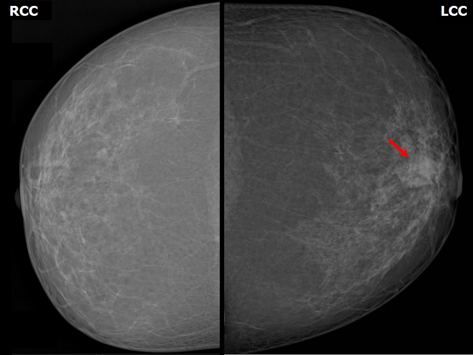 | 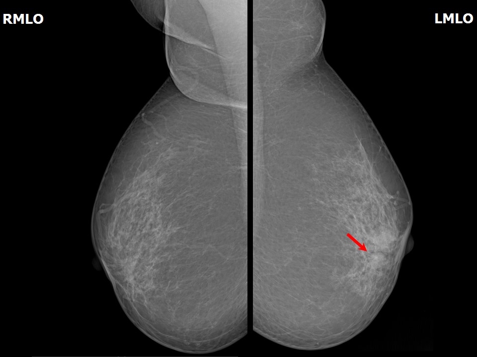 |
| Breast composition: | ACR category b (there are scattered areas of fibroglandular density) | Mammography features: |
| ‣ Location of the lesion: | Left breast, central portion of the breast, central zone, anterior third |
| ‣ Mass: | |
| • Number: | Multiple |
| • Size: | Largest 1.5 cm in greatest dimension |
| • Shape: | Irregular |
| • Margins: | Indistinct |
| • Density: | Equal |
| ‣ Calcifications: | |
| • Typically benign: | None |
| • Suspicious: | Fine pleomorphic |
| • Distribution: | In mass |
| ‣ Architectural distortion: | None |
| ‣ Asymmetry: | None |
| ‣ Intramammary node: | None |
| ‣ Skin lesion: | None |
| ‣ Solitary dilated duct: | Present |
| ‣ Associated features: | Fine pleomorphic microcalcifications in mass |
Ultrasound:
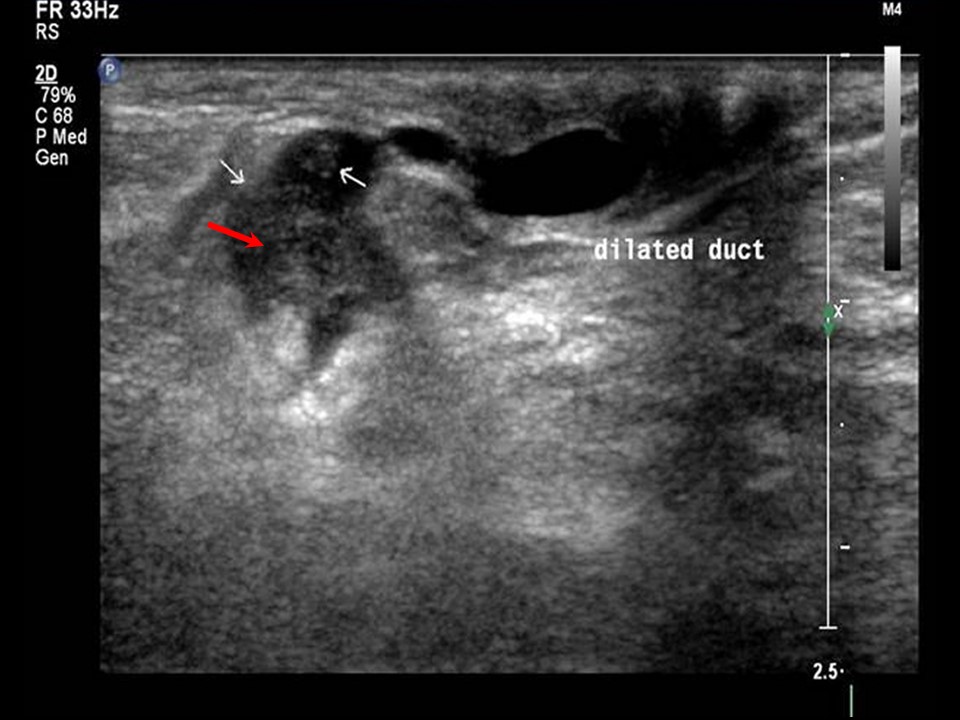 | 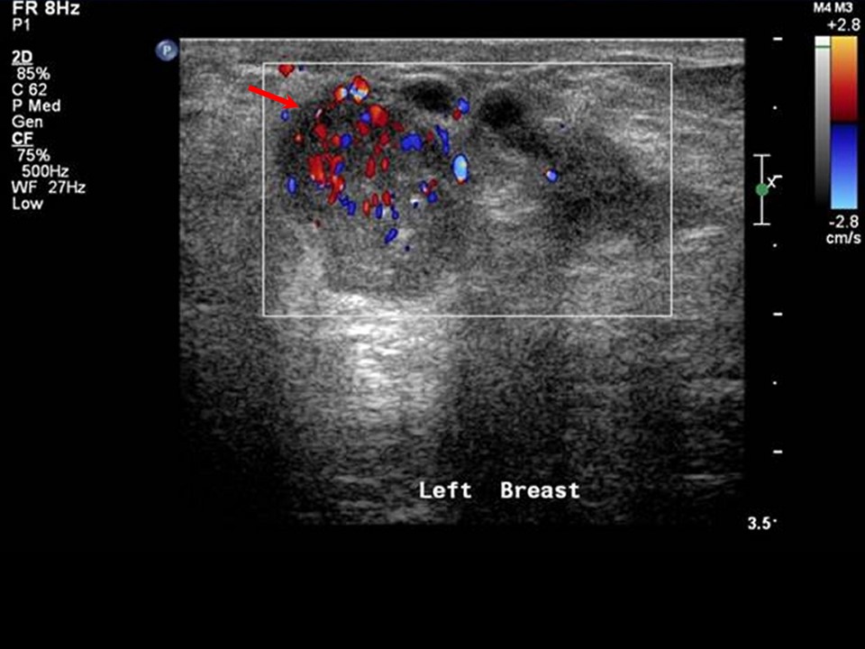 |
| Ultrasound features: Left breast, central portion of the breast, intraductal multiple lesions | |
| ‣ Mass | |
| • Location: | Left breast, central portion of the breast, intraductal multiple lesions |
| • Number: | Multiple intraductal |
| • Size: | Largest 1.8 cm in greatest dimension |
| • Shape: | Irregular |
| • Orientation: | Not parallel |
| • Margins: | Indistinct |
| • Echo pattern: | Heterogeneous |
| • Posterior features: | No posterior features |
| ‣ Calcifications: | None |
| ‣ Associated features: | Internal vascularity, dilated duct |
| ‣ Special cases: | None |
BI-RADS:
BI-RADS Category: 4B (moderate suspicion of malignancy)Further assessment:
Further assessment advised: Referral for cytologyCytology:
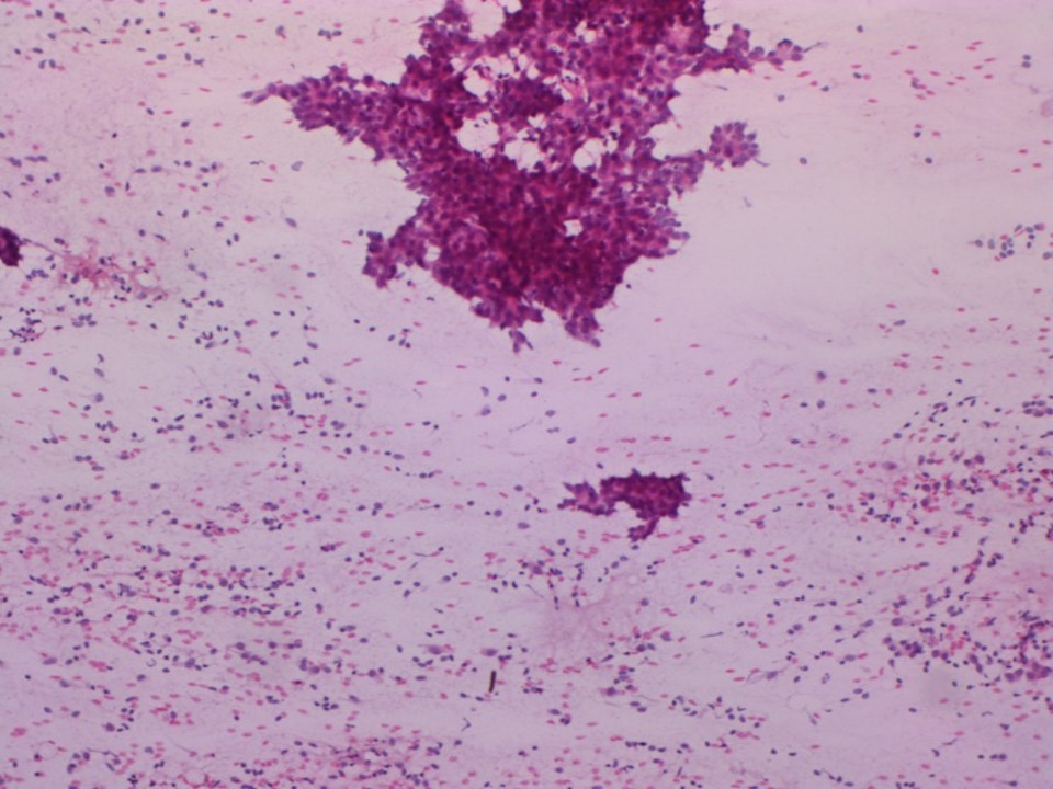 |
| Cytology features: | |
| ‣ Type of sample: | FNAC |
| ‣ Site of biopsy: | |
| • Laterality: | Left |
| • Quadrant: | |
| • Localization technique: | Palpation |
| • Nature of aspirate: | |
| ‣ Cytological description: | Smears reveal a haemorrhagic background with sheets of tightly cohesive ductal epithelial cells suggestive of a proliferative breast lesion. A few cells show a very mild nuclear atypia with karyomegaly and hyperchromasia |
| ‣ Reporting category: | Atypical, probably benign |
| ‣ Diagnosis: | Mild atypia, likely benign |
| ‣ Comments: | A histological evaluation may be more informative |
Case summary:
| Perimenopausal woman presented with left breast nipple discharge. Diagnosed as left breast subareolar solitary dilated duct with intraductal lesions, BI-RADS 4B on imaging, as mild nuclear atypia on cytology, and reported at higher centre as ADH on histopathology. |
Learning points:
|