


Home / Training / Manuals / Atlas of breast cancer early detection / Cases
Atlas of breast cancer early detection
Filter by language: English / Русский
Go back to the list of case studies
.png) Click on the pictures to magnify and display the legends
Click on the pictures to magnify and display the legends
| Case number: | 031 |
| Age: | 45 |
| Clinical presentation: | Premenopausal woman with average risk for developing breast cancer presented with left breast lump of 2 years duration, slowly growing in size. Examination revealed 2 cm diameter lump in upper outer quadrant of left breast. |
Mammography:
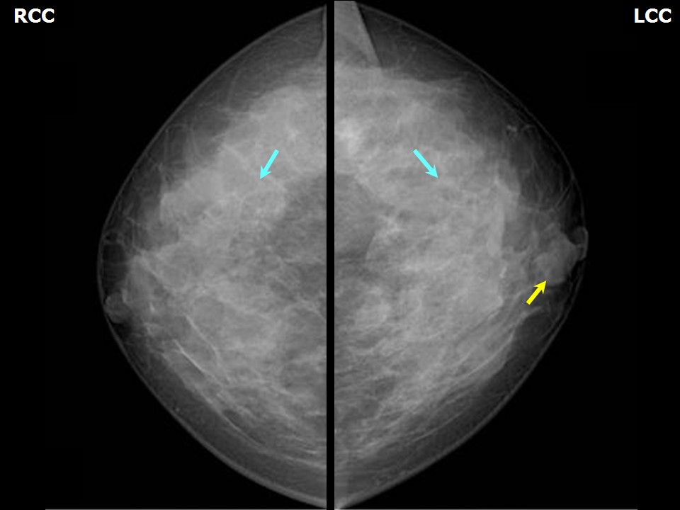 |  |
| Breast composition: | ACR category c (the breasts are heterogeneously dense, which may obscure small masses) | Mammography features: |
| ‣ Location of the lesion: | Left breast, upper quadrants, central zone, anterior third |
| ‣ Mass: | |
| • Number: | 1 |
| • Size: | 2.4 cm in greatest dimension |
| • Shape: | Oval |
| • Margins: | Circumscribed |
| • Density: | Equal |
| ‣ Calcifications: | |
| • Typically benign: | None |
| • Suspicious: | None |
| • Distribution: | None |
| ‣ Architectural distortion: | None |
| ‣ Asymmetry: | None |
| ‣ Intramammary node: | None |
| ‣ Skin lesion: | None |
| ‣ Solitary dilated duct: | None |
| ‣ Associated features: | None |
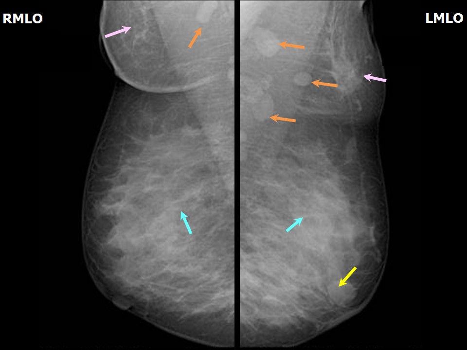
| Breast composition: | ACR category c (the breasts are heterogeneously dense, which may obscure small masses) | Mammography features: |
| ‣ Location of the lesion: | Both axillae |
| ‣ Mass: | |
| • Number: | None |
| • Size: | None |
| • Shape: | None |
| • Margins: | None |
| • Density: | None |
| ‣ Calcifications: | |
| • Typically benign: | None |
| • Suspicious: | None |
| • Distribution: | None |
| ‣ Architectural distortion: | None |
| ‣ Asymmetry: | Present, both axillae |
| ‣ Intramammary node: | None |
| ‣ Skin lesion: | None |
| ‣ Solitary dilated duct: | None |
| ‣ Associated features: | Accessory breast tissue in both axillae, left more than right |
Ultrasound:
 |  |
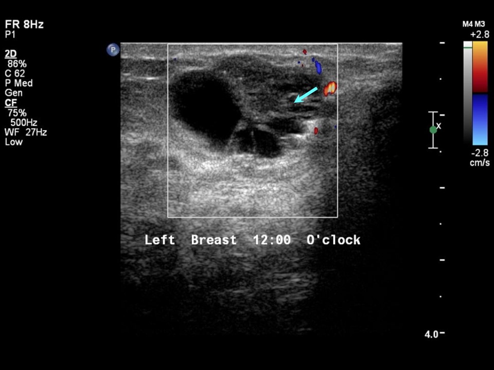 | 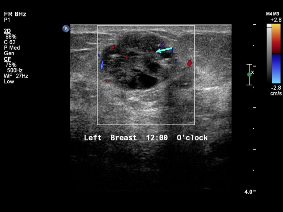 |
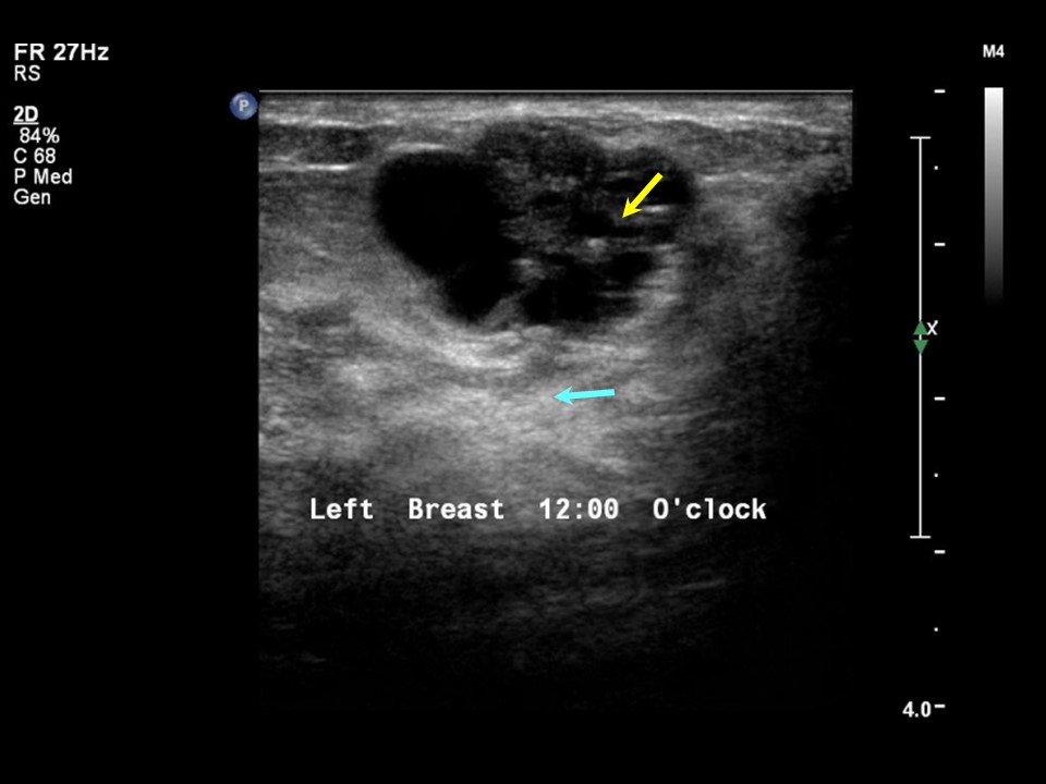
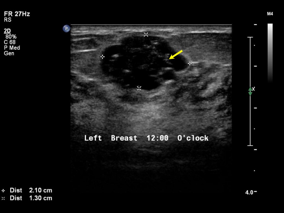
| Ultrasound features: Left breast, central portion of the breast at 12 o'clock | |
| ‣ Mass | |
| • Location: | Left breast, central portion of the breast at 12 o'clock |
| • Number: | 1 |
| • Size: | 2.1 × 1.3 cm |
| • Shape: | Oval |
| • Orientation: | Parallel |
| • Margins: | Circumscribed |
| • Echo pattern: | Heterogeneous |
| • Posterior features: | Posterior Enhancement |
| ‣ Calcifications: | None |
| ‣ Associated features: | Internal vascularity |
| ‣ Special cases: | None |
BI-RADS:
BI-RADS Category: 2 (benign)Further assessment:
Further assessment advised: Referral for cytologyCytology:
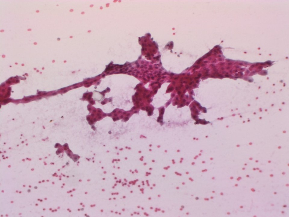 |
| Cytology features: | |
| ‣ Type of sample: | FNAC |
| ‣ Site of biopsy: | |
| • Laterality: | Left |
| • Quadrant: | Subareolar |
| • Localization technique: | Palpation |
| • Nature of aspirate: | 2 mL of clear brownish fluid |
| ‣ Cytological description: | Smears show tightly cohesive sheets of benign ductal epithelial cells with many cells showing apocrine metaplasia. Apocrine cells have abundant granular cytoplasm. Numerous cyst macrophages are seen in the background with proteinaceous cyst fluid |
| ‣ Reporting category: | Benign |
| ‣ Diagnosis: | Fibrocystic change |
| ‣ Comments: | None |
Case summary:
| Premenopausal woman presented with left breast lump. Diagnosed as BI-RADS 2 on imaging and as fibrocystic change on cytology. An incidental finding was accessory breast tissue in both axillae. |
Learning points:
|