


Home / Training / Manuals / Atlas of breast cancer early detection / Cases
Atlas of breast cancer early detection
Filter by language: English / Русский
Go back to the list of case studies
.png) Click on the pictures to magnify and display the legends
Click on the pictures to magnify and display the legends
| Case number: | 140 |
| Age: | 43 |
| Clinical presentation: | Premenopausal woman with average risk of breast cancer presented with a lump in the left breast of recent onset. Examination revealed a left upper quadrant lump. She has had a stable right breast lump for many years, which is proven fibroadenoma on tissue diagnosis. |
Mammography:
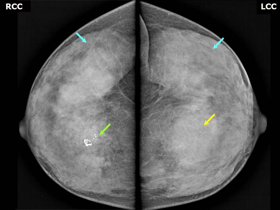 | 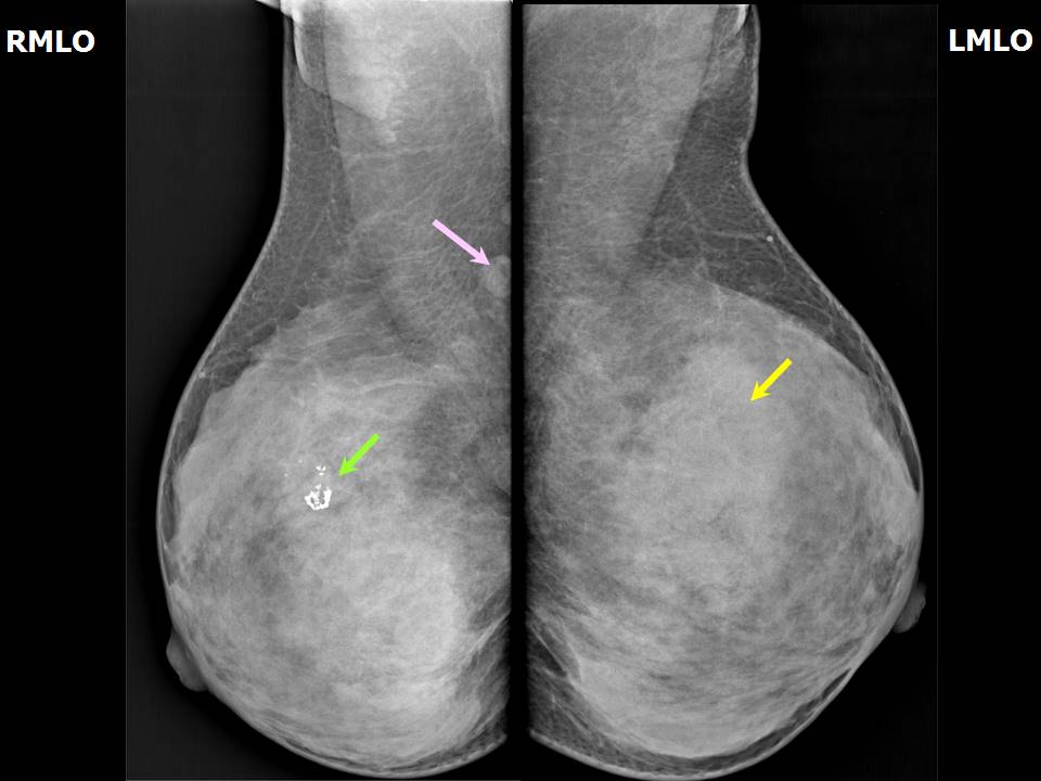 |
| Breast composition: | ACR category d (the breasts are extremely dense, which lowers the sensitivity of mammography) | Mammography features: |
| ‣ Location of the lesion: | Left breast, upper inner quadrant at 1011 oclock, middle third |
| ‣ Mass: | |
| • Number: | 1 |
| • Size: | 4.2 × 4.0 cm |
| • Shape: | Oval |
| • Margins: | Circumscribed |
| • Density: | Equal |
| ‣ Calcifications: | |
| • Typically benign: | None |
| • Suspicious: | None |
| • Distribution: | None |
| ‣ Architectural distortion: | None |
| ‣ Asymmetry: | None |
| ‣ Intramammary node: | None |
| ‣ Skin lesion: | None |
| ‣ Solitary dilated duct: | None |
| ‣ Associated features: | None |
| Breast composition: | ACR category d (the breasts are extremely dense, which lowers the sensitivity of mammography) | Mammography features: |
| ‣ Location of the lesion: | Right breast, upper inner quadrant at 1 oclock, middle third |
| ‣ Mass: | |
| • Number: | 1 |
| • Size: | No |
| • Shape: | None |
| • Margins: | None |
| • Density: | None |
| ‣ Calcifications: | |
| • Typically benign: | Coarse, popcorn-like |
| • Suspicious: | None |
| • Distribution: | None |
| ‣ Architectural distortion: | None |
| ‣ Asymmetry: | None |
| ‣ Intramammary node: | None |
| ‣ Skin lesion: | None |
| ‣ Solitary dilated duct: | None |
| ‣ Associated features: | None |
Ultrasound:
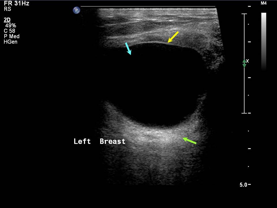 |  |
| Ultrasound features: Left breast, upper inner quadrant at 11 oclock | |
| ‣ Mass | |
| • Location: | Left breast, upper inner quadrant at 11 oclock |
| • Number: | 1 |
| • Size: | 3.5 × 2.5 cm |
| • Shape: | Oval |
| • Orientation: | Parallel |
| • Margins: | Circumscribed |
| • Echo pattern: | Anechoic |
| • Posterior features: | Posterior shadowing |
| ‣ Calcifications: | None |
| ‣ Associated features: | None |
| ‣ Special cases: | Simple cyst |
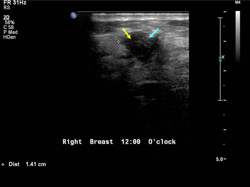
| Ultrasound features: Right breast, upper inner quadrant at 1 oclock | |
| ‣ Mass | |
| • Location: | Right breast, upper inner quadrant at 1 oclock |
| • Number: | 1 |
| • Size: | 1.4 × 1.2 cm |
| • Shape: | Oval |
| • Orientation: | Parallel |
| • Margins: | Circumscribed |
| • Echo pattern: | Hypoechoic |
| • Posterior features: | No posterior features |
| ‣ Calcifications: | Macrocalcifications in mass |
| ‣ Associated features: | Poor vascularity |
| ‣ Special cases: | None |
BI-RADS:
BI-RADS Category: 2 (benign)Further assessment:
Further assessment advised: Referral for cytologyCytology:
 |
| Cytology features: | |
| ‣ Type of sample: | FNAC |
| ‣ Site of biopsy: | |
| • Laterality: | Right |
| • Quadrant: | Lower outer |
| • Localization technique: | Palpation |
| • Nature of aspirate: | Whitish |
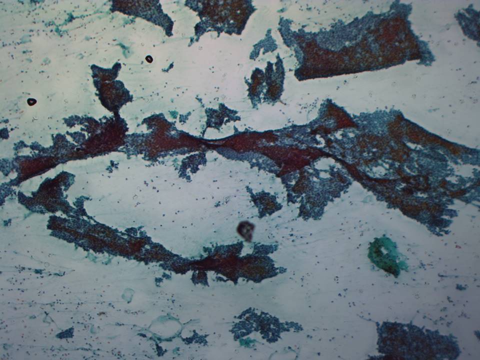
| ‣ Cytological description: | Highly cellular aspirate, showing many cohesive groups of ductal cells with sharply demarcated borders with branching glove-like or antler-horn shapes. Many stripped or bare nuclei are seen in the background |
| ‣ Reporting category: | Benign |
| ‣ Diagnosis: | Fibroadenoma |
| ‣ Comments: | None |
 |
| Cytology features: | |
| ‣ Type of sample: | FNAC |
| ‣ Site of biopsy: | |
| • Laterality: | Left |
| • Quadrant: | Upper inner |
| • Localization technique: | Palpation |
| • Nature of aspirate: | 0.5 mL of brownish fluid |
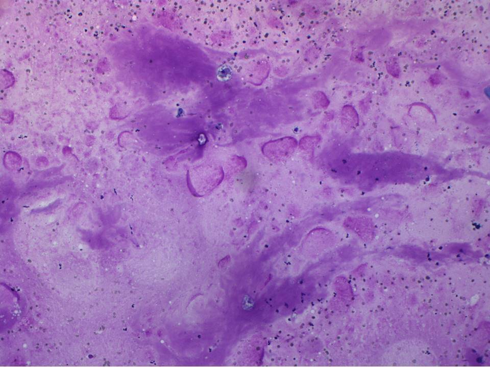
| ‣ Cytological description: | Smears show foamy histiocytes on a thick proteinaceous background. Very a few ductal cells are seen |
| ‣ Reporting category: | Benign |
| ‣ Diagnosis: | Fibrocystic change, non-proliferative |
| ‣ Comments: | None |
Case summary:
| Premenopausal woman presented with left breast lump. Diagnosed as simple cyst, BI-RADS 2 on imaging and as non-proliferative fibrocystic change on cytology. Incidentally noted right breast involuting fibroadenoma, BI-RADS 2 on imaging. |
Learning points:
|