


Home / Training / Manuals / Atlas of breast cancer early detection / Cases
Atlas of breast cancer early detection
Filter by language: English / Русский
Go back to the list of case studies
.png) Click on the pictures to magnify and display the legends
Click on the pictures to magnify and display the legends
| Case number: | 125 |
| Age: | 56 |
| Clinical presentation: | Postmenopausal woman with average risk of developing breast cancer presented with a right breast lump and mastalgia of duration 1 week. Examination revealed a soft palpable right breast lump. |
Mammography:
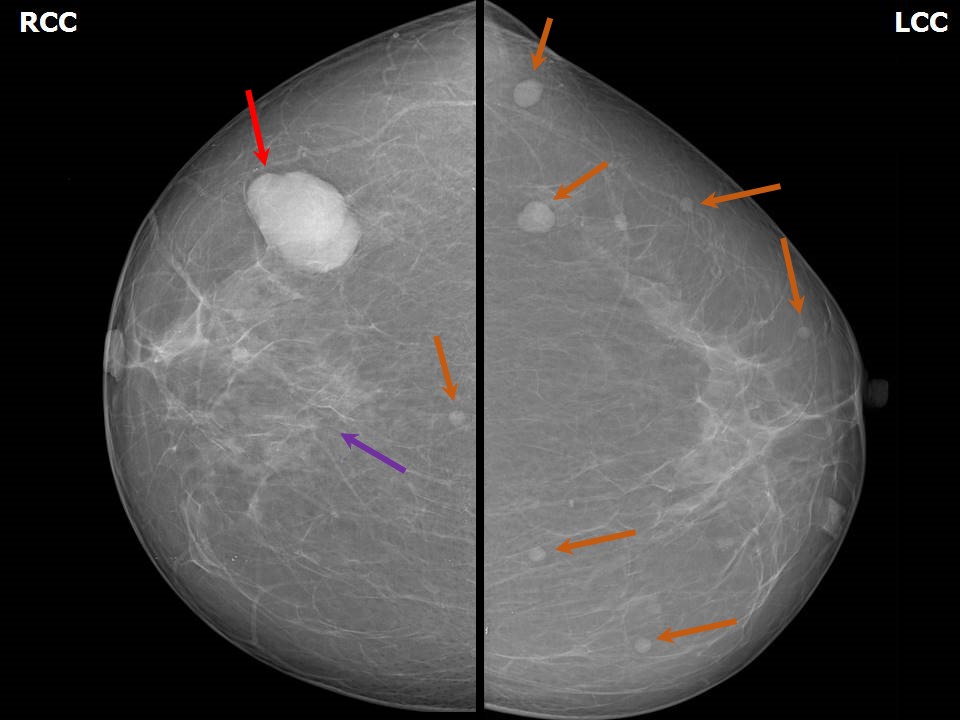 | 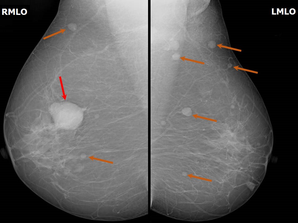 |
| Breast composition: | ACR category a (the breasts are almost entirely fatty) | Mammography features: |
| ‣ Location of the lesion: | Right breast, multiple lesions in all quadrants, largest in upper outer quadrant at 10 oclock, middle third |
| ‣ Mass: | |
| • Number: | Multiple |
| • Size: | Largest 3.8 × 3.0 cm |
| • Shape: | Oval |
| • Margins: | Circumscribed |
| • Density: | High |
| ‣ Calcifications: | |
| • Typically benign: | None |
| • Suspicious: | None |
| • Distribution: | None |
| ‣ Architectural distortion: | None |
| ‣ Asymmetry: | None |
| ‣ Intramammary node: | None |
| ‣ Skin lesion: | None |
| ‣ Solitary dilated duct: | None |
| ‣ Associated features: | None |
| Mammography features: | |
| ‣ Location of the lesion: | Left breast, multiple lesions in all quadrants, largest in upper outer quadrant at 2 oclock, posterior third |
| ‣ Mass: | |
| • Number: | Multiple |
| • Size: | Largest 1.2 × 1.0 cm |
| • Shape: | Round to oval |
| • Margins: | Circumscribed |
| • Density: | High |
| ‣ Calcifications: | |
| • Typically benign: | None |
| • Suspicious: | None |
| • Distribution: | None |
| ‣ Architectural distortion: | None |
| ‣ Asymmetry: | None |
| ‣ Intramammary node: | None |
| ‣ Skin lesion: | None |
| ‣ Solitary dilated duct: | None |
| ‣ Associated features: | None |
Ultrasound:
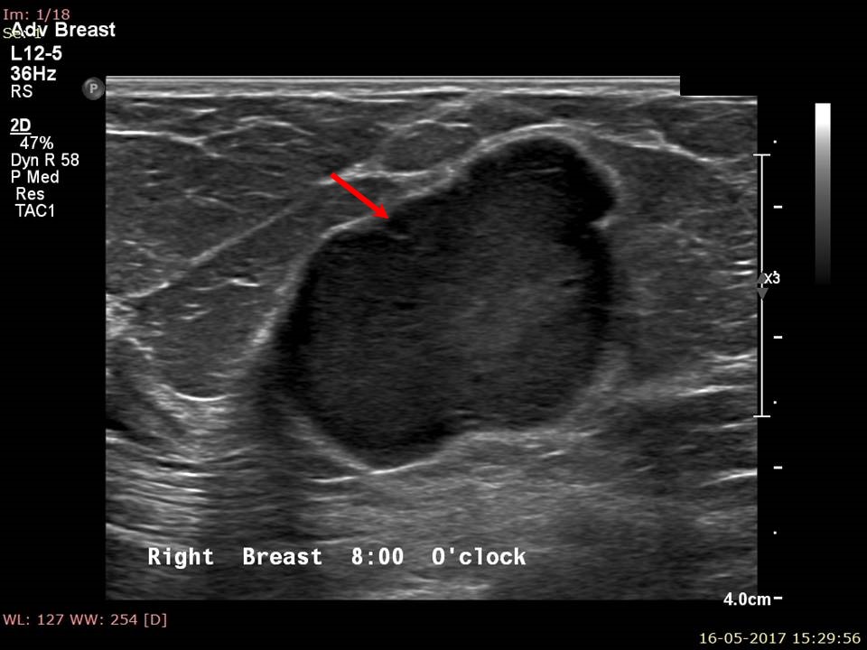 |  |
| Ultrasound features: Right breast, all quadrants | |
| ‣ Mass | |
| • Location: | Right breast, all quadrants |
| • Number: | Multiple |
| • Size: | Largest 3.5 × 3.0 cm at 8 oclock position, 3.5 cm from nipple and at 1.8 cm skin depth |
| • Shape: | Round to oval |
| • Orientation: | Not parallel |
| • Margins: | Circumscribed |
| • Echo pattern: | Hypoechoic |
| • Posterior features: | No posterior features |
| ‣ Calcifications: | None |
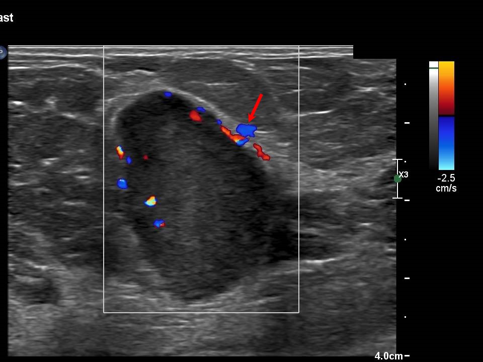
| ‣ Associated features: | Vascularity: in rim and internal |
| ‣ Special cases: | None |
 |  |
 |  |
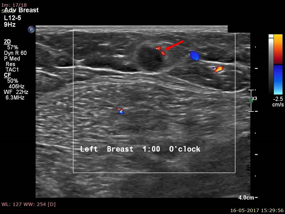 |
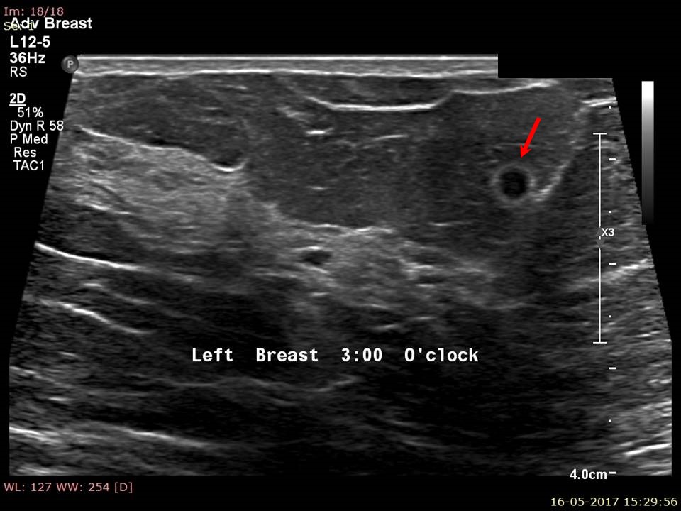
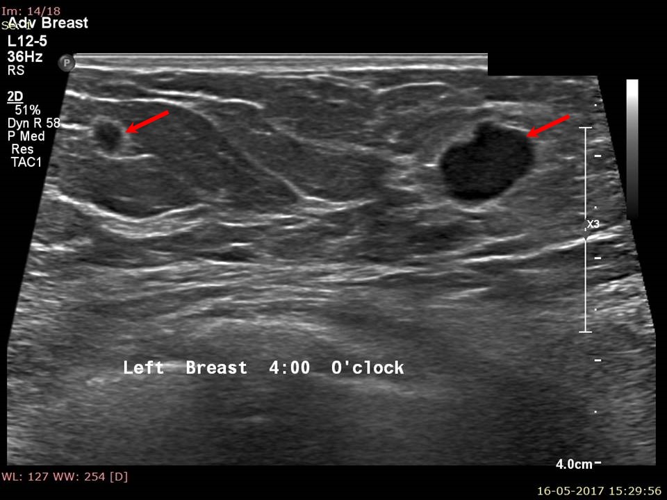
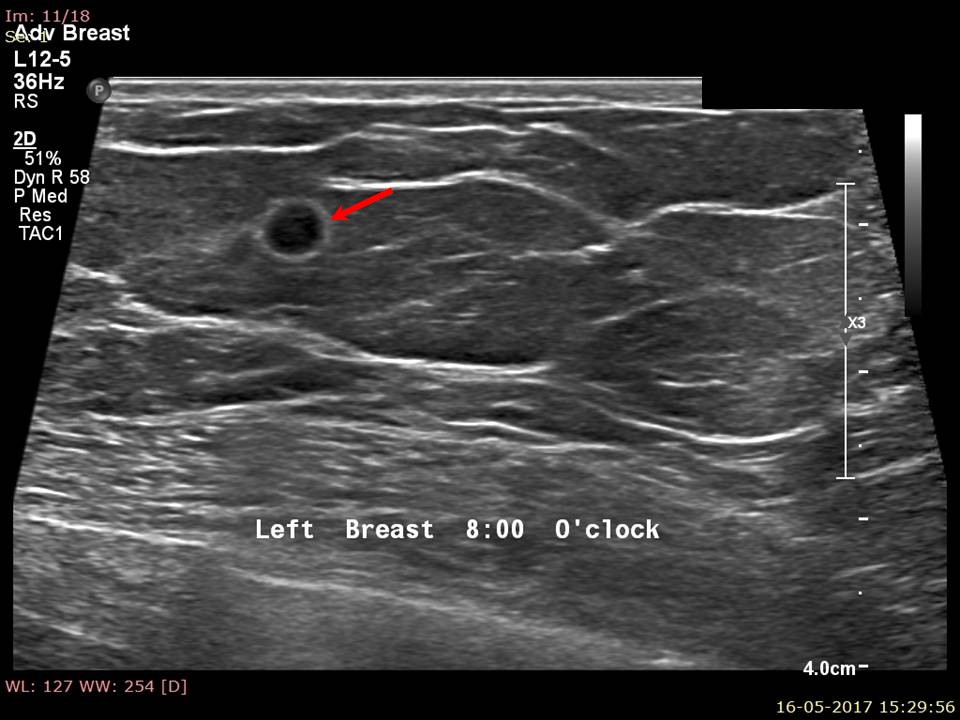
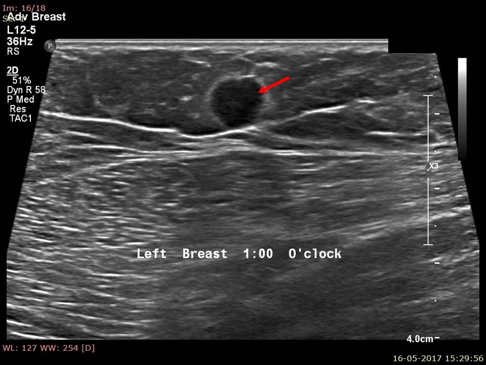
| Ultrasound features: Left breast, all quadrants | |
| ‣ Mass | |
| • Location: | Left breast, all quadrants |
| • Number: | Multiple |
| • Size: | Largest 1.2 × 0.65 cm at 9 oclock position, 1.7 cm from nipple and at 0.3 cm skin depth |
| • Shape: | Round to oval |
| • Orientation: | Not parallel |
| • Margins: | Circumscribed |
| • Echo pattern: | Hypoechoic |
| • Posterior features: | No posterior features |
| ‣ Calcifications: | None |
| ‣ Associated features: | Vascularity: in rim and internal |
| ‣ Special cases: | None |
BI-RADS:
BI-RADS Category: 4C (high suspicion for malignancy)Further assessment:
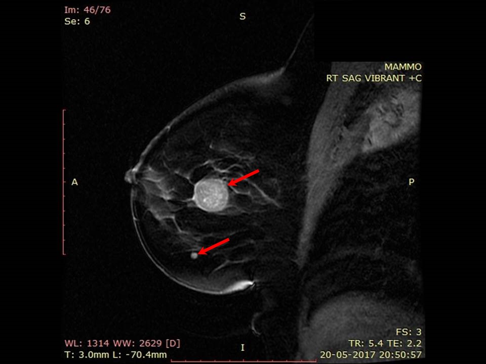
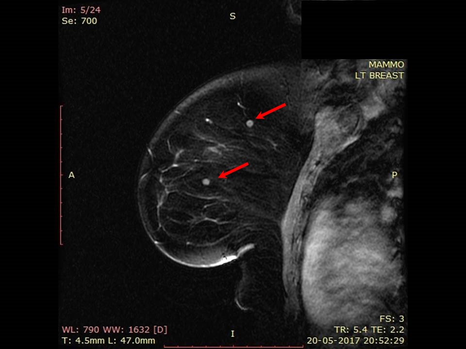
Further assessment advised: Referral for cytology, for core biopsy and further imaging with breast MRIMRI:
 |  |
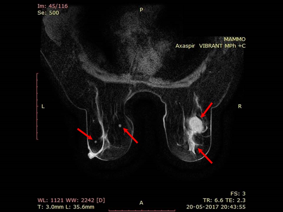 |  |
| MRI features: | ||
| ‣ MRI features: | Amount of fibroglandular tissue: ACR category a (the breasts are almost entirely fatty). Background parenchymal enhancement: Minimal (< 25%), symmetrical | |
| ‣ Location: | Bilateral breasts multiple of varying sizes | |
| ‣ Focus: | No | |
| ‣ Mass: | ||
| • Shape: | Round | |
| • Margin: | Circumscribed | |
| • Internal enhancement: | Homogenous | |
| • Kinetic curve: | Type 3 | |
| ‣ Non-mass enhancement: | ||
| • Distribution: | No | |
| • Internal enhancement: | No | |
| ‣ Non-enhancing findings: | No | |
| ‣ Associated features: | No | |
| ‣ Axillary nodes: | No | |
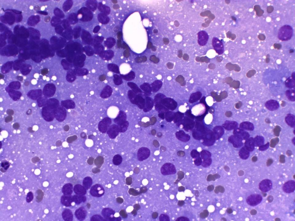
Cytology:
 |
| Cytology features: | |
| ‣ Type of sample: | FNAC |
| ‣ Site of biopsy: | |
| • Laterality: | Right |
| • Quadrant: | 7 oclock |
| • Localization technique: | Palpation |
| • Nature of aspirate: | whitish |
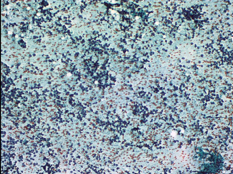
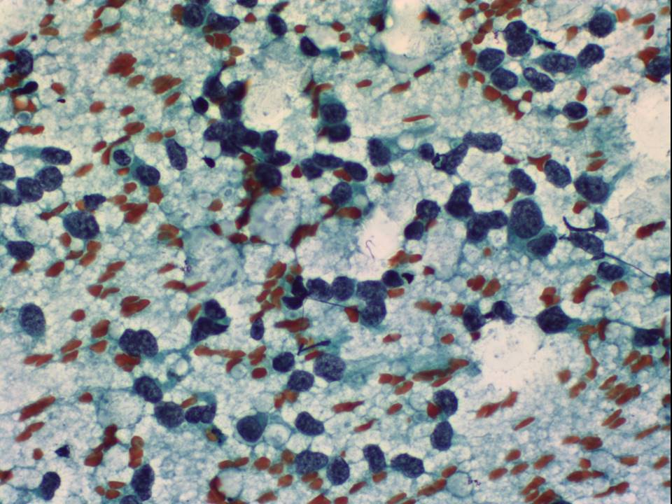
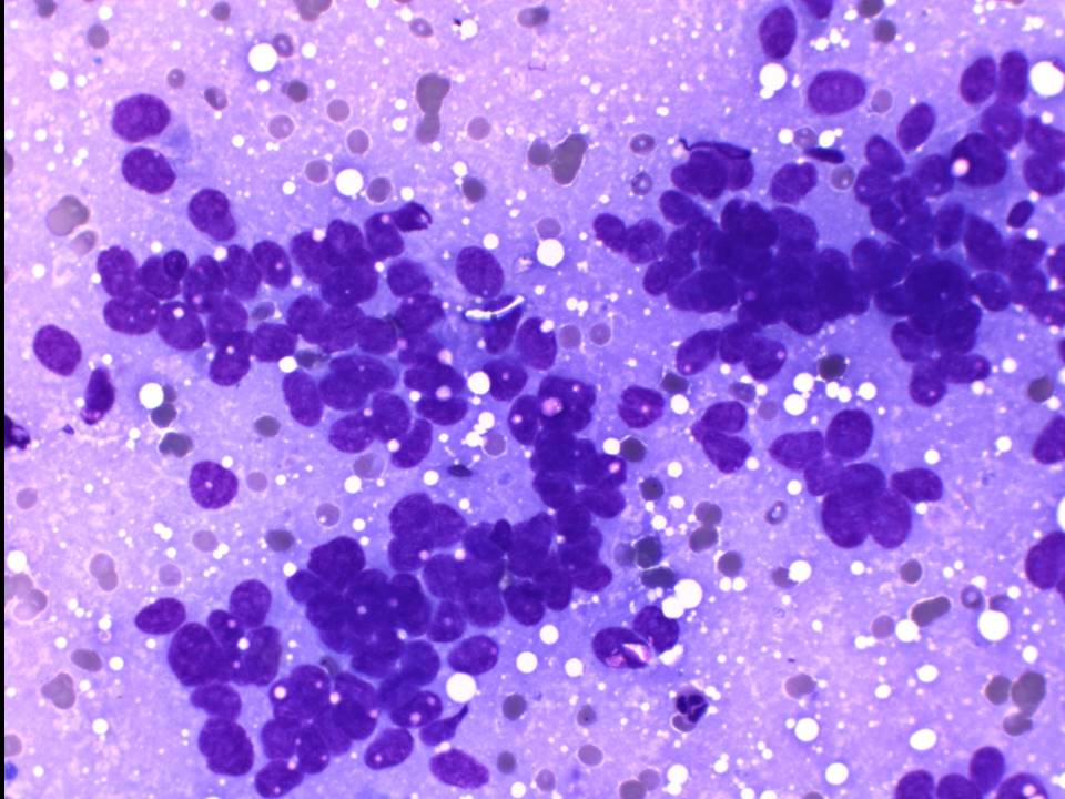
| ‣ Cytological description: | Smears are very cellular and show a pleomorphic population of malignant ductal cells arranged as dyscohesive cell clusters. Single isolated malignant cells are also seen |
| ‣ Reporting category: | Malignant |
| ‣ Diagnosis: | Carcinoma |
| ‣ Comments: | None |
 |  |
 |
| Cytology features: | |
| ‣ Type of sample: | FNAC |
| ‣ Site of biopsy: | |
| • Laterality: | Left |
| • Quadrant: | Lesion 1: 4o clock lesion. Lesion 2: 1 oclock near axillary tail. Lesion 3: 9 oclock |
| • Localization technique: | Ultrasound-guided |
| • Nature of aspirate: | Thick whitish material obtained from all three areas |
| ‣ Cytological description: | Smears from all three lesions reveal very cellular smears with loosely cohesive cells, which are pleomorphic and hyperchromatic with a high N:C ratio. Background shows RBCs |
| ‣ Reporting category: | Malignant |
| ‣ Diagnosis: | Carcinoma |
| ‣ Comments: | None |
Case summary:
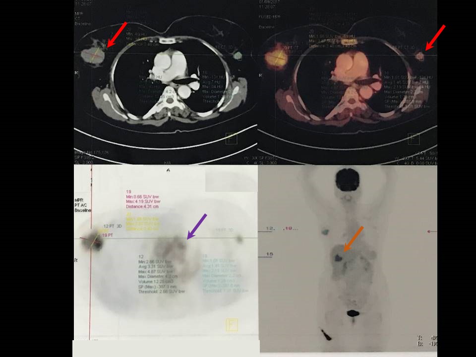
| Postmenopausal woman presented with recent-onset right breast lump with mastalgia. Multiple variable-sized lesions of suspicious morphology are seen in both breasts, diagnosed as BI-RADS category 4C on imaging and carcinoma bilateral breast on cytology. Further detailed evaluation was done at a higher centre. Right breast core biopsy revealed moderately differentiated neuroendocrine carcinoma, favour metastasis. PET scan performed to look for primary site of malignancy revealed mass lesion at tail of pancreas; biopsy proved neuroendocrine tumour of pancreas. PET scan also revealed metastatic foci in liver and vertebrae. Liver biopsy revealed high-grade neuroendocrine carcinoma, primary likely from gastrointestinal tract, thyroid, or pancreatic tumour. Final diagnosis: Pancreatic neuroendocrine tumour with distant metastasis to liver, vertebrae, and bilateral breasts. |
Learning points:
|