


Home / Training / Manuals / Atlas of breast cancer early detection / Cases
Atlas of breast cancer early detection
Filter by language: English / Русский
Go back to the list of case studies
.png) Click on the pictures to magnify and display the legends
Click on the pictures to magnify and display the legends
| Case number: | 156 |
| Age: | 54 |
| Clinical presentation: | Postmenopausal woman with average risk of developing breast cancer presented for mammography screening. CBE revealed no significant findings. |
Mammography:
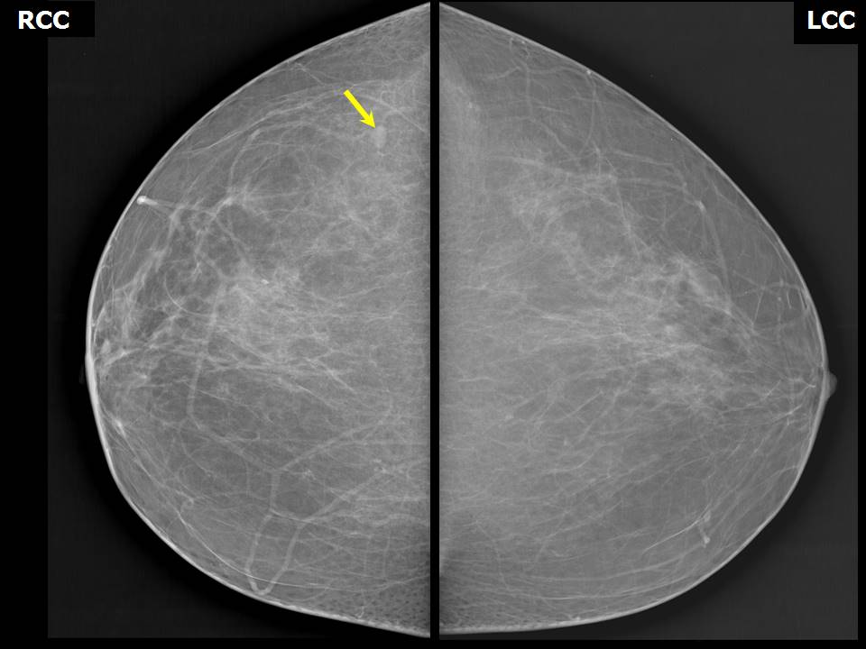 | 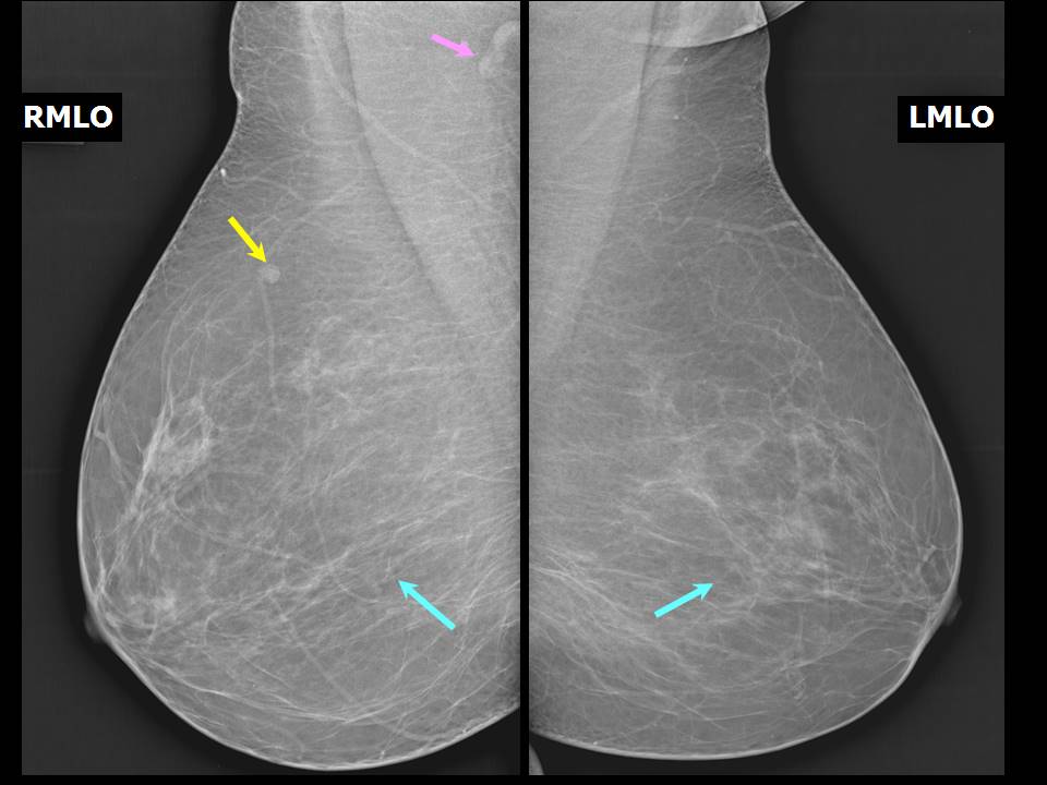 |
| Breast composition: | ACR category a (the breasts are almost entirely fatty) | Mammography features: |
| ‣ Location of the lesion: | Right breast, upper outer quadrant at 10 oclock, posterior third |
| ‣ Mass: | |
| • Number: | 1 |
| • Size: | 1.1 cm in greatest dimension |
| • Shape: | Oval |
| • Margins: | Circumscribed |
| • Density: | Equal |
| ‣ Calcifications: | |
| • Typically benign: | None |
| • Suspicious: | None |
| • Distribution: | None |
| ‣ Architectural distortion: | None |
| ‣ Asymmetry: | None |
| ‣ Intramammary node: | Present |
| ‣ Skin lesion: | None |
| ‣ Solitary dilated duct: | None |
| ‣ Associated features: | None |
Ultrasound:
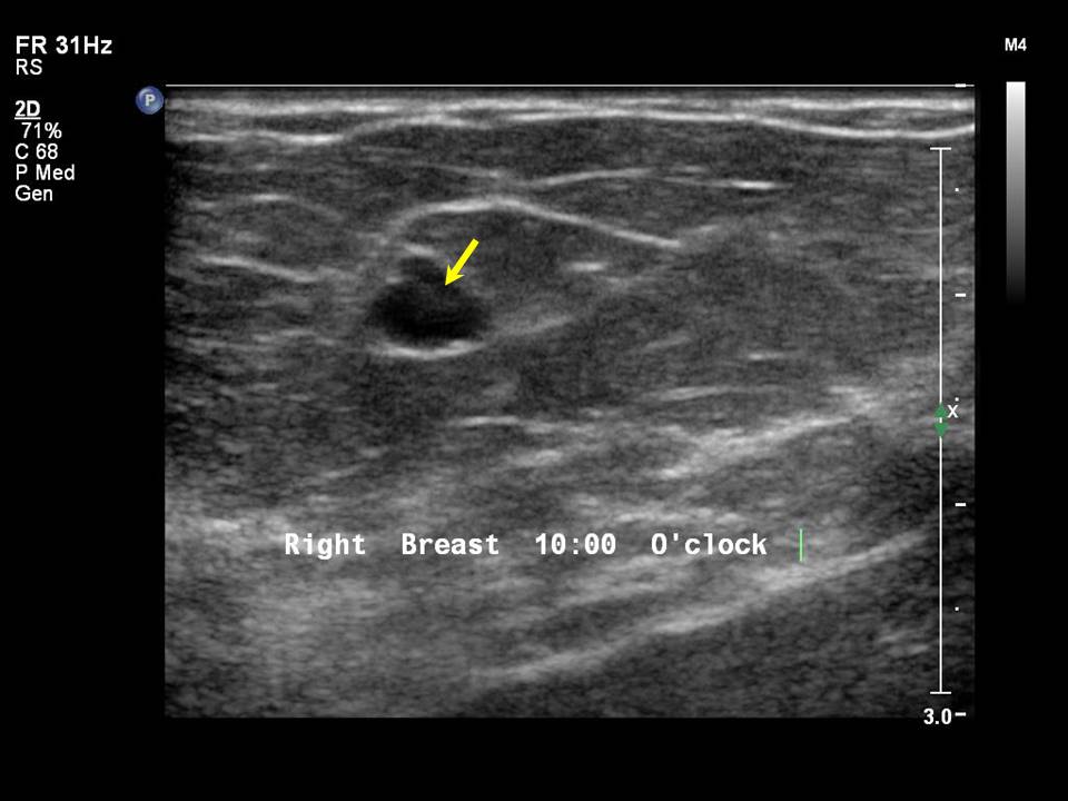 |
| Ultrasound features: Right breast, upper outer quadrant at 10 oclock | |
| ‣ Mass | |
| • Location: | Right breast, upper outer quadrant at 10 oclock |
| • Number: | 1 |
| • Size: | 0.7 cm in greatest dimension |
| • Shape: | Oval |
| • Orientation: | Not parallel |
| • Margins: | Circumscribed |
| • Echo pattern: | Hypoechoic with central sinus |
| • Posterior features: | No posterior features |
| ‣ Calcifications: | None |
| ‣ Associated features: | Hilar vascularity |
| ‣ Special cases: | Lymph nodes, intramammary |
BI-RADS:
BI-RADS Category: 2 (benign)Case summary:
| Postmenopausal woman with average risk of developing breast cancer came for mammography screening. Diagnosed as intramammary node in the right breast, BI-RADS 2 on imaging. |
Learning points:
|