


Home / Training / Manuals / Atlas of breast cancer early detection / Cases
Atlas of breast cancer early detection
Filter by language: English / Русский
Go back to the list of case studies
.png) Click on the pictures to magnify and display the legends
Click on the pictures to magnify and display the legends
| Case number: | 045 |
| Age: | 67 |
| Clinical presentation: | Postmenopausal woman with average risk of developing breast cancer presented with a lump in the right breast. Examination revealed a 4 × 3 cm lump in the upper inner quadrant of the right breast. It was freely mobile and not fixed to the skin or chest wall. |
Mammography:
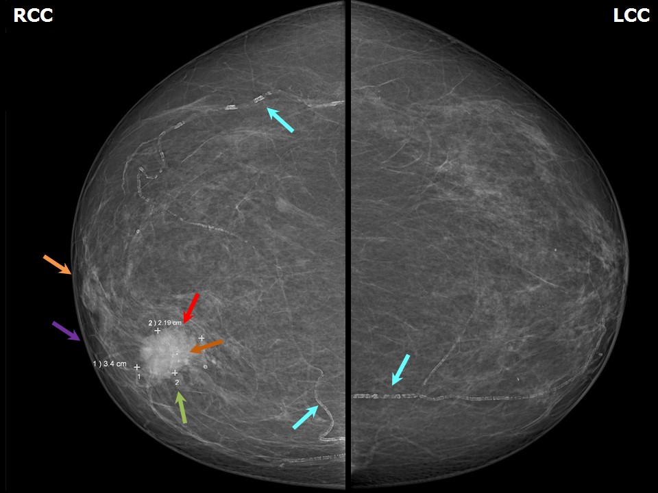 | 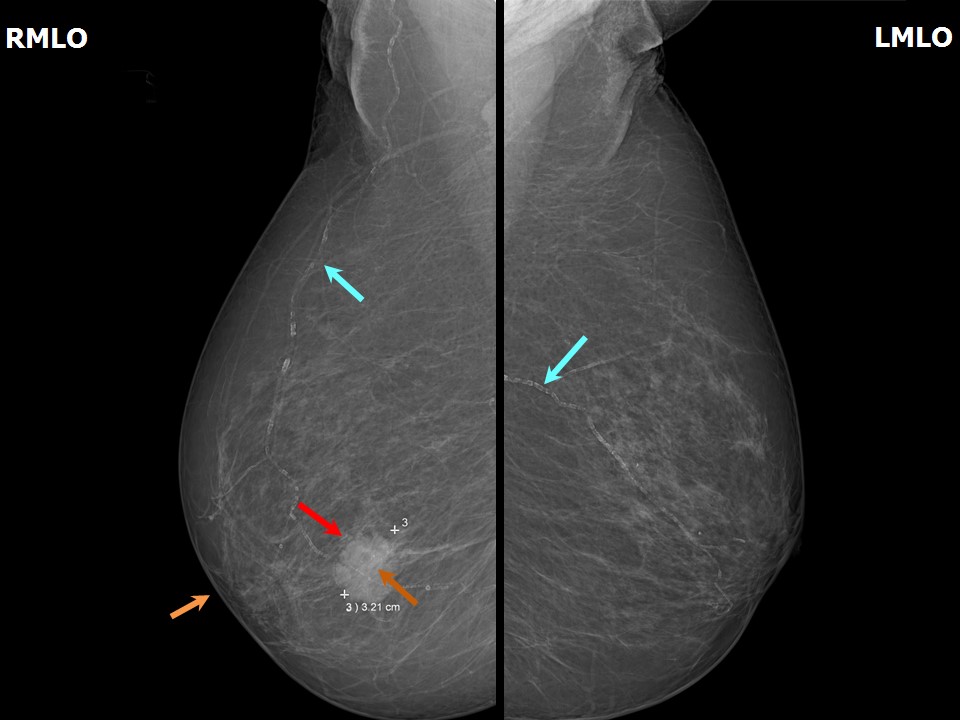 |
| Breast composition: | ACR category a (the breasts are almost entirely fatty) | Mammography features: |
| ‣ Location of the lesion: | Right breast, inner quadrant at 121 oclock, middle third |
| ‣ Mass: | |
| • Number: | 1 |
| • Size: | 3.4 × 3.2 × 2.2 cm |
| • Shape: | Irregular |
| • Margins: | Angular |
| • Density: | High |
| ‣ Calcifications: | |
| • Typically benign: | Vascular calcification |
| • Suspicious: | Fine pleomorphic |
| • Distribution: | In mass |
| ‣ Architectural distortion: | None |
| ‣ Asymmetry: | None |
| ‣ Intramammary node: | None |
| ‣ Skin lesion: | None |
| ‣ Solitary dilated duct: | None |
| ‣ Associated features: | Calcifications and axillary lymphadenopathy |
Ultrasound:
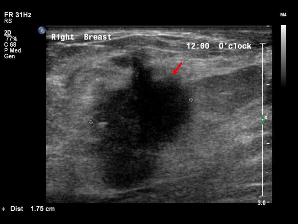 | 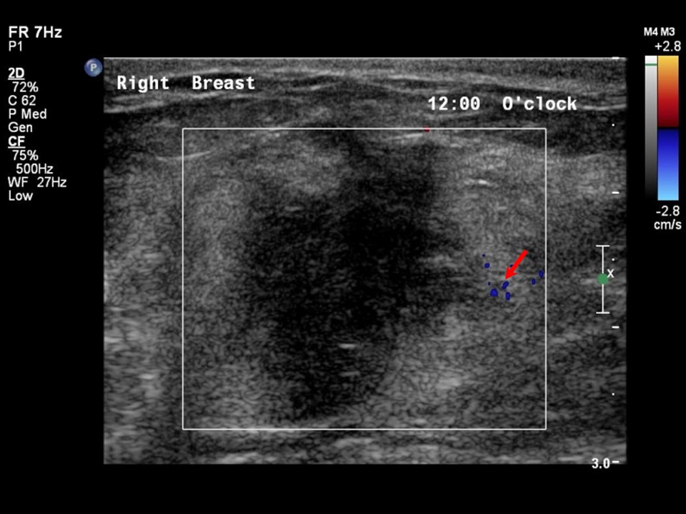 |
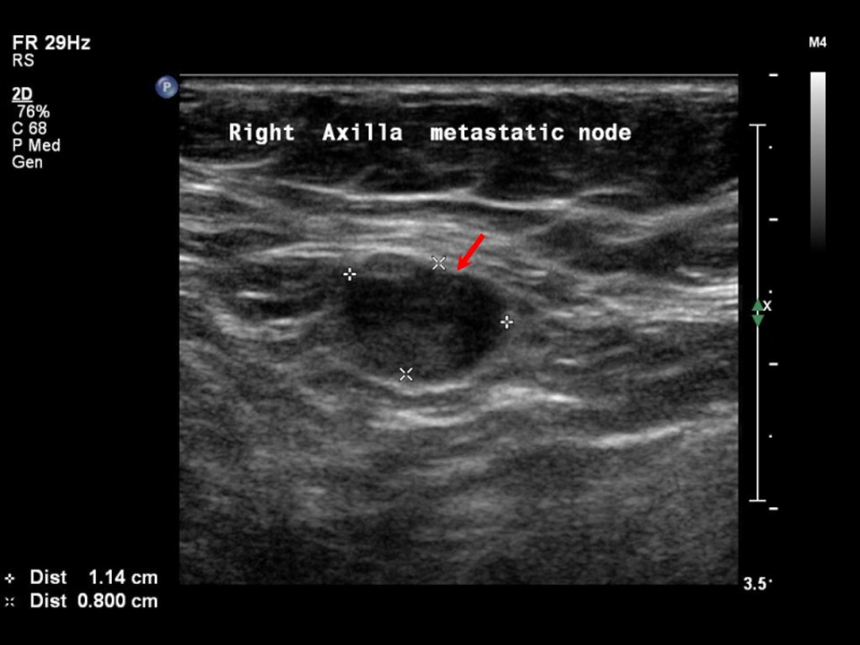 | 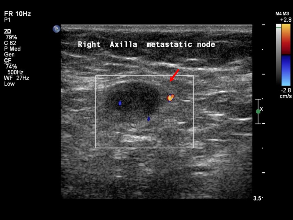 |
| Ultrasound features: Right breast, upper inner quadrant at 121 oclock | |
| ‣ Mass | |
| • Location: | Right breast, upper inner quadrant at 121 oclock |
| • Number: | 1 |
| • Size: | 3.2 × 2.0 cm |
| • Shape: | Irregular |
| • Orientation: | Not parallel |
| • Margins: | Angular |
| • Echo pattern: | Hypoechoic |
| • Posterior features: | Posterior shadowing |
| ‣ Calcifications: | Macrocalcifications in mass |
| ‣ Associated features: | Vessels in rim and axillary lymphadenopathy |
| ‣ Special cases: | None |
BI-RADS:
BI-RADS Category: 5 (highly suggestive of malignancy)Further assessment:
Further assessment advised: Referral for cytologyCytology:
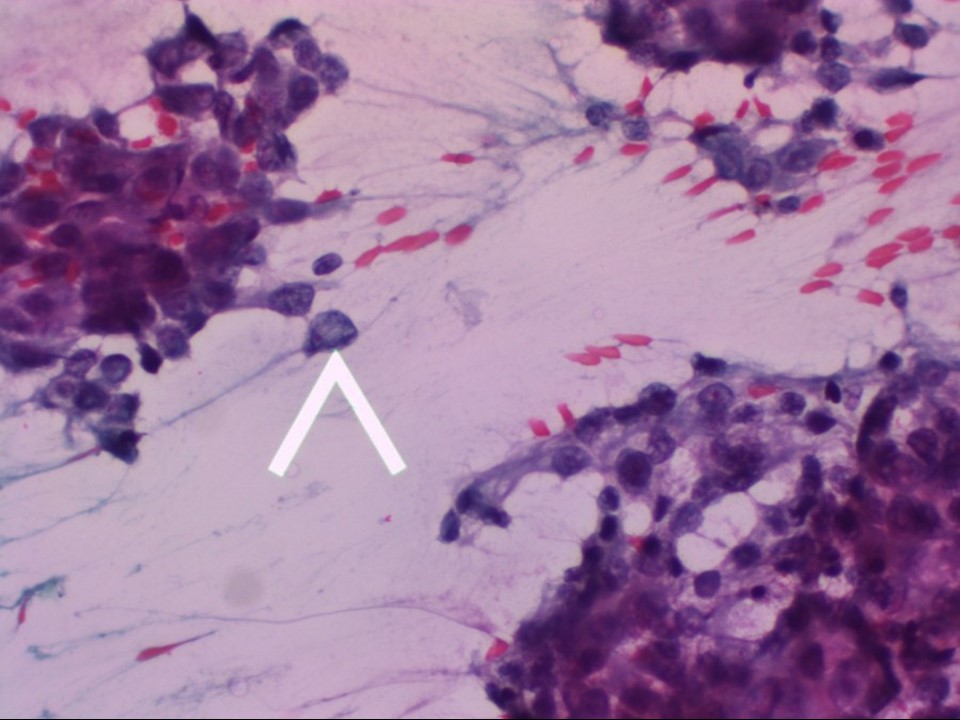 |
| Cytology features: | |
| ‣ Type of sample: | FNAC |
| ‣ Site of biopsy: | |
| • Laterality: | Right |
| • Quadrant: | |
| • Localization technique: | Palpation |
| • Nature of aspirate: | 0.5 mL of thick whitish material |
| ‣ Cytological description: | Smear shows tumour cells with overt malignant characteristics, such as dissociated cells (arrow), marked nuclear enlargement, nuclear hyperchromasia, and loosely cohesive cell clusters. (Pap; 400×) |
| ‣ Reporting category: | Malignant |
| ‣ Diagnosis: | Carcinoma |
| ‣ Comments: | None |
Histopathology:
BCS
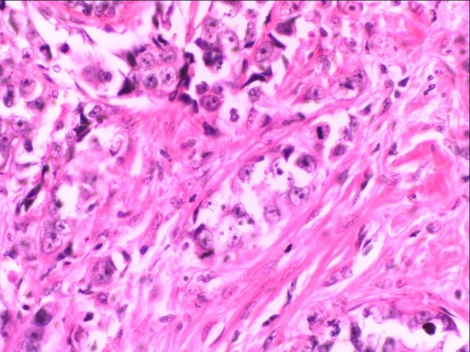 |
| Histopathology features: | |
| ‣ Specimen type: | BCS |
| ‣ Laterality: | Right |
| ‣ Macroscopy: | Right breast lumpectomy specimen (11.0 × 5.0 × 4.5 cm) with an elliptical flap of skin (5.0 × 1.5 cm). Cut surface shows a greyish white tumour (3.4 × 2.3 × 2.5 cm). The tumour is 2.5 cm below the skin, 3.0 cm from the base; 3.0 cm from the lateral margin, 2.3 cm from the medial margin, 2.0 cm from the superior margin, and 1.5 cm from the inferior margin. There is a smooth-walled cyst (2.5 cm in diameter) located inferior to the tumour |
| ‣ Histological type: | Invasive breast carcinoma of no special type |
| ‣ Histological grade: | Grade 3 (3 + 3 + 2 = 8) |
| ‣ Mitosis: | 15 |
| ‣ Maximum invasive tumour size: | 3.4 cm in greatest dimension |
| ‣ Lymph node status: | 2/14 |
| ‣ Peritumoural lymphovascular invasion: | Absent |
| ‣ DCIS/EIC: | Comedo type DCIS of high grade; EIC absent |
| ‣ Margins: | Free of tumour |
| ‣ Pathological stage: | pT2N1 |
| ‣ Biomarkers: | |
| ‣ Comments: | Sections from the cyst inferior to the tumour show changes of fibrocystic change |
Case summary:
| Postmenopausal woman presented with right breast lump. Diagnosed as right breast carcinoma with coarse heterogeneous and fine microcalcifications within and nipple and skin retraction, BI-RADS 5 on imaging, as breast carcinoma on cytology, and as invasive breast carcinoma of no special type, pT2N1 on histopathology. |
Learning points:
|