


Home / Training / Manuals / Atlas of breast cancer early detection / Cases
Atlas of breast cancer early detection
Filter by language: English / Русский
Go back to the list of case studies
.png) Click on the pictures to magnify and display the legends
Click on the pictures to magnify and display the legends
| Case number: | 051 |
| Age: | 78 |
| Clinical presentation: | Postmenopausal woman with average risk of developing breast cancer presented with pain and a lump in the left breast of duration 1 month. Examination revealed a hard lump 4 cm in diameter in the left breast. |
Mammography:
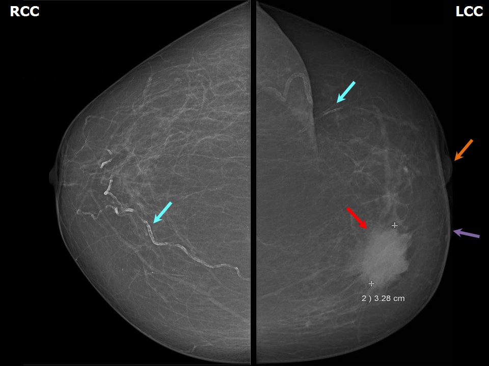 |  |
| Breast composition: | ACR category a (the breasts are almost entirely fatty) | Mammography features: |
| ‣ Location of the lesion: | Left breast, upper inner quadrant at 10 oclock, middle third |
| ‣ Mass: | |
| • Number: | 1 |
| • Size: | 3.2 × 3.2 cm |
| • Shape: | Irregular |
| • Margins: | Indistinct |
| • Density: | High |
| ‣ Calcifications: | |
| • Typically benign: | Vascular calcification |
| • Suspicious: | None |
| • Distribution: | None |
| ‣ Architectural distortion: | None |
| ‣ Asymmetry: | None |
| ‣ Intramammary node: | None |
| ‣ Skin lesion: | None |
| ‣ Solitary dilated duct: | None |
| ‣ Associated features: | Skin thickening and nipple retraction |
Ultrasound:
 |  |
 |
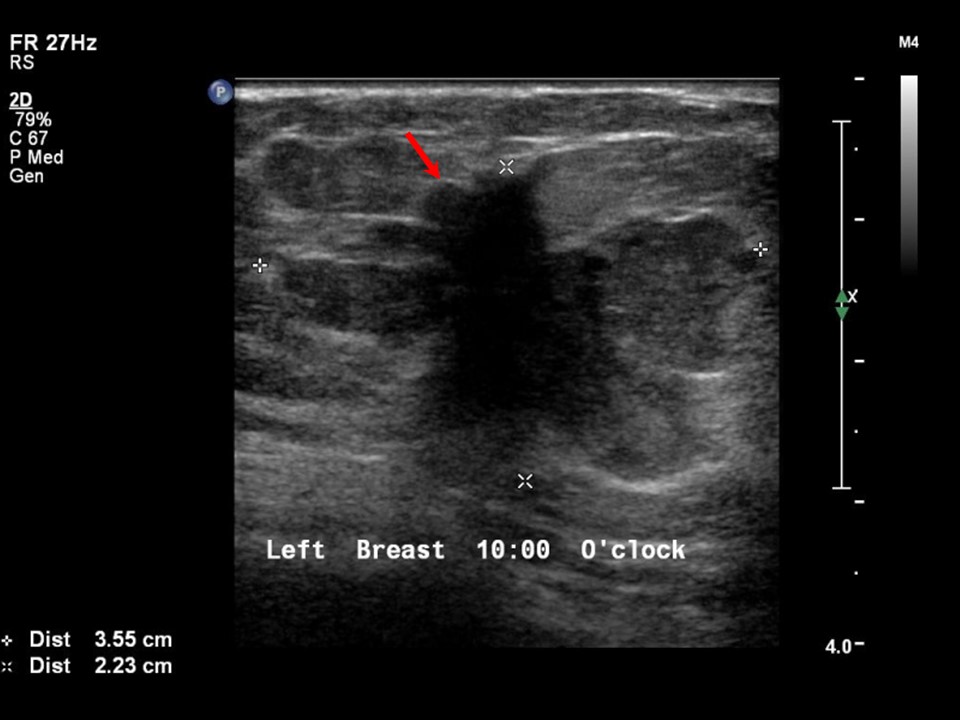
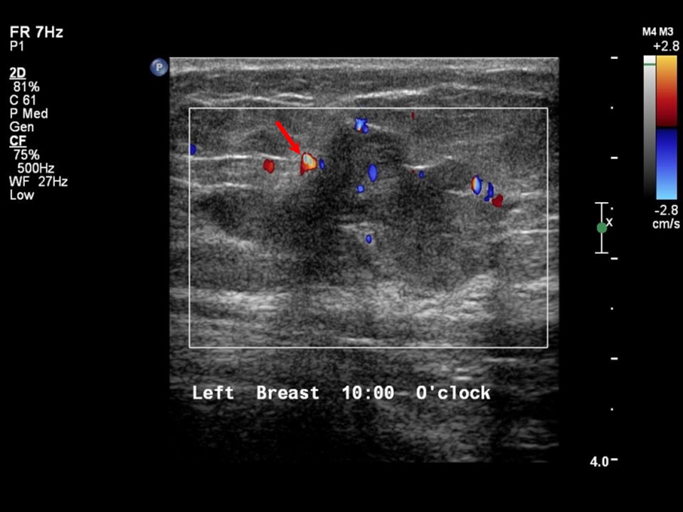
| Ultrasound features: Left breast, upper inner quadrant at 10 oclock | |
| ‣ Mass | |
| • Location: | Left breast, upper inner quadrant at 10 oclock |
| • Number: | 1 |
| • Size: | 3.6 × 2.2 cm |
| • Shape: | Irregular |
| • Orientation: | Not parallel |
| • Margins: | Angular |
| • Echo pattern: | Heteroechoic |
| • Posterior features: | No posterior features |
| ‣ Calcifications: | None |
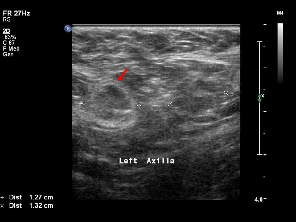
| ‣ Associated features: | Internal vascularity in mass and enlarged left axillary lymph nodes with altered morphology |
| ‣ Special cases: | None |
BI-RADS:
BI-RADS Category: 5 (highly suggestive of malignancy)Further assessment:
Further assessment advised: Referral for cytologyCytology:
 |
| Cytology features: | |
| ‣ Type of sample: | FNAC |
| ‣ Site of biopsy: | |
| • Laterality: | Left |
| • Quadrant: | Upper inner |
| • Localization technique: | Palpation |
| • Nature of aspirate: | whitish |
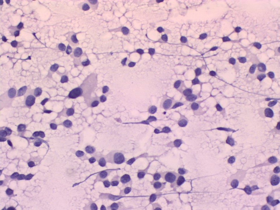
| ‣ Cytological description: | Smears are cellular with poorly cohesive sheets of malignant ductal cells. Cells show features of a low-grade malignancy |
| ‣ Reporting category: | Malignant |
| ‣ Diagnosis: | Carcinoma low grade |
| ‣ Comments: | None |
Histopathology:
MRM
 |
| Histopathology features: | |
| ‣ Specimen type: | MRM |
| ‣ Laterality: | Left |
| ‣ Macroscopy: | A greyish white, irregular tumour mass (3.5 × 3.0 × 3.0 cm) in the inner quadrant. A few tiny cystic areas are seen on the cut surface of the tumour |
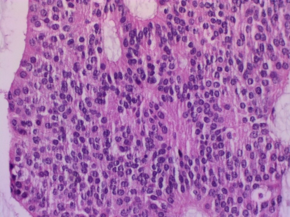
| ‣ Histological type: | Infiltrating duct carcinoma with extensive neuroendocrine differentiation. Extracellular mucin is present, at places forming lakes |
| ‣ Histological grade: | Grade 1 (2 + 1 + 1 = 4) |
| ‣ Mitosis: | 2 |
| ‣ Maximum invasive tumour size: | 3.5 cm in greatest dimension |
| ‣ Lymph node status: | 0/15 |
| ‣ Peritumoural lymphovascular invasion: | Present |
| ‣ DCIS/EIC: | Cribriform and solid DCIS of low grade |
| ‣ Margins: | Free of tumour, 2.0 cm from the base |
| ‣ Pathological stage: | pT2N0 |
| ‣ Biomarkers: | |
| ‣ Comments: | IHC is confirmatory for neuroendocrine differentiation. Neuron-specific enolase and chromogranin are positive |
Case summary:
| Postmenopausal woman presented with left breast lump. Diagnosed as left breast carcinoma, BI-RADS 5 on imaging, as breast carcinoma on cytology, and as invasive breast carcinoma with neuroendocrine differentiation, pT2N0 on histopathology. |
Learning points:
|