


Home / Training / Manuals / Atlas of breast cancer early detection / Cases
Atlas of breast cancer early detection
Filter by language: English / Русский
Go back to the list of case studies
.png) Click on the pictures to magnify and display the legends
Click on the pictures to magnify and display the legends
BI-RADS Category (2012): 5 (highly suggestive of malignancy)
| Case number: | 102 |
| Age: | 58 |
| Clinical presentation: | Postmenopausal woman with increased risk of developing breast cancer in view of family history of breast cancer in paternal aunt and maternal grandmother. She presented with a lump in the right breast. Examination revealed a 2.5 cm right breast lump at the 12 oclock position. |
Mammography:
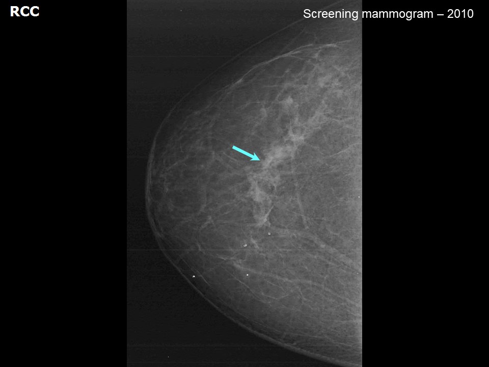 | 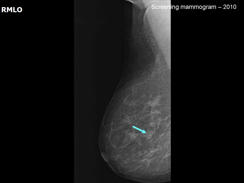 |
| Breast composition: | ACR category a (the breasts are almost entirely fatty) | Mammography features: |
| ‣ Location of the lesion: | November 2010: No mass |
| ‣ Mass: | |
| • Number: | 0 |
| • Size: | None |
| • Shape: | None |
| • Margins: | None |
| • Density: | None |
| ‣ Calcifications: | |
| • Typically benign: | None |
| • Suspicious: | None |
| • Distribution: | None |
| ‣ Architectural distortion: | None |
| ‣ Asymmetry: | None |
| ‣ Intramammary node: | None |
| ‣ Skin lesion: | None |
| ‣ Solitary dilated duct: | None |
| ‣ Associated features: | None |
 |  |
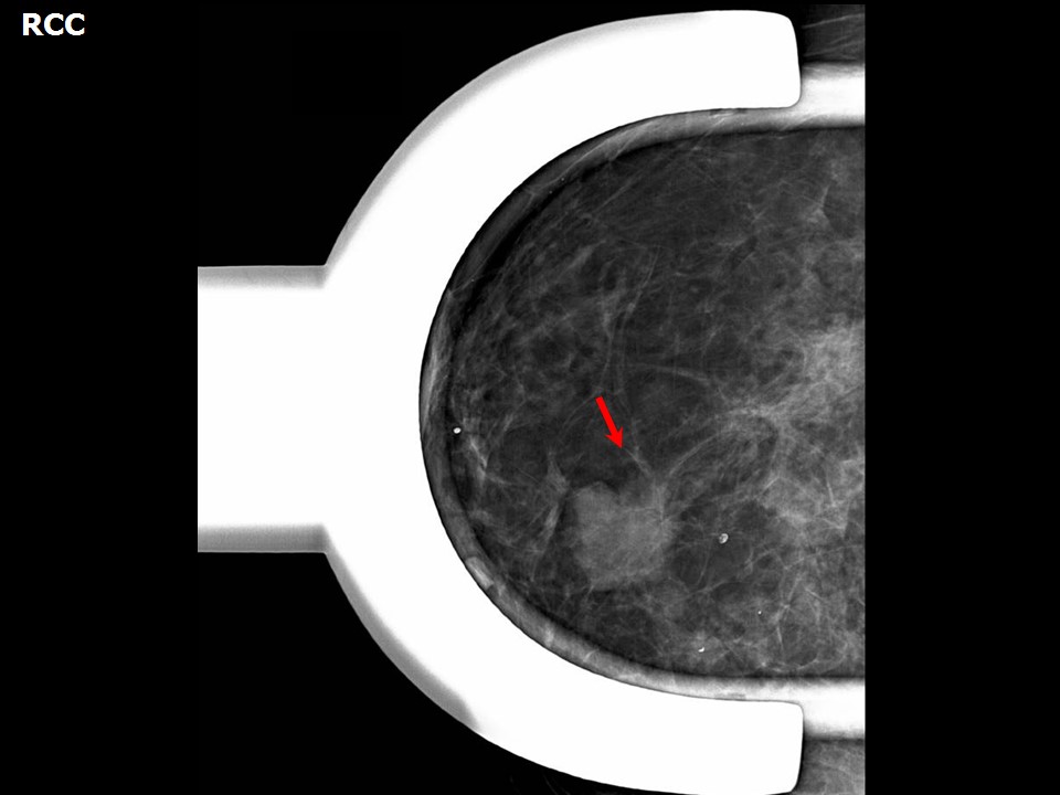 |
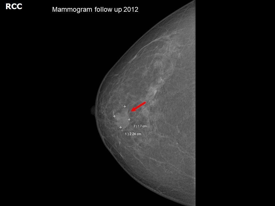
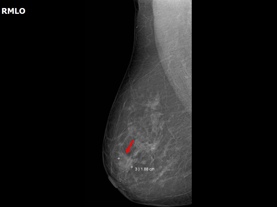
| Breast composition: | ACR category a (the breasts are almost entirely fatty) | Mammography features: |
| ‣ Location of the lesion: | October 2012: Right breast, central portion of the breast, upper inner quadrant at 121 oclock, anterior third |
| ‣ Mass: | |
| • Number: | 1 (this is the developing asymmetry) |
| • Size: | 2.2 × 1.7 cm |
| • Shape: | Irregular |
| • Margins: | Spiculated |
| • Density: | High |
| ‣ Calcifications: | |
| • Typically benign: | None |
| • Suspicious: | None |
| • Distribution: | None |
| ‣ Architectural distortion: | Perilesional |
| ‣ Asymmetry: | Focal |
| ‣ Intramammary node: | None |
| ‣ Skin lesion: | None |
| ‣ Solitary dilated duct: | None |
| ‣ Associated features: | Architectural distortion |
Ultrasound:
 |  |
| Ultrasound features: October 2012: Right breast, central portion of the breast at 12 oclock | |
| ‣ Mass | |
| • Location: | October 2012: Right breast, central portion of the breast at 12 oclock |
| • Number: | 1, developing asymmetry |
| • Size: | 2.6 × 1.6 cm |
| • Shape: | Irregular |
| • Orientation: | Not parallel |
| • Margins: | Indistinct |
| • Echo pattern: | Heteroechoic |
| • Posterior features: | Posterior shadowing |
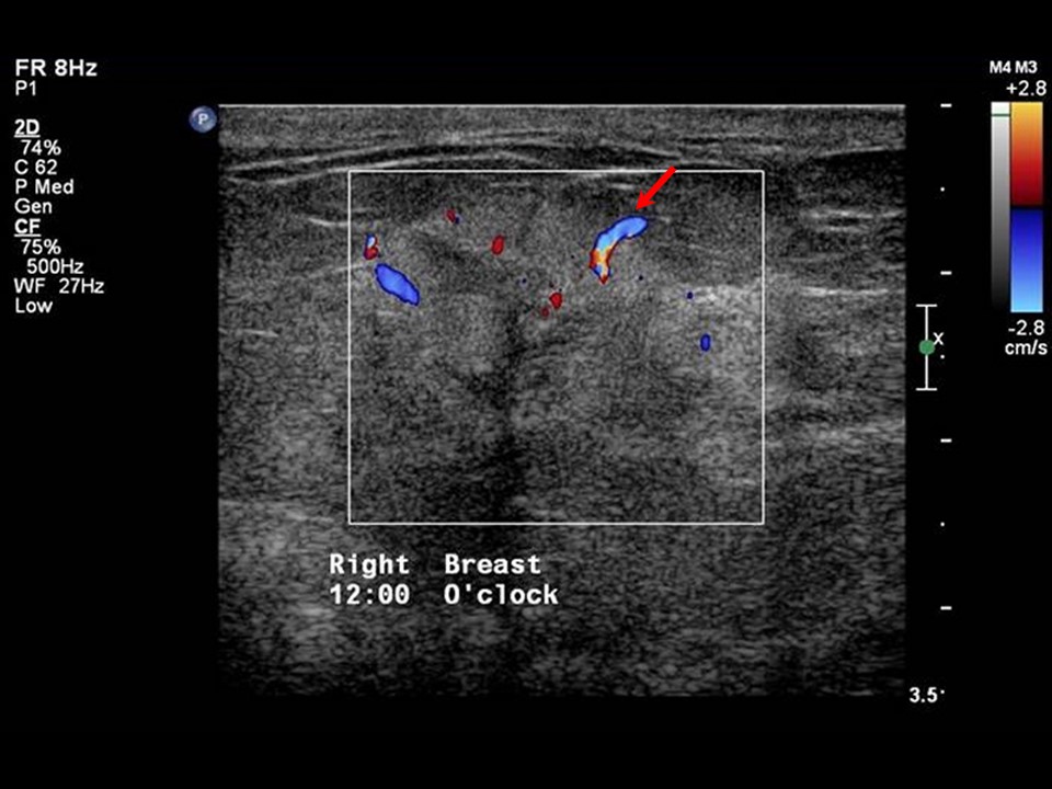
| ‣ Calcifications: | None |
| ‣ Associated features: | Internal vascularity |
| ‣ Special cases: | None |
BI-RADS:
BI-RADS Category (2010): 1 (negative)BI-RADS Category (2012): 5 (highly suggestive of malignancy)
Further assessment:
Further assessment advised: Referral for cytologyCytology:
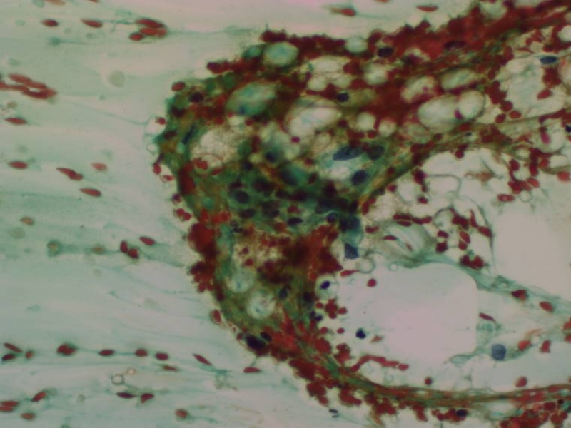 |
| Cytology features: | |
| ‣ Type of sample: | FNAC |
| ‣ Site of biopsy: | |
| • Laterality: | Right |
| • Quadrant: | 12 oclock, superior areolar margin |
| • Localization technique: | Palpation |
| • Nature of aspirate: | Whitish |
| ‣ Cytological description: | Cellular smears with pleomorphic malignant cells with prominent nucleoli within many stromal fibrous fragments |
| ‣ Reporting category: | Malignant |
| ‣ Diagnosis: | Carcinoma |
| ‣ Comments: | None |
Histopathology:
Breast-conserving surgery
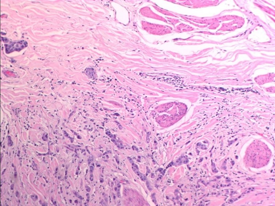 |
| Histopathology features: | |
| ‣ Specimen type: | Breast-conserving surgery |
| ‣ Laterality: | Right |
| ‣ Macroscopy: | Cut surface shows a fairly well-circumscribed tumour (2.5 × 1.5 × 2.5 cm) |
| ‣ Histological type: | Invasive carcinoma of no special type |
| ‣ Histological grade: | Grade 3 (3 + 3 + 2 = 8) |
| ‣ Mitosis: | 15 |
| ‣ Maximum invasive tumour size: | 2.5 cm in greatest dimension |
| ‣ Lymph node status: | 5/20 |
| ‣ Peritumoural lymphovascular invasion: | Present |
| ‣ DCIS/EIC: | Solid and comedo DCIS high grade |
| ‣ Margins: | Free of tumour |
| ‣ Pathological stage: | pT2N2 |
| ‣ Biomarkers: | |
| ‣ Comments: |
Case summary:
| Postmenopausal woman with increased risk of developing breast cancer presented with right breast lump. Mammography and breast ultrasound dated 2012 reveals a new irregular lesion in the right breast. Compared with an earlier mammogram dated 2010, follow-up reveals developing asymmetry in the right breast. Interval change is seen. Diagnosed as highly suggestive of malignancy, BI-RADS 5 on imaging, as carcinoma on cytology, and as invasive breast carcinoma of no special type, pT2N2 on histopathology. |
Learning points:
|