


Home / Training / Manuals / Atlas of breast cancer early detection / Cases
Atlas of breast cancer early detection
Filter by language: English / Русский
Go back to the list of case studies
.png) Click on the pictures to magnify and display the legends
Click on the pictures to magnify and display the legends
BI-RADS Category (right breast): 2 (benign)
| Case number: | 183 |
| Age: | 71 |
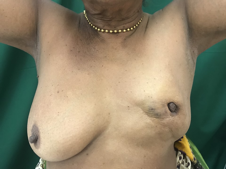
| Clinical presentation: | Postmenopausal woman with average risk of developing breast cancer presented with a left breast lump noticed 6 months ago. On examination, abnormal unilateral reduction in the size of the left breast was noted. A hard lump was palpable in the lower inner quadrant of the left breast with skin retraction seen. The left nipple was retracted. |
 |
Mammography:
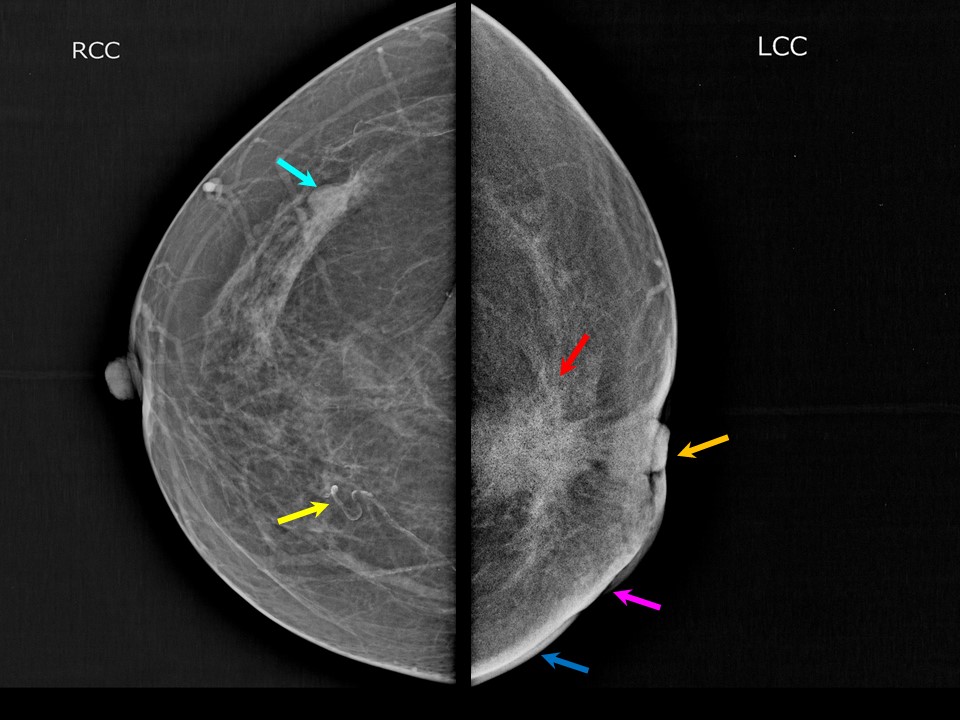 |  |
| Breast composition: | ACR category a (the breasts are almost entirely fatty) | Mammography features: |
| ‣ Location of the lesion: | Left breast, central portion of the breast at 8-10 o'clock, anterior, middle, and posterior thirds |
| ‣ Mass: | |
| • Number: | 1 |
| • Size: | 2.6 × 2.0 cm |
| • Shape: | Irregular |
| • Margins: | Spiculated |
| • Density: | High |
| ‣ Calcifications: | |
| • Typically benign: | Vascular calcification in both breasts |
| • Suspicious: | None |
| • Distribution: | None |
| ‣ Architectural distortion: | Present |
| ‣ Asymmetry: | None |
| ‣ Intramammary node: | Present |
| ‣ Skin lesion: | None |
| ‣ Solitary dilated duct: | None |
| ‣ Associated features: | Left nipple retraction, skin thickening, skin retraction, and axillary lymphadenopathy |
| Breast composition: | ACR category a (the breasts are almost entirely fatty) | Mammography features: |
| ‣ Location of the lesion: | Right breast, upper outer quadrant at 10 oclock, middle third |
| ‣ Mass: | |
| • Number: | 1 |
| • Size: | 1 cm |
| • Shape: | Round |
| • Margins: | Circumscribed |
| • Density: | Equal |
| ‣ Calcifications: | |
| • Typically benign: | Vascular calcification in both breasts |
| • Suspicious: | None |
| • Distribution: | None |
| ‣ Architectural distortion: | None |
| ‣ Asymmetry: | None |
| ‣ Intramammary node: | None |
| ‣ Skin lesion: | None |
| ‣ Solitary dilated duct: | None |
| ‣ Associated features: | None |
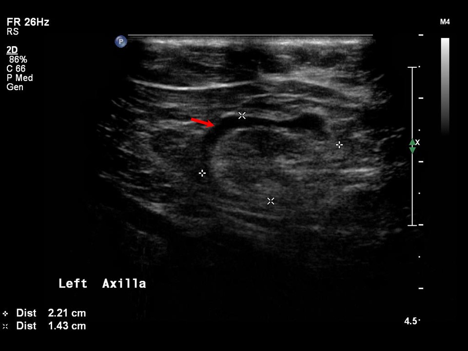
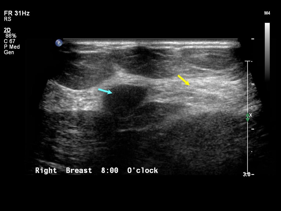
Ultrasound:
 |  |
 | 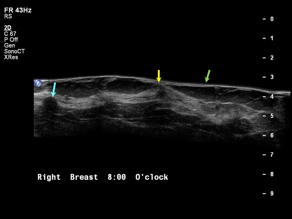 |
| Ultrasound features: Left breast, lower inner quadrant at 8 oclock | |
| ‣ Mass | |
| • Location: | Left breast, lower inner quadrant at 8 oclock |
| • Number: | 1 |
| • Size: | 2.8 × 2.5 cm |
| • Shape: | Irregular |
| • Orientation: | Not parallel |
| • Margins: | Spiculated |
| • Echo pattern: | Hypoechoic |
| • Posterior features: | Strong posterior shadowing |
| ‣ Calcifications: | None |
| ‣ Associated features: | Skin thickening and enlarged left axillary node |
| ‣ Special cases: | None |
| Ultrasound features: Right breast, upper outer quadrant at 10 oclock | |
| ‣ Mass | |
| • Location: | Right breast, upper outer quadrant at 10 oclock |
| • Number: | 1 |
| • Size: | 0.8 cm |
| • Shape: | Round |
| • Orientation: | Parallel |
| • Margins: | Circumscribed |
| • Echo pattern: | Hypoechoic |
| • Posterior features: | No posterior features |
| ‣ Calcifications: | None |
| ‣ Associated features: | None |
| ‣ Special cases: | None |
BI-RADS:
BI-RADS Category (left breast): 5 (highly suggestive of malignancy)BI-RADS Category (right breast): 2 (benign)
Further assessment:
Further assessment advised: Referral for cytology and for core biopsyCytology:
 |
| Cytology features: | |
| ‣ Type of sample: | FNAC |
| ‣ Site of biopsy: | |
| • Laterality: | Left |
| • Quadrant: | Inner lower |
| • Localization technique: | Palpation |
| • Nature of aspirate: | Whitish |
| ‣ Cytological description: | Smears are very cellular. They show loosely cohesive clusters and single isolated pleomorphic malignant ductal epithelial cells |
| ‣ Reporting category: | Malignant |
| ‣ Diagnosis: | Malignant |
| ‣ Comments: | None |
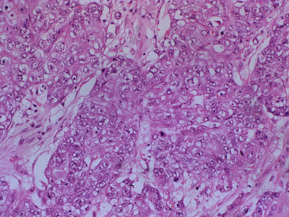
Histopathology:
Core needle biopsy
 |
| Histopathology features: | |
| ‣ Specimen type: | Core needle biopsy |
| ‣ Laterality: | Right |
| ‣ Macroscopy: | Three whitish cores (1.5 cm, 1.5 cm, and 1 cm long) |
| ‣ Histological type: | Invasive breast carcinoma of no special type |
| ‣ Histological grade: | Grade 3 (3 + 3 + 2 = 8) |
| ‣ Mitosis: | 6 |
| ‣ Maximum invasive tumour size: | |
| ‣ Lymph node status: | |
| ‣ Peritumoural lymphovascular invasion: | |
| ‣ DCIS/EIC: | |
| ‣ Margins: | |
| ‣ Pathological stage: | |
| ‣ Biomarkers: | |
| ‣ Comments: |
Case summary:
| Postmenopausal woman presented with left breast lump. On examination, abnormal unilateral reduction in the size of the left breast was noted, with a hard lump in the left breast, skin retraction, and left nipple retraction. Diagnosed as left breast carcinoma with skin thickening and retraction, and left nipple retraction, BI-RADS 5 on imaging and as invasive breast carcinoma of no special type on histopathology. |
Learning points:
|