


Home / Training / Manuals / Atlas of breast cancer early detection / Cases
Atlas of breast cancer early detection
Filter by language: English / Русский
Go back to the list of case studies
.png) Click on the pictures to magnify and display the legends
Click on the pictures to magnify and display the legends
| Case number: | 054 |
| Age: | 62 |
| Clinical presentation: | Postmenopausal woman with average risk of developing breast cancer presented with right nipple retraction of duration 4 months. She had also noted a lump in the right breast a month before presentation. Examination revealed a hard lump, 6 cm in diameter, fixed to the rest of the breast tissue but not to the underlying muscles. Axilla was unremarkable. |
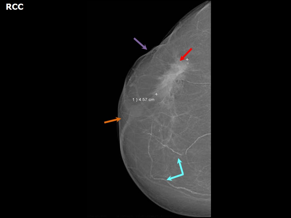
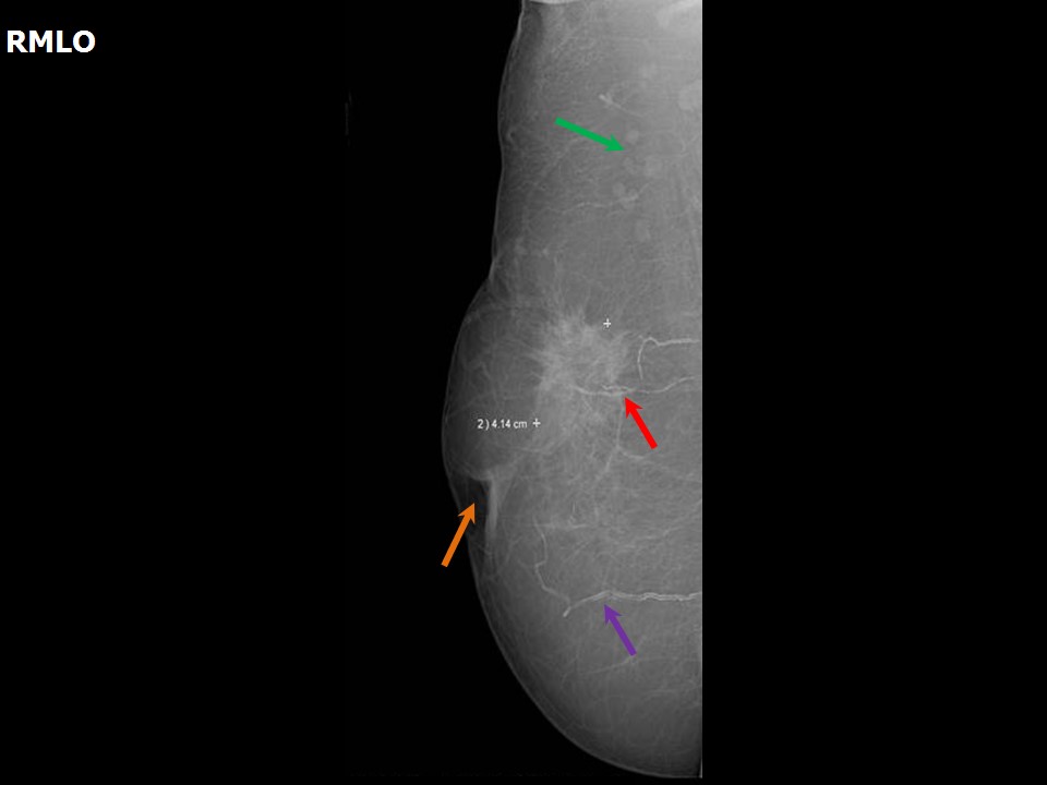
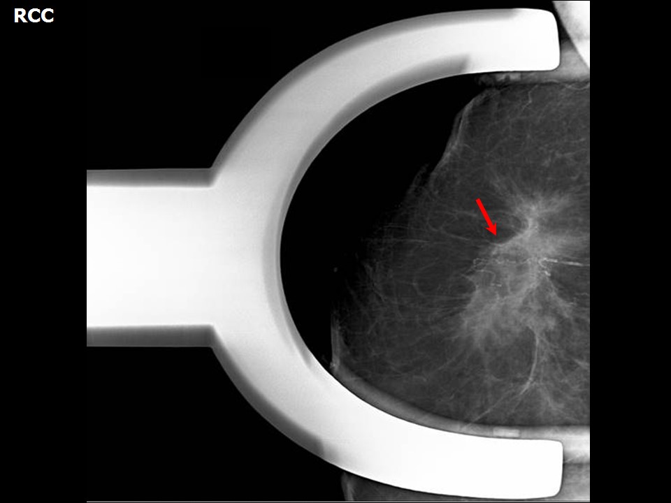
Mammography:
 |  |
 |
| Breast composition: | ACR category a (the breasts are almost entirely fatty) | Mammography features: |
| ‣ Location of the lesion: | Right breast, upper outer quadrant at 1011 oclock, middle third |
| ‣ Mass: | |
| • Number: | 1 |
| • Size: | 4.6 × 4.1 cm |
| • Shape: | Irregular |
| • Margins: | Spiculated |
| • Density: | High |
| ‣ Calcifications: | |
| • Typically benign: | Vascular calcification |
| • Suspicious: | None |
| • Distribution: | None |
| ‣ Architectural distortion: | Present |
| ‣ Asymmetry: | None |
| ‣ Intramammary node: | None |
| ‣ Skin lesion: | None |
| ‣ Solitary dilated duct: | None |
| ‣ Associated features: | Skin retraction and nipple retraction |
Ultrasound:
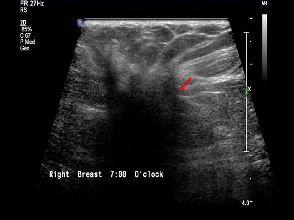 | 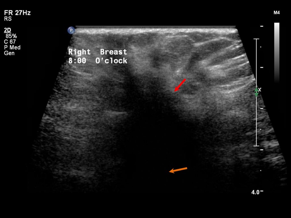 |
| Ultrasound features: Right breast, upper outer quadrant at 10 oclock position | |
| ‣ Mass | |
| • Location: | Right breast, upper outer quadrant at 10 oclock position |
| • Number: | 1 |
| • Size: | > 3.0 cm in greatest dimension |
| • Shape: | Irregular |
| • Orientation: | Not parallel |
| • Margins: | Spiculated |
| • Echo pattern: | Hypoechoic |
| • Posterior features: | Posterior shadowing |
| ‣ Calcifications: | None |
| ‣ Associated features: | Architectural distortion and skin retraction |
| ‣ Special cases: | None |
BI-RADS:
BI-RADS Category: 5 (highly suggestive of malignancy)Further assessment:
Further assessment advised: Referral for cytologyCytology:
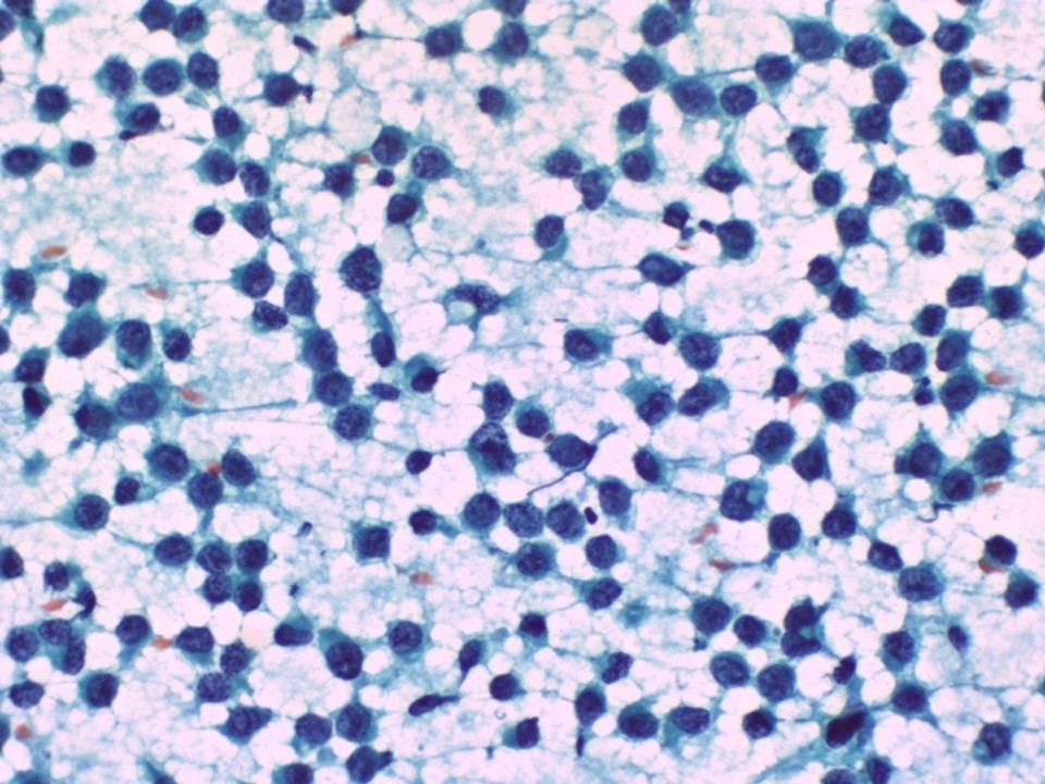 |
| Cytology features: | |
| ‣ Type of sample: | FNAC |
| ‣ Site of biopsy: | |
| • Laterality: | Right |
| • Quadrant: | Upper outer |
| • Localization technique: | Palpation |
| • Nature of aspirate: | whitish |
| ‣ Cytological description: | Smears show mainly dispersed malignant cells. These cells have a high N:C ratio. Many cells show small cytoplasmic vacuoles in the cytoplasm |
| ‣ Reporting category: | Malignant |
| ‣ Diagnosis: | Carcinoma |
| ‣ Comments: | None |
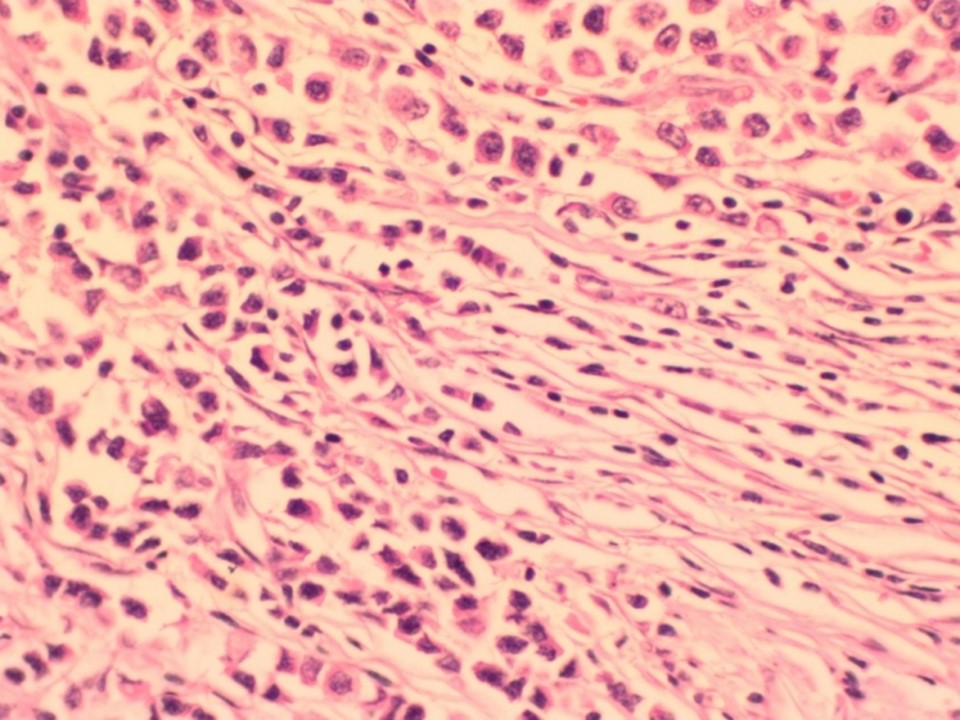
Histopathology:
MRM
 |
| Histopathology features: | |
| ‣ Specimen type: | MRM |
| ‣ Laterality: | Right |
| ‣ Macroscopy: | Cut surface shows a solid, whitish, firm to hard tumour (4.0 × 3.0 × 1.5 cm) with infiltrating margins. Tumour is situated at a depth of 2.0 cm below the skin. The base is 0.5 cm away from the tumour. A separate specimen labelled muscle from base was a single nodular brownish firm tissue bit (2.0 × 1.5 × 1.0 cm) |
| ‣ Histological type: | Infiltrating lobular carcinoma right breast |
| ‣ Histological grade: | Grade 1 (3 + 1 + 1 = 5) |
| ‣ Mitosis: | 4 |
| ‣ Maximum invasive tumour size: | 4.0 cm in greatest dimension |
| ‣ Lymph node status: | 5/30 |
| ‣ Peritumoural lymphovascular invasion: | Present |
| ‣ DCIS/EIC: | Absent; lobular carcinoma in situ not seen |
| ‣ Margins: | Free of tumour. Sections from the muscle from base show only skeletal muscle fibres and are free of tumour invasion |
| ‣ Pathological stage: | pT2N2 |
| ‣ Biomarkers: | |
| ‣ Comments: |
Case summary:
| Postmenopausal woman presented with right breast lump and right nipple retraction. Diagnosed as right breast carcinoma with skin and nipple retraction, BI-RADS 5 on imaging, as breast carcinoma on cytology, and as invasive lobular carcinoma, pT2N2 on histopathology. |
Learning points:
|