


Home / Training / Manuals / Atlas of breast cancer early detection / Cases
Atlas of breast cancer early detection
Filter by language: English / Русский
Go back to the list of case studies
.png) Click on the pictures to magnify and display the legends
Click on the pictures to magnify and display the legends
BI-RADS Category (2016): 4C (high suspicion for malignancy)
| Case number: | 113 |
| Age: | 51 |
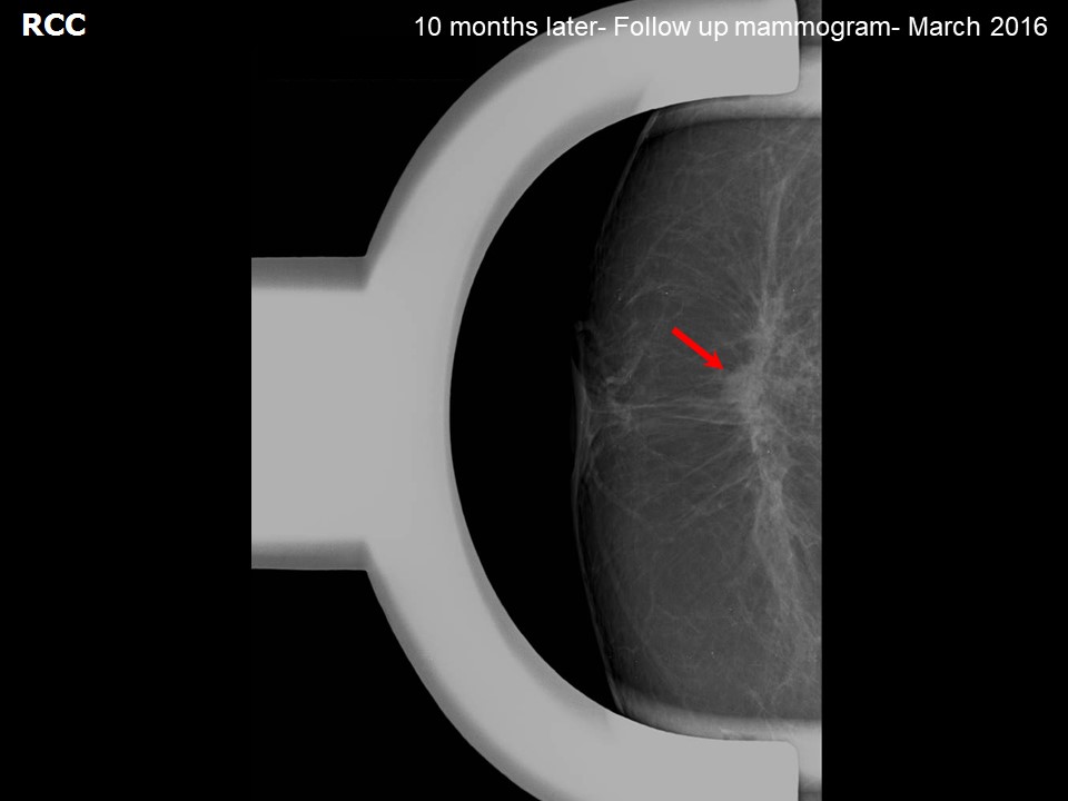
| Clinical presentation: | Postmenopausal woman with increased risk of developing breast cancer presents for a follow-up because she feels pain in her right breast. She has a strong family history of breast carcinoma. Clinical examination reveals different para-areolar consistencies on the right and left breasts. Comparison was made with a previous mammogram dated May 2015 (BI-RADS 2). |
Mammography:
 |  |
| Breast composition: | ACR category b (there are scattered areas of fibroglandular density) | Mammography features: |
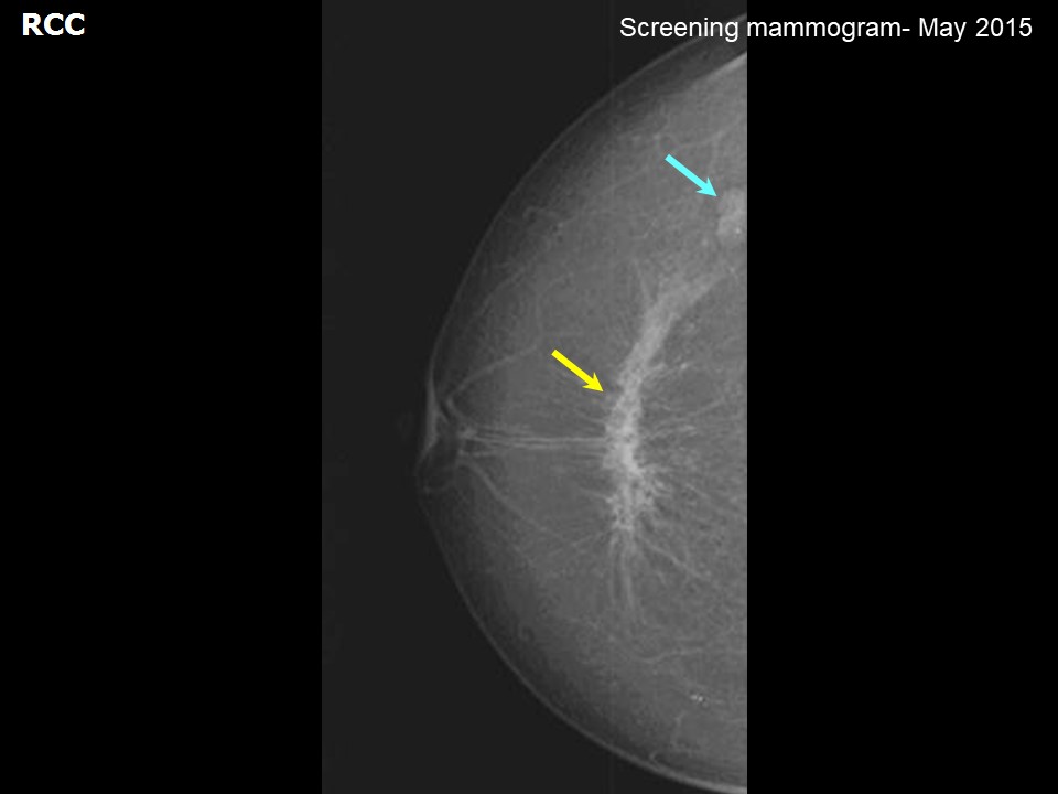
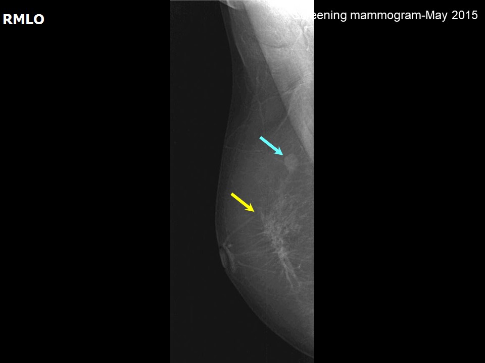
| ‣ Location of the lesion: | May 2015: Right breast, upper outer quadrant at 10 oclock, posterior third |
| ‣ Mass: | |
| • Number: | 1 |
| • Size: | 1.2 cm in greatest dimension |
| • Shape: | Oval |
| • Margins: | Circumscribed |
| • Density: | Equal |
| ‣ Calcifications: | |
| • Typically benign: | None |
| • Suspicious: | None |
| • Distribution: | None |
| ‣ Architectural distortion: | None |
| ‣ Asymmetry: | None |
| ‣ Intramammary node: | None |
| ‣ Skin lesion: | None |
| ‣ Solitary dilated duct: | None |
| ‣ Associated features: | None |
 |  |
 |  |
 |
| Breast composition: | ACR category b (there are scattered areas of fibroglandular density) | Mammography features: |
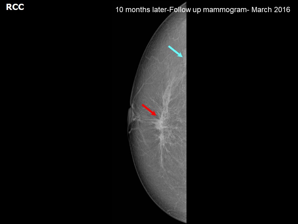
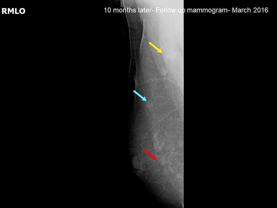
| ‣ Location of the lesion: | 2016: Right breast, upper outer quadrant at 10 oclock, posterior third |
| ‣ Mass: | |
| • Number: | 1 |
| • Size: | 1.2 cm in greatest dimension |
| • Shape: | Oval (stable lesion since 2015) |
| • Margins: | Circumscribed |
| • Density: | Equal |
| ‣ Calcifications: | |
| • Typically benign: | None |
| • Suspicious: | None |
| • Distribution: | None |
| ‣ Architectural distortion: | None |
| ‣ Asymmetry: | None |
| ‣ Intramammary node: | None |
| ‣ Skin lesion: | None |
| ‣ Solitary dilated duct: | None |
| ‣ Associated features: | None |
| Breast composition: | ACR category b (there are scattered areas of fibroglandular density) | Mammography features: |
| ‣ Location of the lesion: | 2016: Right breast, lower inner quadrant at 5 oclock, middle third |
| ‣ Mass: | |
| • Number: | 1 (this is a new finding, developing asymmetry) |
| • Size: | 0.9 cm in greatest dimension |
| • Shape: | Irregular |
| • Margins: | Indistinct |
| • Density: | Equal |
| ‣ Calcifications: | |
| • Typically benign: | None |
| • Suspicious: | None |
| • Distribution: | None |
| ‣ Architectural distortion: | None |
| ‣ Asymmetry: | None |
| ‣ Intramammary node: | None |
| ‣ Skin lesion: | None |
| ‣ Solitary dilated duct: | None |
| ‣ Associated features: | None |
Ultrasound:
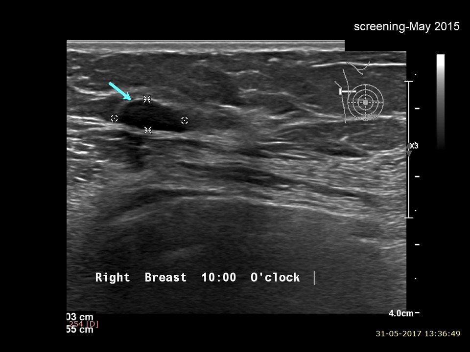 |
| Ultrasound features: May 2015: Right breast, upper outer quadrant at 10 oclock | |
| ‣ Mass | |
| • Location: | May 2015: Right breast, upper outer quadrant at 10 oclock |
| • Number: | 1 |
| • Size: | 1.0 × 0.5 cm |
| • Shape: | Oval |
| • Orientation: | Parallel |
| • Margins: | Circumscribed |
| • Echo pattern: | Hypoechoic |
| • Posterior features: | No posterior features |
| ‣ Calcifications: | None |
| ‣ Associated features: | None |
| ‣ Special cases: | None |
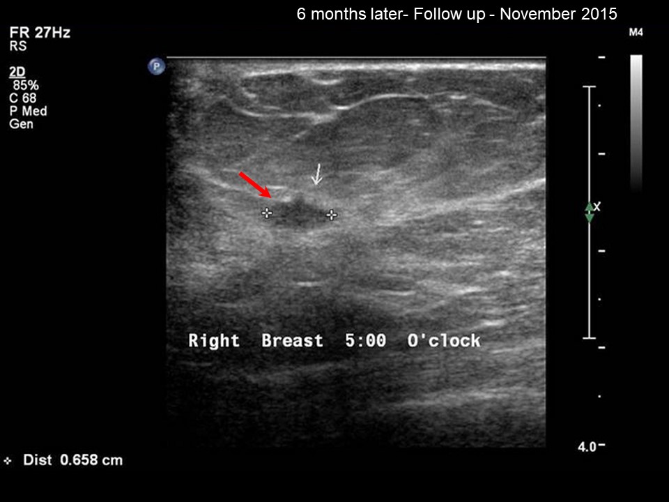 | 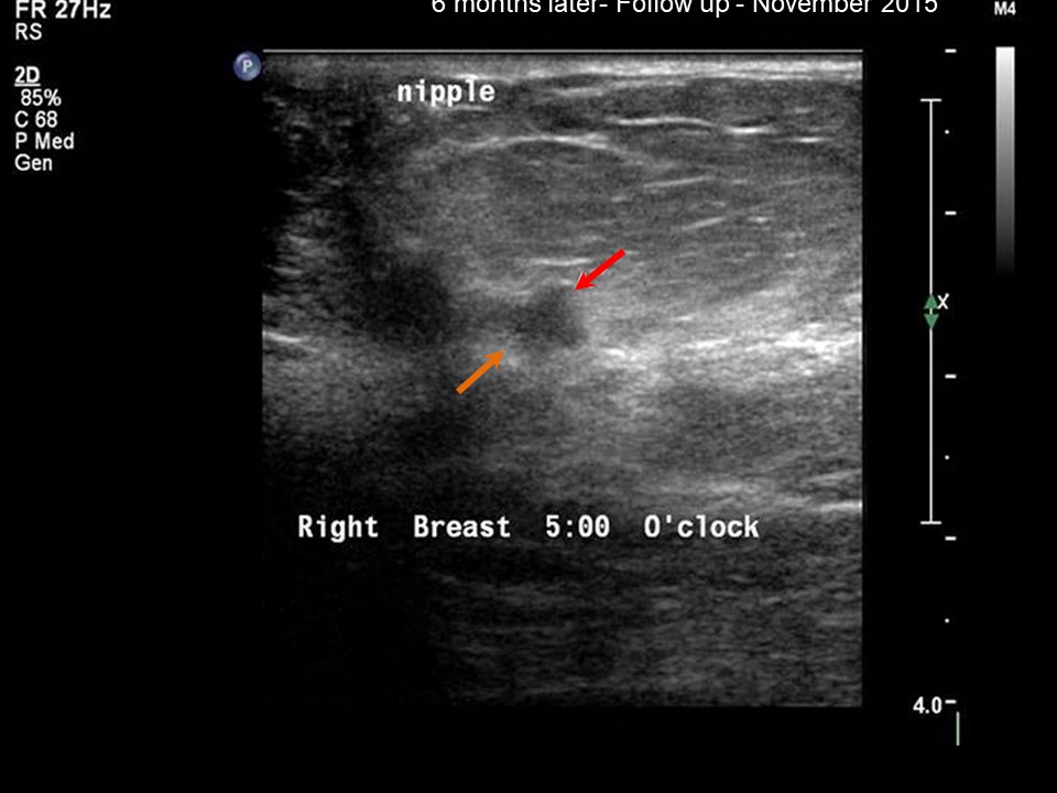 |
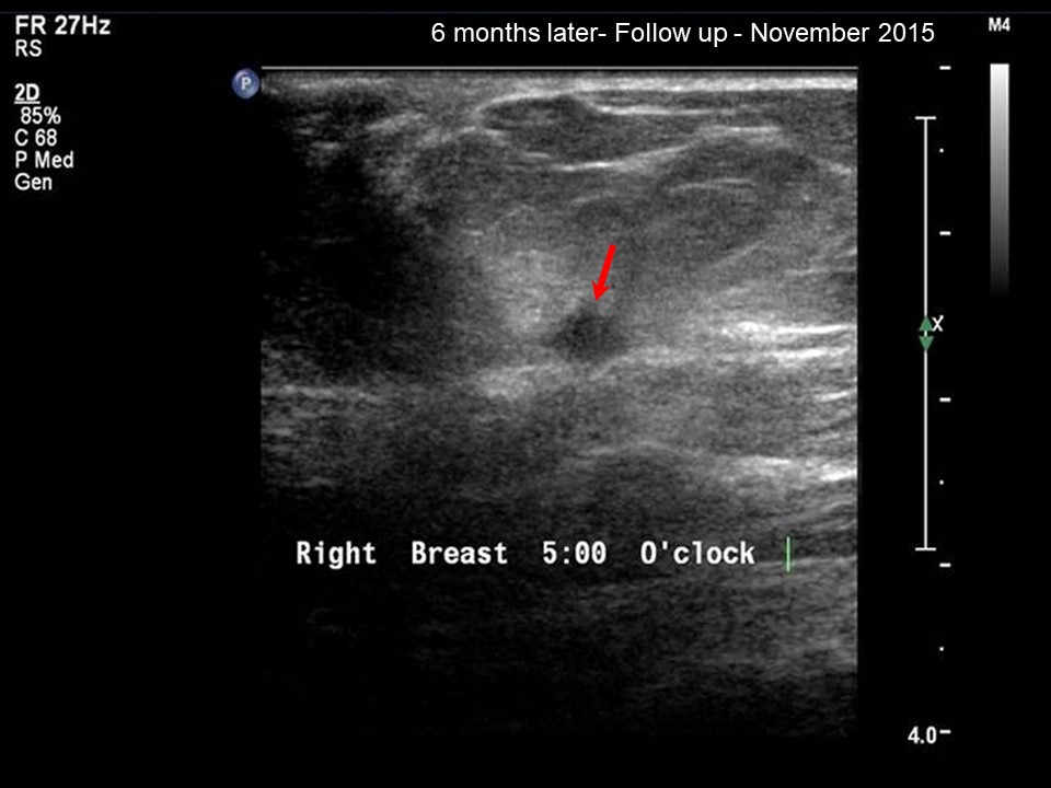 |
| Ultrasound features: November 2015: Right breast, lower inner quadrant at 5 oclock | |
| ‣ Mass | |
| • Location: | November 2015: Right breast, lower inner quadrant at 5 oclock |
| • Number: | 1 (suspicious interval change) |
| • Size: | 0.6 cm in greatest dimension |
| • Shape: | Irregular |
| • Orientation: | Parallel |
| • Margins: | Angular |
| • Echo pattern: | Hypoechoic |
| • Posterior features: | No posterior features |
| ‣ Calcifications: | None |
| ‣ Associated features: | None |
| ‣ Special cases: | None |
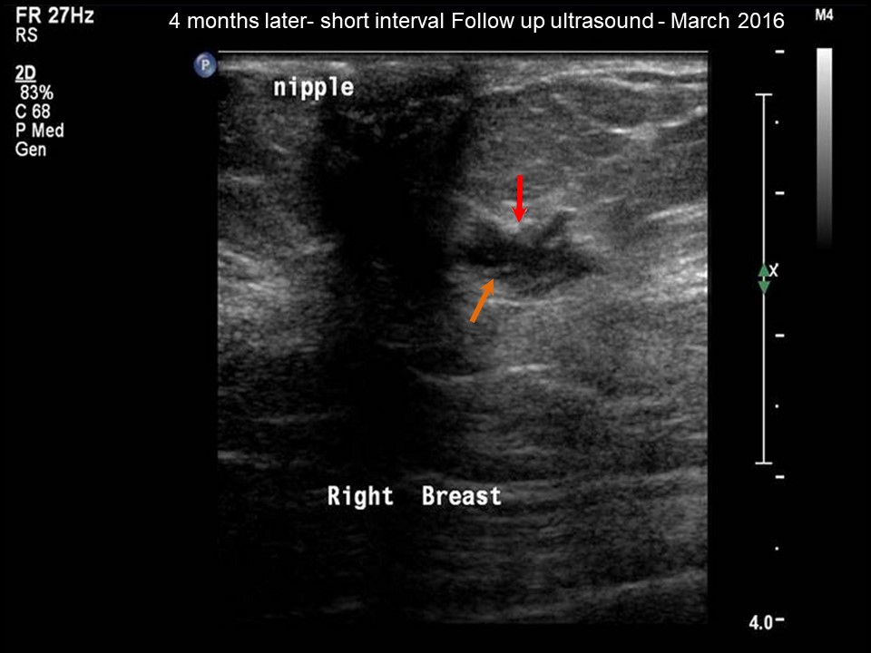 | 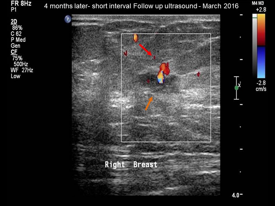 |
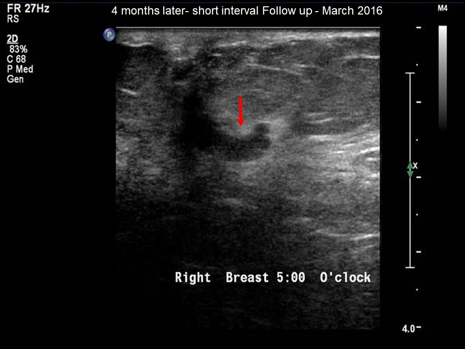 |  |
| Ultrasound features: March 2016: Right breast, lower inner quadrant at 5 oclock | |
| ‣ Mass | |
| • Location: | March 2016: Right breast, lower inner quadrant at 5 oclock |
| • Number: | 1 |
| • Size: | 0.9 cm in greatest dimension |
| • Shape: | Irregular |
| • Orientation: | Parallel |
| • Margins: | Angular and spiculated |
| • Echo pattern: | Hypoechoic |
| • Posterior features: | No posterior features |
| ‣ Calcifications: | None |
| ‣ Associated features: | Dilated duct |
| ‣ Special cases: | Developing asymmetry interval cancer |
BI-RADS:
BI-RADS Category (2015): 2 (benign)BI-RADS Category (2016): 4C (high suspicion for malignancy)
Further assessment:
Further assessment advised: Referral for cytologyCytology:
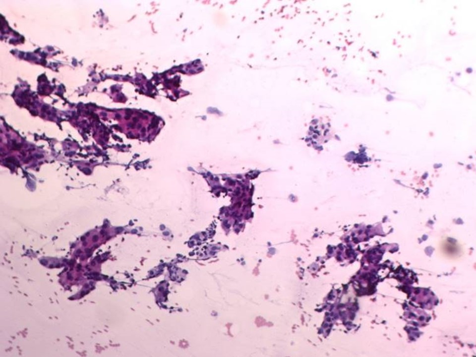 |
| Cytology features: | |
| ‣ Type of sample: | Ultrasound-guided FNAC |
| ‣ Site of biopsy: | |
| • Laterality: | Right |
| • Quadrant: | 5 oclock |
| • Localization technique: | 1.5 |
| • Nature of aspirate: | Whitish |
| ‣ Cytological description: | Smears reveal many dyscohesive clusters and many singly scattered large malignant cells with marked nuclear polymorphism, hyperchromasia, and abundant cytoplasm |
| ‣ Reporting category: | Malignant |
| ‣ Diagnosis: | Ductal carcinoma high grade |
| ‣ Comments: | None |
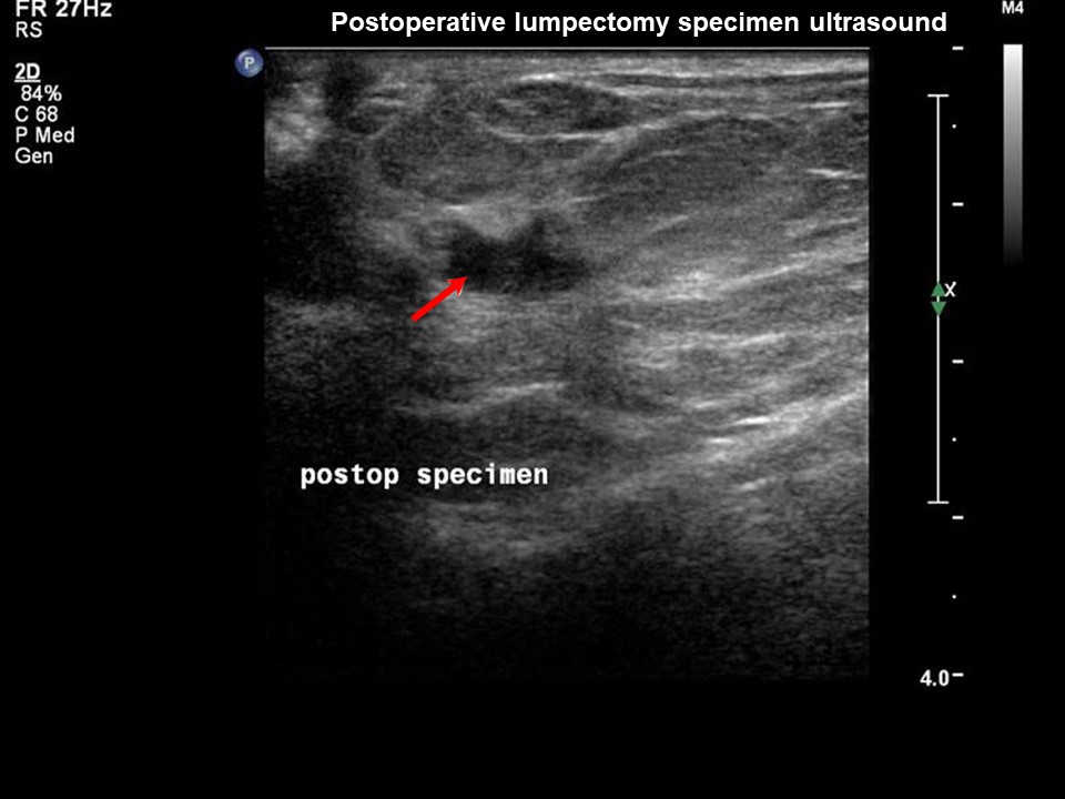
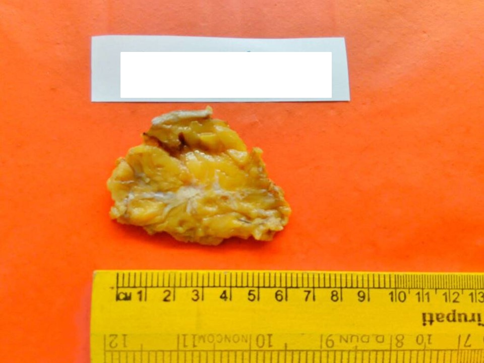
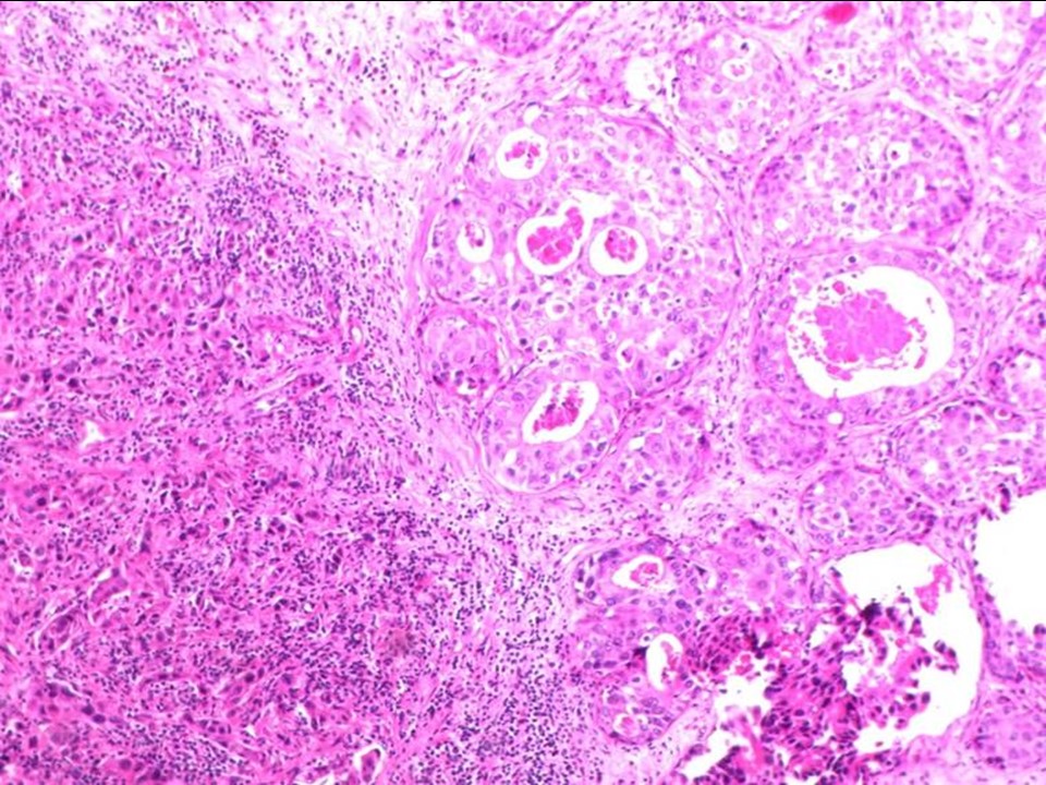
Histopathology:
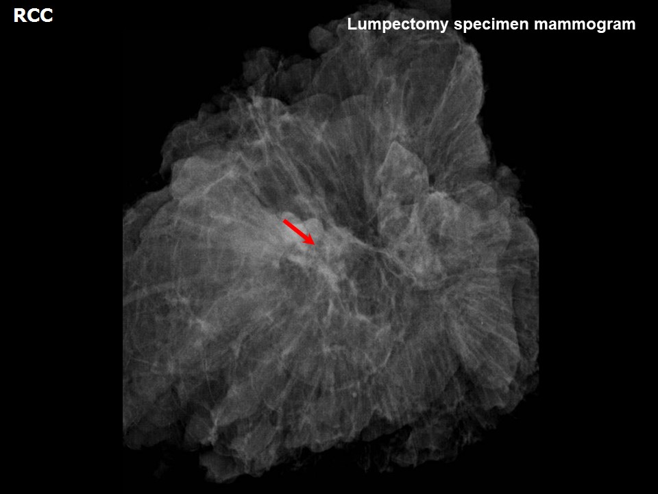
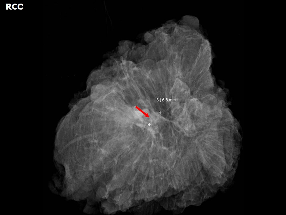
Breast-conserving surgery
 |  |
 |  |
| Histopathology features: | |
| ‣ Specimen type: | Breast-conserving surgery |
| ‣ Laterality: | |
| ‣ Macroscopy: | |
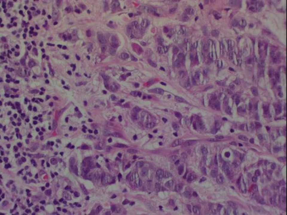
| ‣ Histological type: | Invasive breast carcinoma of no special type |
| ‣ Histological grade: | Grade 3 |
| ‣ Mitosis: | |
| ‣ Maximum invasive tumour size: | 3 |
| ‣ Lymph node status: | 1/15 |
| ‣ Peritumoural lymphovascular invasion: | Present |
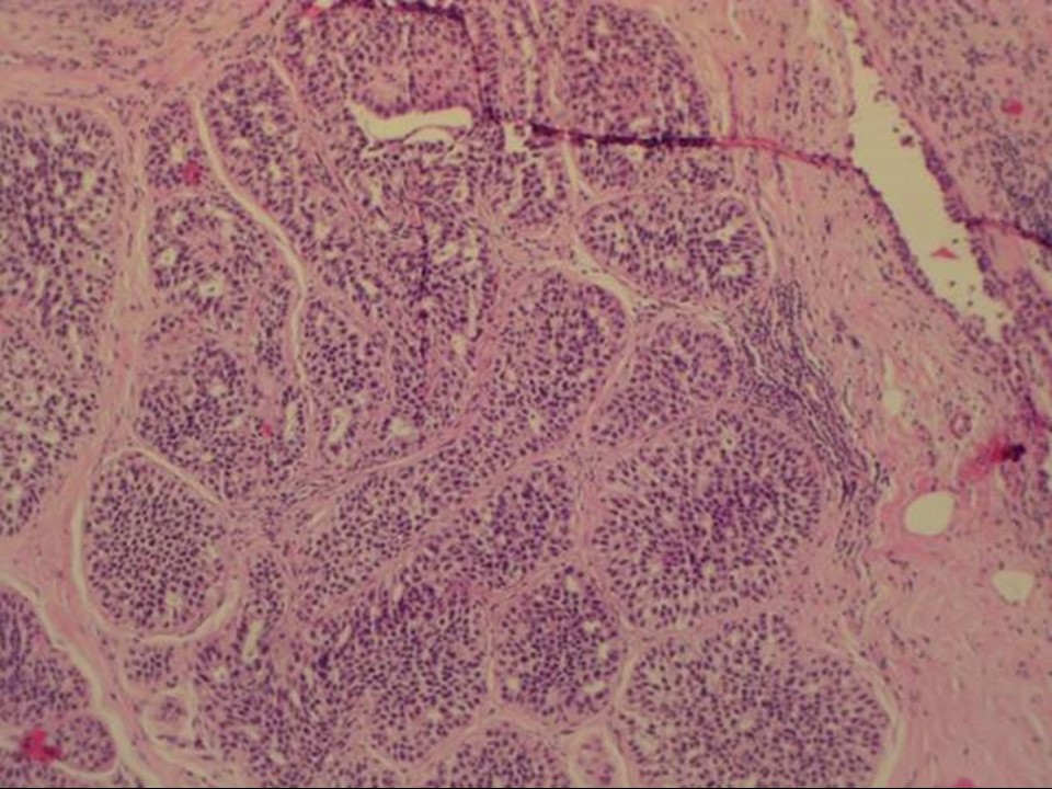
| ‣ DCIS/EIC: | EIC present high grade with necrosis |
| ‣ Margins: | Free of tumour |
| ‣ Pathological stage: | pT2N1 |
| ‣ Biomarkers: | |
| ‣ Comments: |
Case summary:
| Postmenopausal woman with increased risk of developing breast cancer presented for a follow-up because of pain in her right breast. Clinical examination reveals different para-areolar consistencies in the right and left breasts. Mammography and breast ultrasound dated 2016 reveal a new irregular lesion in right breast. Compared with a previous mammogram dated 2015, follow-up reveals developing asymmetry in the right breast. Interval change is seen. Diagnosed as highly suspicious for malignancy, BI-RADS 4C on imaging, as carcinoma on cytology, and as invasive breast carcinoma of no special type, pT2N1 on histopathology. |
Learning points:
|