


Home / Training / Manuals / Atlas of breast cancer early detection / Cases
Atlas of breast cancer early detection
Filter by language: English / Русский
Go back to the list of case studies
.png) Click on the pictures to magnify and display the legends
Click on the pictures to magnify and display the legends
BI-RADS Category (2014): 5 (highly suggestive of malignancy)
| Case number: | 100 |
| Age: | 66 |
| Clinical presentation: | Postmenopausal woman with increased risk of developing breast cancer because of a family history of breast cancer presented for follow-up. Previous mammography was normal. Examination revealed symmetrical diffuse thickening in the upper outer quadrants of both breasts. |
Mammography:
 |
| Breast composition: | ACR category a (the breasts are almost entirely fatty) | Mammography features: |
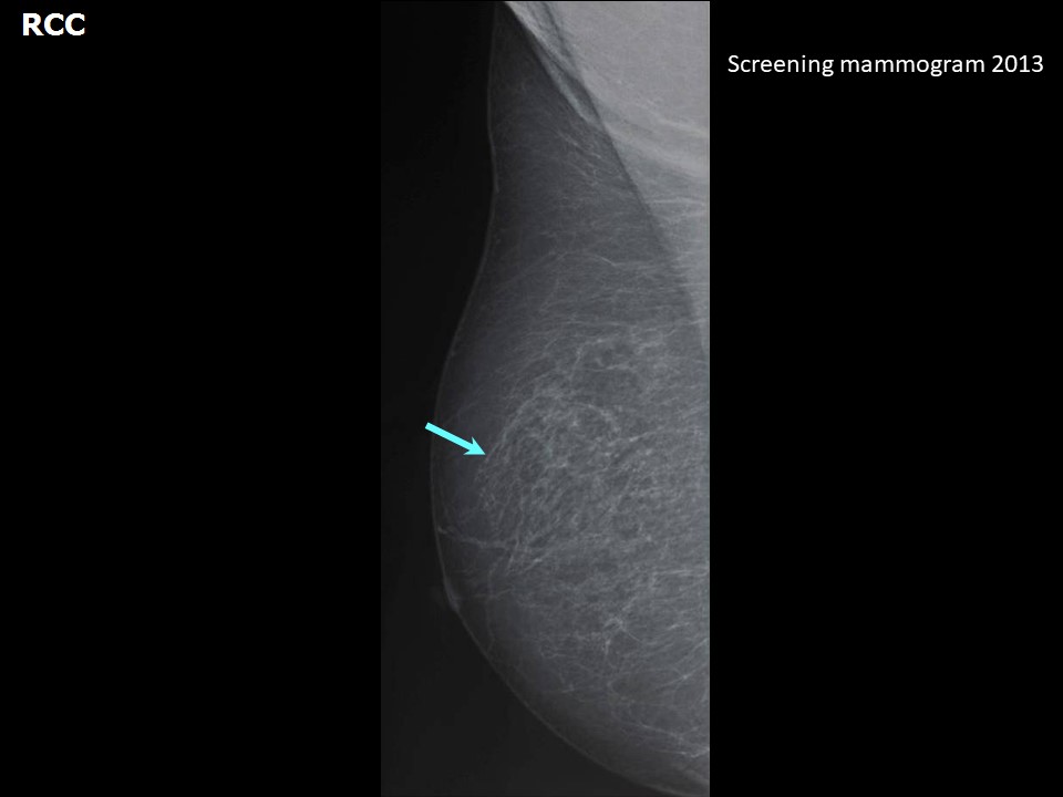
| ‣ Location of the lesion: | October 2013: Right breast |
| ‣ Mass: | |
| • Number: | 0 |
| • Size: | None |
| • Shape: | None |
| • Margins: | None |
| • Density: | None |
| ‣ Calcifications: | |
| • Typically benign: | None |
| • Suspicious: | None |
| • Distribution: | None |
| ‣ Architectural distortion: | None |
| ‣ Asymmetry: | None |
| ‣ Intramammary node: | None |
| ‣ Skin lesion: | None |
| ‣ Solitary dilated duct: | None |
| ‣ Associated features: | None |
 |  |
| Breast composition: | ACR category a (the breasts are almost entirely fatty) | Mammography features: |
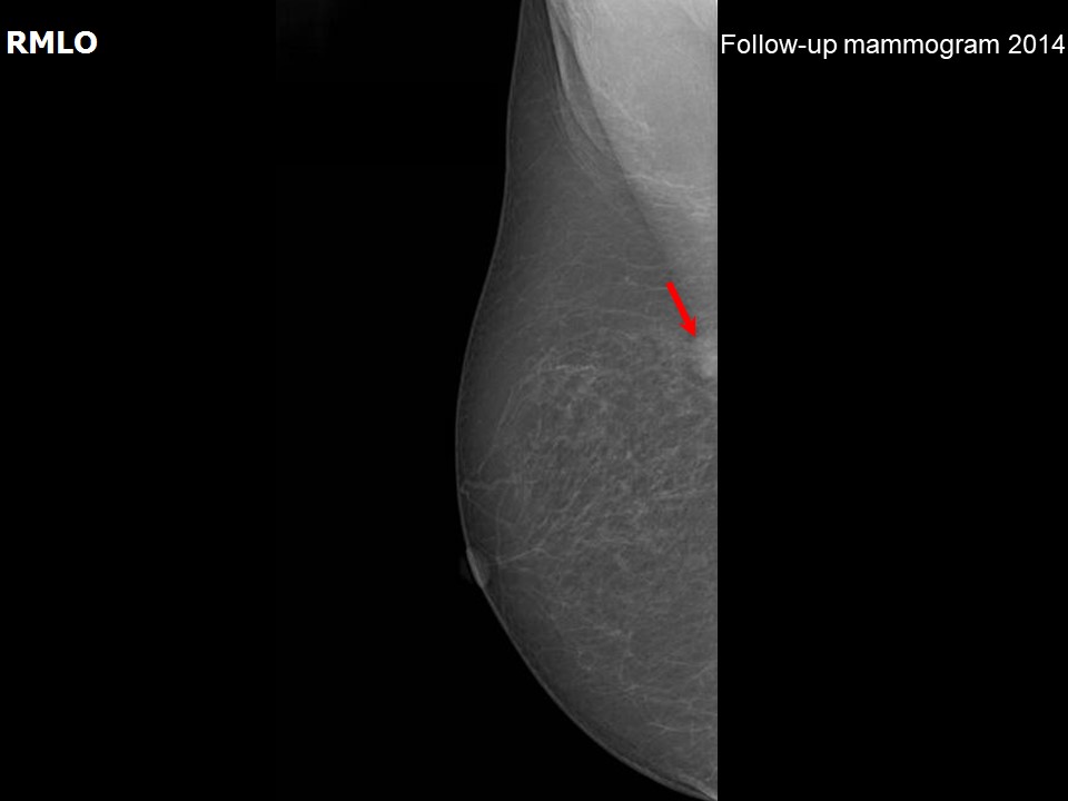
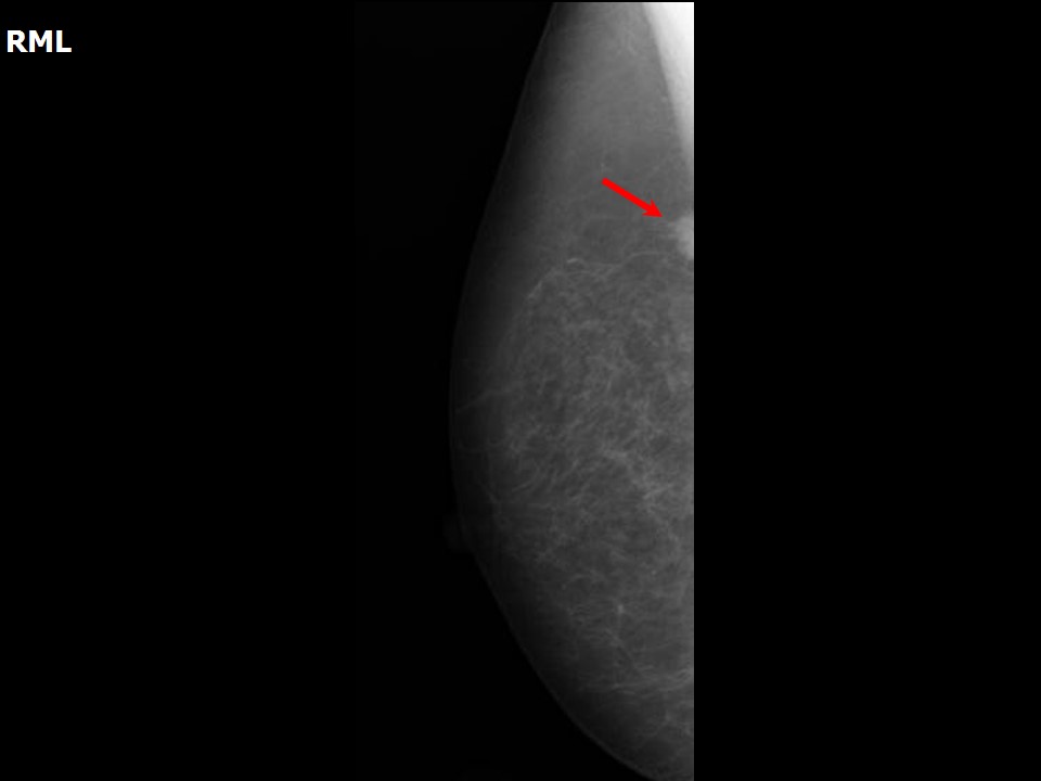
| ‣ Location of the lesion: | April 2014: Right breast, upper quadrants at 12 oclock, posterior third |
| ‣ Mass: | |
| • Number: | 1 (this is a new finding, developing asymmetry) |
| • Size: | 1.0 cm in greatest dimension |
| • Shape: | Irregular |
| • Margins: | Spiculated |
| • Density: | High |
| ‣ Calcifications: | |
| • Typically benign: | None |
| • Suspicious: | None |
| • Distribution: | None |
| ‣ Architectural distortion: | None |
| ‣ Asymmetry: | None |
| ‣ Intramammary node: | None |
| ‣ Skin lesion: | None |
| ‣ Solitary dilated duct: | None |
| ‣ Associated features: | None |
Ultrasound:
 |  |
 |
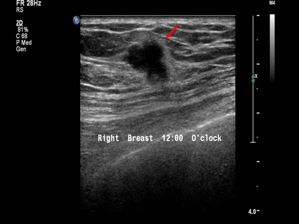
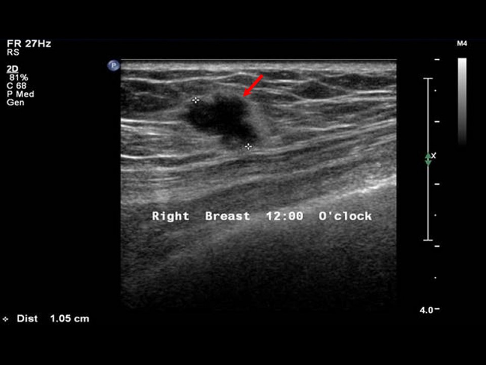
| Ultrasound features: April 2014, Right breast, upper quadrants at 12 oclock | |
| ‣ Mass | |
| • Location: | April 2014, Right breast, upper quadrants at 12 oclock |
| • Number: | 1 |
| • Size: | 1.0 cm in greatest dimension |
| • Shape: | Irregular |
| • Orientation: | Not parallel |
| • Margins: | Spiculated |
| • Echo pattern: | Hypoechoic |
| • Posterior features: | No posterior features |
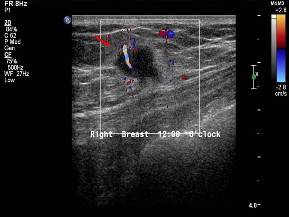
| ‣ Calcifications: | None |
| ‣ Associated features: | Internal vascularity |
| ‣ Special cases: | None |
BI-RADS:
BI-RADS Category (2013): 1 (negative)BI-RADS Category (2014): 5 (highly suggestive of malignancy)
Further assessment:
Further assessment advised: Referral for cytologyCytology:
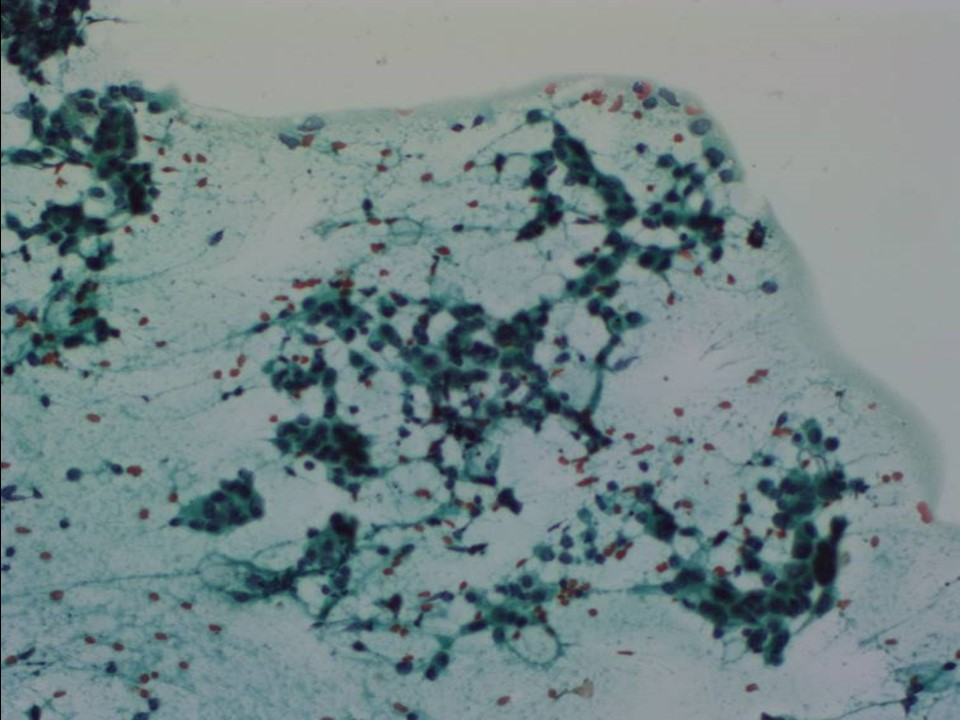 |
| Cytology features: | |
| ‣ Type of sample: | FNAC |
| ‣ Site of biopsy: | |
| • Laterality: | Right |
| • Quadrant: | Upper quadrant at 12 oclock |
| • Localization technique: | Palpation |
| • Nature of aspirate: | Whitish |
| ‣ Cytological description: | Cellular smears with dyscohesive clusters of malignant ductal cells. Single isolated malignant cells also seen |
| ‣ Reporting category: | Malignant |
| ‣ Diagnosis: | Carcinoma low grade |
| ‣ Comments: | None |
Histopathology:
Breast-conserving surgery
 |
| Histopathology features: | |
| ‣ Specimen type: | Breast-conserving surgery |
| ‣ Laterality: | Right |
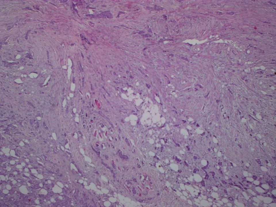
| ‣ Macroscopy: | Tumour (1.5 × 1.5 × 1.0 cm) with an irregular stellate outline |
| ‣ Histological type: | Invasive carcinoma of no special type |
| ‣ Histological grade: | Grade 2 (3 + 2 + 1 = 6) |
| ‣ Mitosis: | 5 |
| ‣ Maximum invasive tumour size: | 1.5 cm |
| ‣ Lymph node status: | 0/22 |
| ‣ Peritumoural lymphovascular invasion: | Not identified |
| ‣ DCIS/EIC: | Cribriform DCIS low grade |
| ‣ Margins: | Free of tumour |
| ‣ Pathological stage: | pT1cN0 |
| ‣ Biomarkers: | |
| ‣ Comments: | Extensive hyalinization of the stroma in the centre of the tumour |
Case summary:
| Postmenopausal woman with increased risk of developing breast cancer presented for follow-up screening. Examination revealed symmetrical diffuse thickening in the upper outer quadrants of both breasts. Mammography and breast ultrasound dated 2014 revealed a new suspicious irregular lesion in the right breast. Compared with previous mammograms dated 2013, follow-up reveals developing asymmetry in the right breast. Interval change is seen. Diagnosed as highly suggestive of malignancy, BI-RADS 5 on imaging, as carcinoma on cytology, and as invasive breast carcinoma of no special type, pT1cN0 on histopathology. |
Learning points:
|