


Home / Training / Manuals / Atlas of breast cancer early detection / Cases
Atlas of breast cancer early detection
Filter by language: English / Русский
Go back to the list of case studies
.png) Click on the pictures to magnify and display the legends
Click on the pictures to magnify and display the legends
| Case number: | 099 |
| Age: | 77 |
| Clinical presentation: | Woman was diagnosed with right breast carcinoma and underwent MRM in 2000. She noticed a left breast lump 68 months ago. On clinical examination, a hard lump was palpable in the upper quadrant of the left breast. |
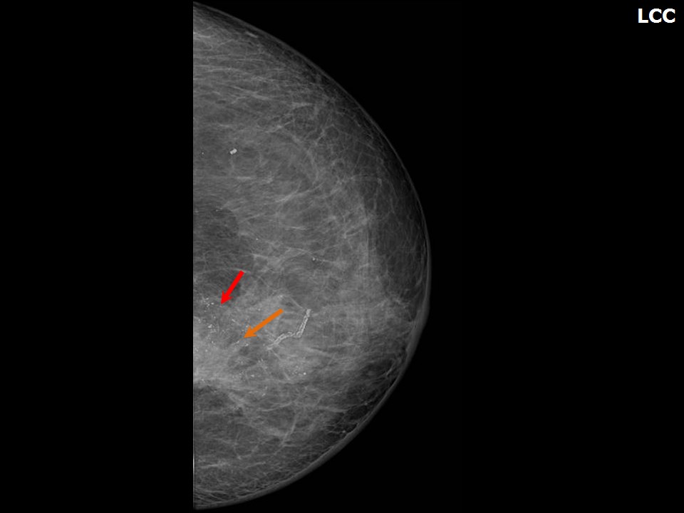
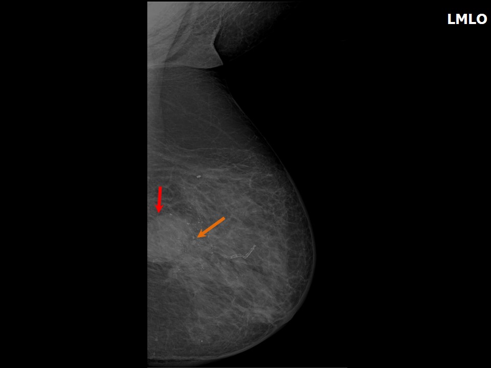
Mammography:
 |  |
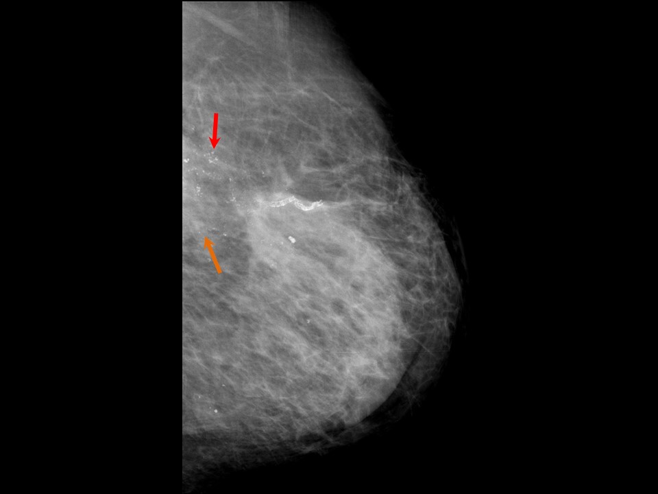 |
| Breast composition: | ACR category b (there are scattered areas of fibroglandular density) | Mammography features: |
| ‣ Location of the lesion: | Left breast, upper inner quadrant at 1011 oclock, posterior third |
| ‣ Mass: | |
| • Number: | 1 |
| • Size: | 4.5 × 3.4 cm |
| • Shape: | Irregular |
| • Margins: | Indistinct |
| • Density: | High |
| ‣ Calcifications: | |
| • Typically benign: | None |
| • Suspicious: | Fine pleomorphic |
| • Distribution: | Regional |
| ‣ Architectural distortion: | Present |
| ‣ Asymmetry: | Focal |
| ‣ Intramammary node: | None |
| ‣ Skin lesion: | None |
| ‣ Solitary dilated duct: | None |
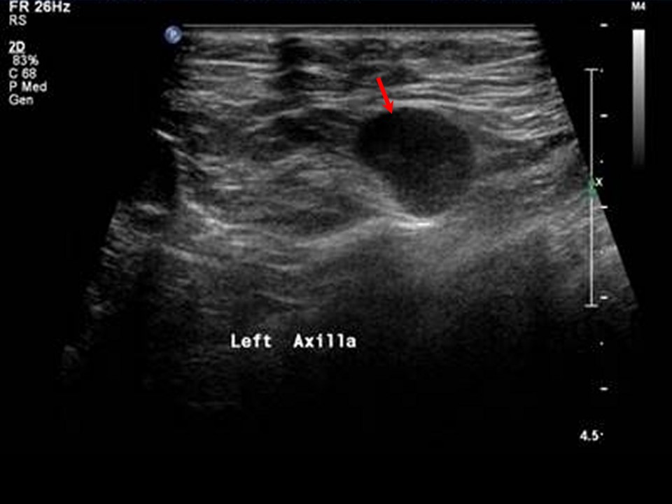
| ‣ Associated features: | Trabecular thickening, architectural distortion, pleomorphic microcalcifications, skin thickening, and axillary adenopathy nodes with thickened cortex and loss of fatty hilum |
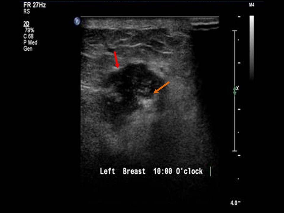
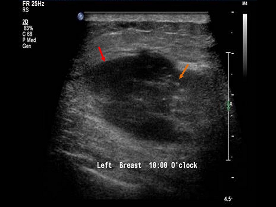
Ultrasound:
 |  |
 |
| Ultrasound features: Left breast, upper inner quadrant at 1011 oclock position | |
| ‣ Mass | |
| • Location: | Left breast, upper inner quadrant at 1011 oclock position |
| • Number: | 23, grouped together |
| • Size: | Largest 4.5 × 2.5 cm |
| • Shape: | Irregular |
| • Orientation: | Not parallel |
| • Margins: | Angular |
| • Echo pattern: | Hypoechoic |
| • Posterior features: | No posterior features |
| ‣ Calcifications: | Present in mass |
| ‣ Associated features: | Architectural distortion, skin thickening, internal vascularity, oedema, and axillary lymphadenopathy |
| ‣ Special cases: | None |
BI-RADS:
BI-RADS Category (Right MRM): 5 (highly suggestive of malignancy)Further assessment:
Further assessment advised: Referral for cytology and for core biopsyCytology:
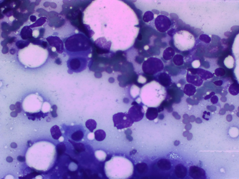 |
| Cytology features: | |
| ‣ Type of sample: | FNAC |
| ‣ Site of biopsy: | |
| • Laterality: | Left |
| • Quadrant: | Upper inner |
| • Localization technique: | Palpation |
| • Nature of aspirate: | Whitish |
| ‣ Cytological description: | Loosely cohesive sheets of large malignant cells with marked nuclear pleomorphism |
| ‣ Reporting category: | Malignant |
| ‣ Diagnosis: | Carcinoma high grade |
| ‣ Comments: | None |
Histopathology:
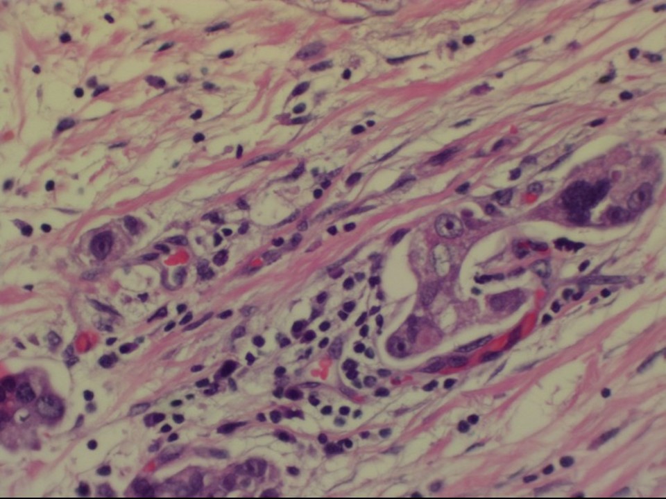
Core needle biopsy
 |
| Histopathology features: | |
| ‣ Specimen type: | Core needle biopsy |
| ‣ Laterality: | Left |
| ‣ Macroscopy: | 5 cores |
| ‣ Histological type: | Invasive carcinoma of no special type |
| ‣ Histological grade: | Grade 3 (3 + 3 + 2 = 8) |
| ‣ Mitosis: | 18 |
| ‣ Maximum invasive tumour size: | |
| ‣ Lymph node status: | |
| ‣ Peritumoural lymphovascular invasion: | |
| ‣ DCIS/EIC: | |
| ‣ Margins: | |
| ‣ Pathological stage: | |
| ‣ Biomarkers: | |
| ‣ Comments: |
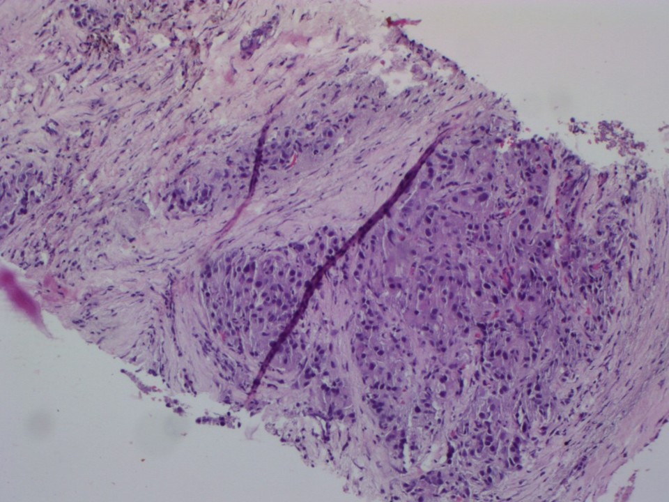
MRM
 | 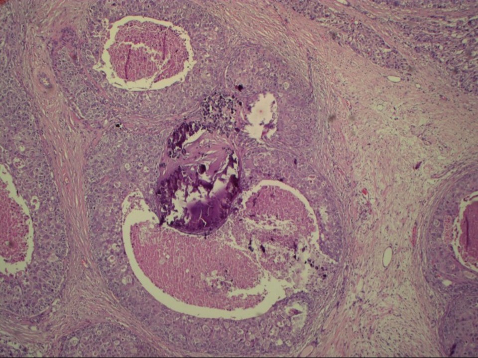 |
| Histopathology features: | |
| ‣ Specimen type: | MRM |
| ‣ Laterality: | Left |
| ‣ Macroscopy: | On serial sectioning, a firm greyish white tumour (6.8 × 4.5 × 2.8 cm) is identified. It is located 1.4 cm below the skin and 0.2 cm from the base |
| ‣ Histological type: | Invasive carcinoma of no special type |
| ‣ Histological grade: | Grade 3 (3 + 3 + 3 = 9) |
| ‣ Mitosis: | 22 |
| ‣ Maximum invasive tumour size: | 6.8 cm |
| ‣ Lymph node status: | 3/19 |
| ‣ Peritumoural lymphovascular invasion: | Present |
| ‣ DCIS/EIC: | Comedo DCIS high grade |
| ‣ Margins: | Free of tumour |
| ‣ Pathological stage: | pT3N3 |
| ‣ Biomarkers: | |
| ‣ Comments: | The remaining breast tissue shows extensive areas of fibrosis |
Case summary:
| Postmenopausal woman, operated 15 years ago for right breast carcinoma, presented with a large, hard left breast lump. Diagnosed as left breast carcinoma with clustered pleomorphic microcalcifications, BI-RADS 5 on imaging, as carcinoma on cytology, and as invasive breast carcinoma of no special type, pT3N3 on histopathology. |
Learning points:
|