


Home / Training / Manuals / Atlas of breast cancer early detection / Cases
Atlas of breast cancer early detection
Filter by language: English / Русский
Go back to the list of case studies
.png) Click on the pictures to magnify and display the legends
Click on the pictures to magnify and display the legends
| Case number: | 186 |
| Age: | 74 |
| Clinical presentation: | Postmenopausal woman with average risk of developing breast cancer presented with a right breast lump noticed more than 2 years ago. She now also had right nipple retraction. |
 |
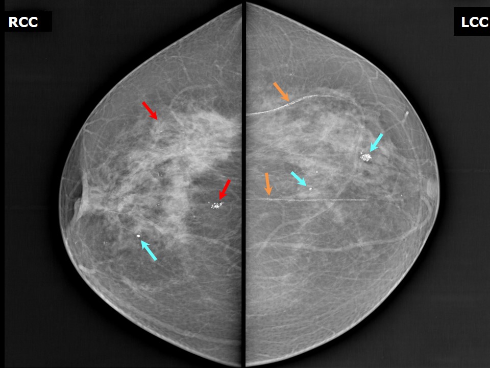
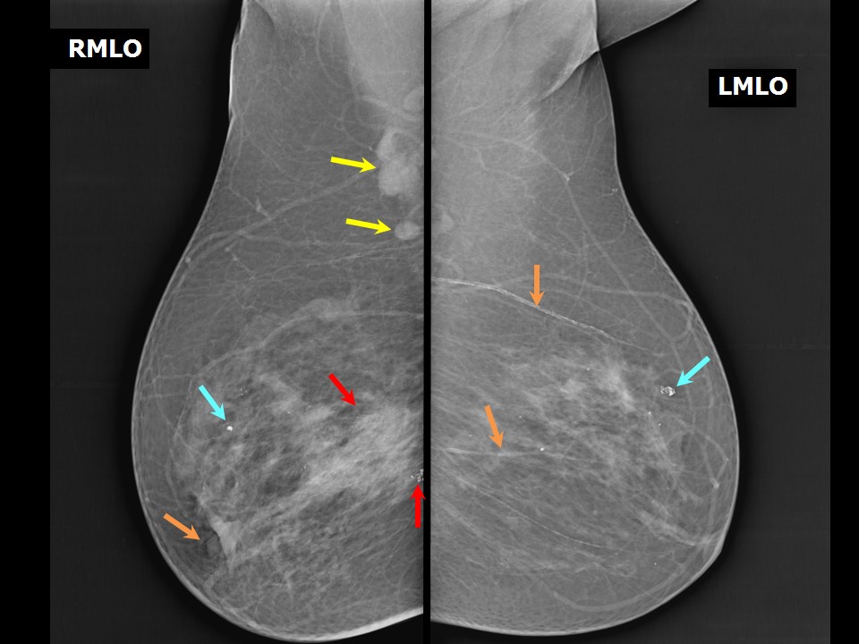
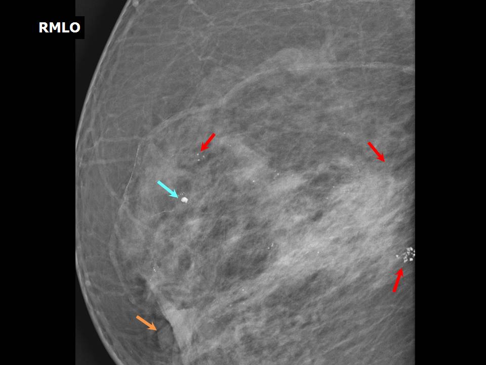
Mammography:
 |  |
 |  |
| Breast composition: | ACR category b (there are scattered areas of fibroglandular density) | Mammography features: |
| ‣ Location of the lesion: | Right breast, upper outer quadrant at 910 oclock, anterior, middle, and posterior thirds |
| ‣ Mass: | |
| • Number: | 0 |
| • Size: | None |
| • Shape: | None |
| • Margins: | None |
| • Density: | None |
| ‣ Calcifications: | |
| • Typically benign: | None |
| • Suspicious: | Fine microcalcifications and clustered pleomorphic microcalcifications |
| • Distribution: | Segmental, grouped |
| ‣ Architectural distortion: | None |
| ‣ Asymmetry: | Focal |
| ‣ Intramammary node: | None |
| ‣ Skin lesion: | None |
| ‣ Solitary dilated duct: | None |
| ‣ Associated features: | Enlarged axillary nodes, focal asymmetry, trabecular thickening, fine microcalcifications, and clustered pleomorphic microcalcifications |
| Breast composition: | ACR category b (there are scattered areas of fibroglandular density) | Mammography features: |
| ‣ Location of the lesion: | Right breast, upper inner quadrant at 1 oclock, middle third |
| ‣ Mass: | |
| • Number: | 0 |
| • Size: | None |
| • Shape: | None |
| • Margins: | None |
| • Density: | None |
| ‣ Calcifications: | |
| • Typically benign: | Yes, round |
| • Suspicious: | None |
| • Distribution: | Diffuse |
| ‣ Architectural distortion: | None |
| ‣ Asymmetry: | None |
| ‣ Intramammary node: | None |
| ‣ Skin lesion: | None |
| ‣ Solitary dilated duct: | None |
| ‣ Associated features: | None |
| Breast composition: | ACR category b (there are scattered areas of fibroglandular density) | Mammography features: |
| ‣ Location of the lesion: | Left breast, upper outer quadrant at 1 oclock, anterior and middle thirds |
| ‣ Mass: | |
| • Number: | 0 |
| • Size: | None |
| • Shape: | None |
| • Margins: | None |
| • Density: | None |
| ‣ Calcifications: | |
| • Typically benign: | Yes, round, coarse heterogeneous, and vascular calcifications |
| • Suspicious: | None |
| • Distribution: | Diffuse |
| ‣ Architectural distortion: | None |
| ‣ Asymmetry: | None |
| ‣ Intramammary node: | None |
| ‣ Skin lesion: | None |
| ‣ Solitary dilated duct: | None |
| ‣ Associated features: | None |
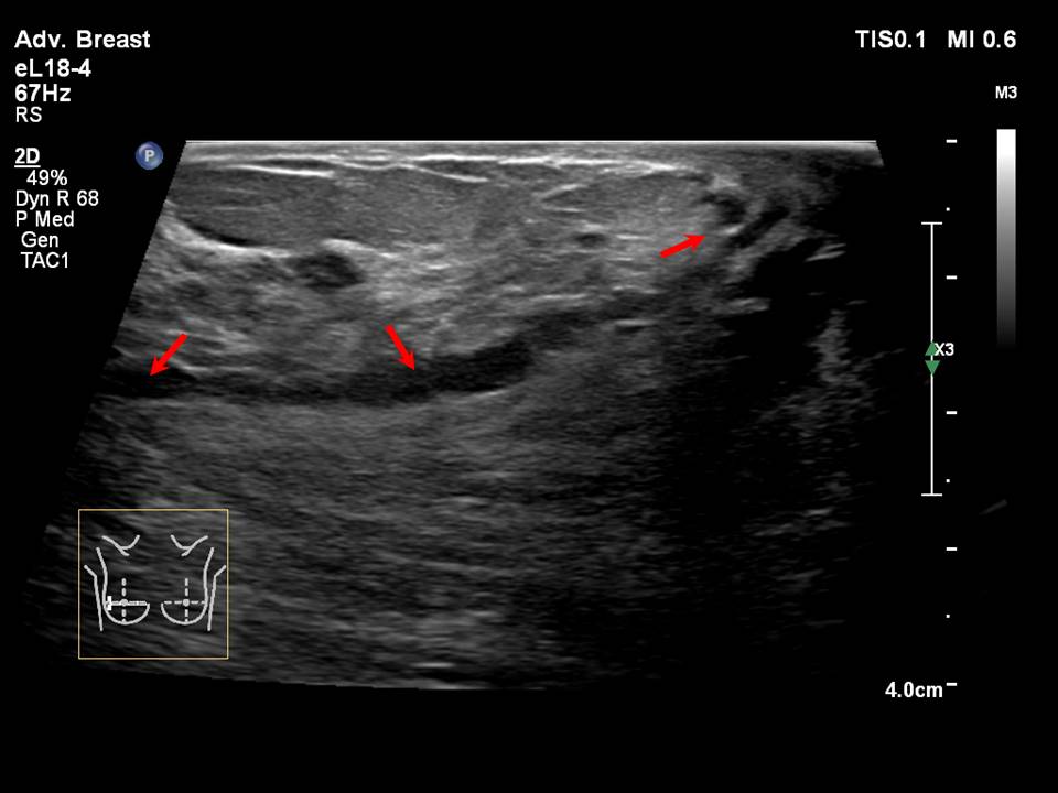
Ultrasound:
 |  |
 | 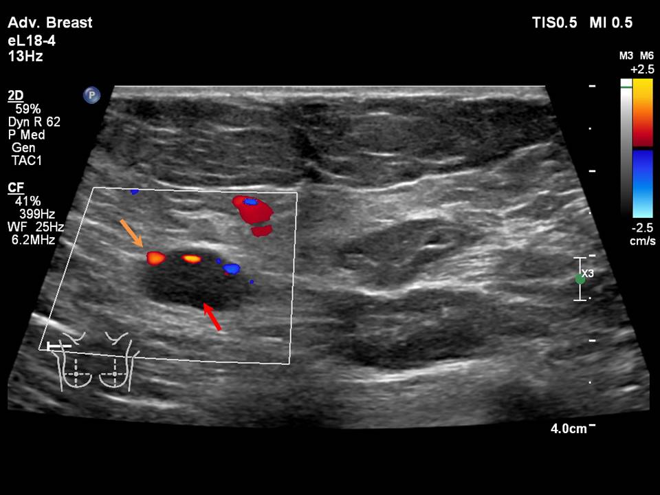 |
| Ultrasound features: None | |
| ‣ Mass | |
| • Location: | None |
| • Number: | 0 |
| • Size: | None |
| • Shape: | None |
| • Orientation: | None |
| • Margins: | None |
| • Echo pattern: | None |
| • Posterior features: | No posterior features |
| ‣ Calcifications: | Intraductal microcalcifications |
| ‣ Associated features: | Focal asymmetry, dilated ducts, axillary dysmorphic lymphadenopathy with non-hilar vascularity on colour flow mapping |
| ‣ Special cases: | None |
BI-RADS:
BI-RADS Category: 5 (highly suggestive of malignancy)Further assessment:
Further assessment advised: Referral for cytologyCytology:
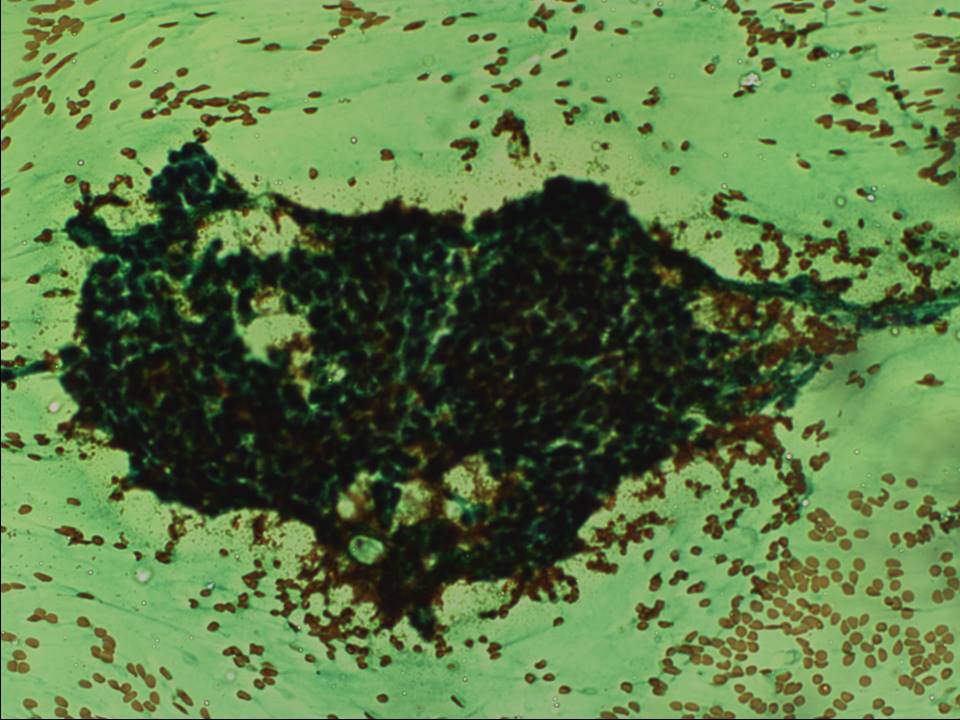 | 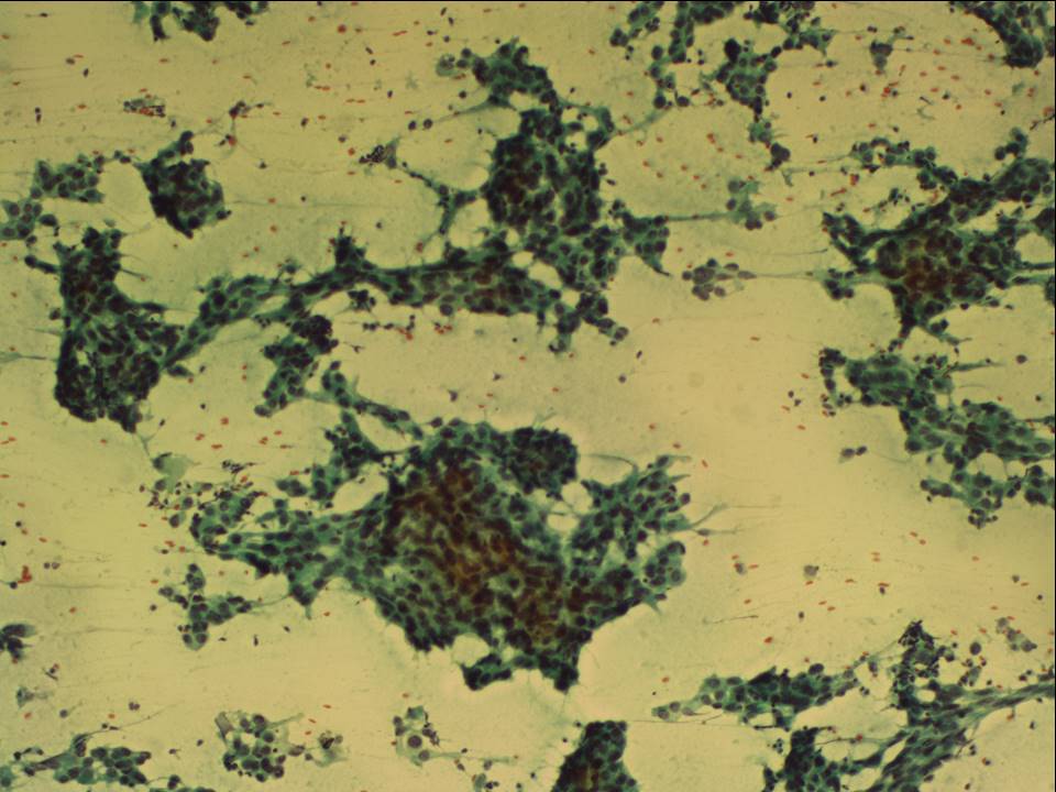 |
| Cytology features: | |
| ‣ Type of sample: | FNAC |
| ‣ Site of biopsy: | |
| • Laterality: | Right |
| • Quadrant: | Upper half lumpish feel on palpation |
| • Localization technique: | Palpation |
| • Nature of aspirate: | Whitish |
| ‣ Cytological description: | Smears from first pass show proteinaceous fluid, foamy macrophages, and a few cohesive benign ductal epithelial cells. Smears from second pass show loosely cohesive sheets of ductal epithelial cells with nuclear pleomorphism, high N:C ratio, and hyperchromasia. Myoepithelial cells are absent |
| ‣ Reporting category: | Suspicious, probably in situ or invasive carcinoma |
| ‣ Diagnosis: | Suspicious for malignancy, probably in situ or invasive carcinoma |
| ‣ Comments: | None |
Histopathology:
MRM
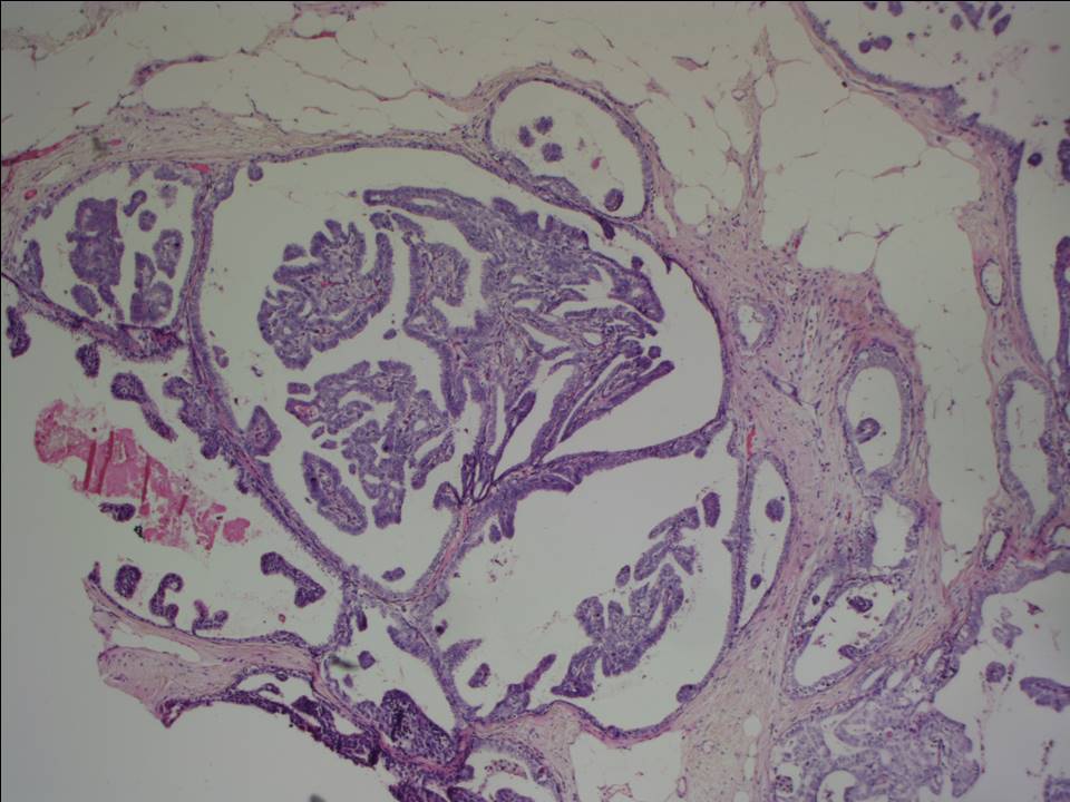 |  |
 |  |
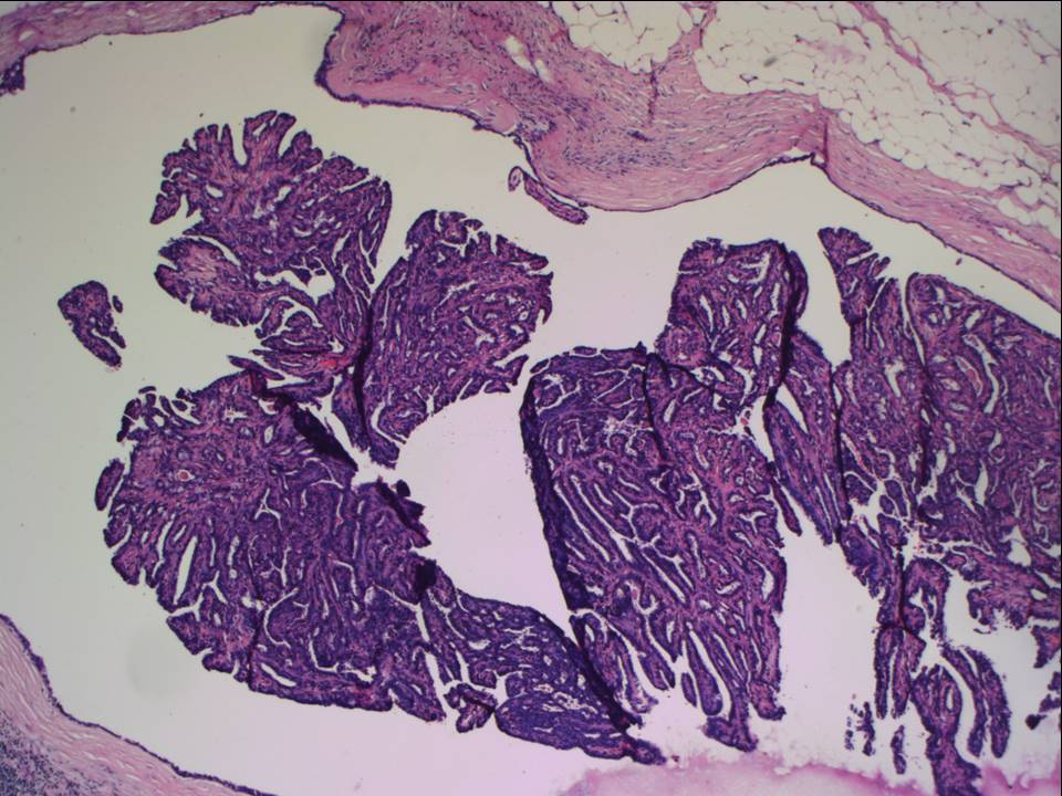 |
| Histopathology features: | |
| ‣ Specimen type: | MRM |
| ‣ Laterality: | Right |
| ‣ Macroscopy: | Right MRM specimen (27.5 × 12.5 × 3.5 cm) with skin flap (21.0 × 7.5 cm). The nipple is retracted. On serial sectioning, multiple firm greyish white areas (7.0 × 3.0 × 3.0 cm, 3.0 × 2.0 × 2.5 cm, and 2.5 × 2.5 × 2.0 cm) are seen over the upper half of the breast parenchyma. Distance from base (posterior margin of resection) is 2 cm. Largest node (2.5 × 1.5 × 1.5 cm) was dissected. The cut surface is whitish |
| ‣ Histological type: | Invasive breast carcinoma of no special type |
| ‣ Histological grade: | Grade 2 (3 + 2 + 2 = 7) |
| ‣ Mitosis: | 12 |
| ‣ Maximum invasive tumour size: | 5.5 cm (other tumours 3 cm and 2.6 cm in greatest dimension) |
| ‣ Lymph node status: | 5/20 |
| ‣ Peritumoural lymphovascular invasion: | Present |
| ‣ DCIS/EIC: | DCIS: cribriform, solid, papillary, and micropapillary type, moderate nuclear grade mainly, no necrosis. DCIS also involves the large lactiferous ducts. Paget disease of nipple present. EIC present |
| ‣ Margins: | Free of tumour |
| ‣ Pathological stage: | pT3(3)N2 |
| ‣ Biomarkers: | ER positive, PR positive, and HER2 negative |
| ‣ Comments: | The adjacent breast shows fibrocystic change, apocrine metaplasia, papilloma, and multiple areas with UDH |
Case summary:
| Postmenopausal woman with average risk of developing breast cancer presented with right breast lump noticed more than 2 years ago. Now with right nipple retraction also. Diagnosed as right breast outer quadrant focal asymmetry with calcifications of suspicious morphology, BI-RADS 5 (highly suggestive of malignancy) on imaging, as suspicious for malignancy on cytology, and as invasive breast carcinoma of no special type with EIC pT3(3)N2 with Paget disease on histopathology. |
Learning points:
|