


Home / Training / Manuals / Atlas of breast cancer early detection / Cases
Atlas of breast cancer early detection
Filter by language: English / Русский
Go back to the list of case studies
.png) Click on the pictures to magnify and display the legends
Click on the pictures to magnify and display the legends
| Case number: | 184 |
| Age: | 70 |
| Clinical presentation: | Postmenopausal woman with average risk of developing breast cancer presented with a left breast lump. Examination revealed a hard lump in the outer quadrant of the left breast. |
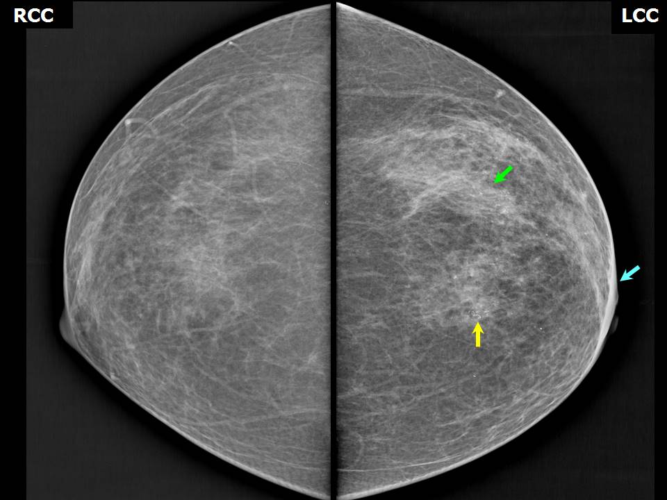
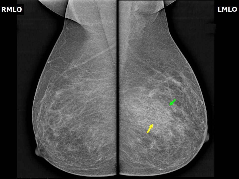
Mammography:
 |  |
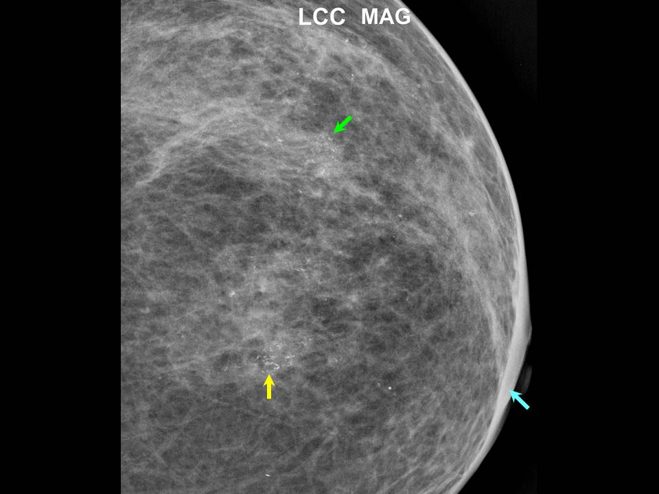 | 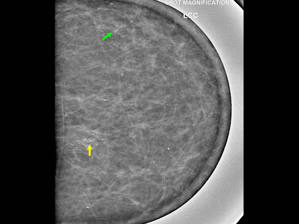 |
| Breast composition: | ACR category a (the breasts are almost entirely fatty) | Mammography features: |
| ‣ Location of the lesion: | Left breast, upper outer quadrant at 122 oclock, anterior and middle thirds |
| ‣ Mass: | |
| • Number: | None |
| • Size: | None |
| • Shape: | None |
| • Margins: | None |
| • Density: | None |
| ‣ Calcifications: | |
| • Typically benign: | None |
| • Suspicious: | Fine pleomorphic microcalcifications in segmental distribution and in clusters and in linear branching pattern |
| • Distribution: | None |
| ‣ Architectural distortion: | None |
| ‣ Asymmetry: | Present |
| ‣ Intramammary node: | None |
| ‣ Skin lesion: | None |
| ‣ Solitary dilated duct: | None |
| ‣ Associated features: | Skin thickening |
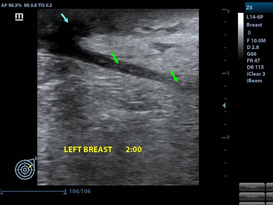
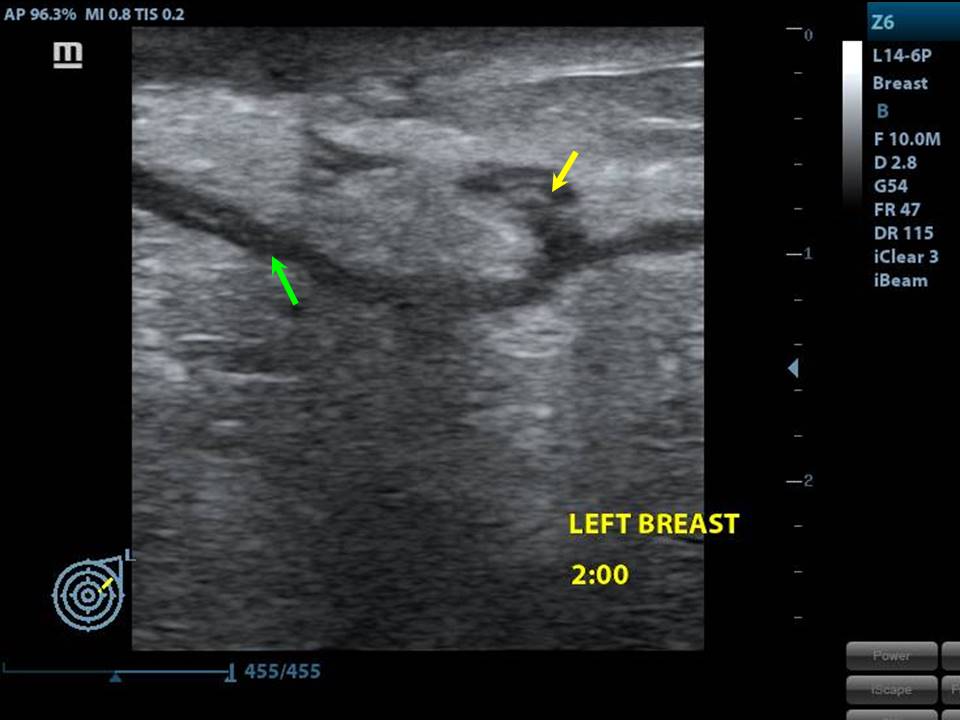
Ultrasound:
 |  |
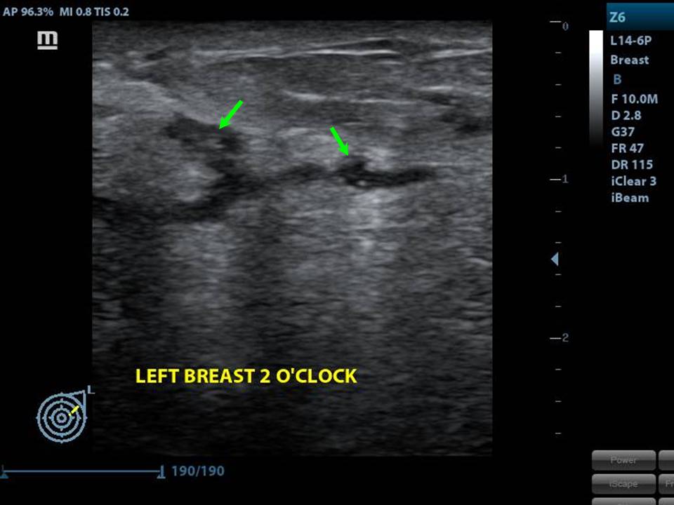 | 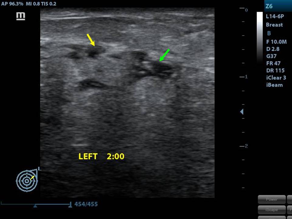 |
| Ultrasound features: Left breast, upper outer quadrant at 122 oclock | |
| ‣ Mass | |
| • Location: | Left breast, upper outer quadrant at 122 oclock |
| • Number: | Multiple small |
| • Size: | < 1 cm |
| • Shape: | Irregular |
| • Orientation: | Not parallel |
| • Margins: | Indistinct and angular |
| • Echo pattern: | Heteroechoic |
| • Posterior features: | No posterior features |
| ‣ Calcifications: | Intraductal microcalcifications |
| ‣ Associated features: | Axillary lymphadenopathy |
| ‣ Special cases: | Solitary dilated duct |
BI-RADS:
BI-RADS Category: 5 (highly suggestive of malignancy)Further assessment:
Further assessment advised: Referral for cytologyCytology:
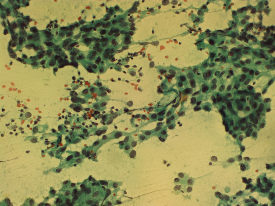 |
| Cytology features: | |
| ‣ Type of sample: | FNAC |
| ‣ Site of biopsy: | |
| • Laterality: | Left |
| • Quadrant: | Upper outer quadrant |
| • Localization technique: | Palpation |
| • Nature of aspirate: | Whitish, blood tinged |
| ‣ Cytological description: | Smears are very cellular and show dyscohesive clusters of pleomorphic malignant cells |
| ‣ Reporting category: | Malignant |
| ‣ Diagnosis: | Carcinoma breast |
| ‣ Comments: | None |
Histopathology:
MRM
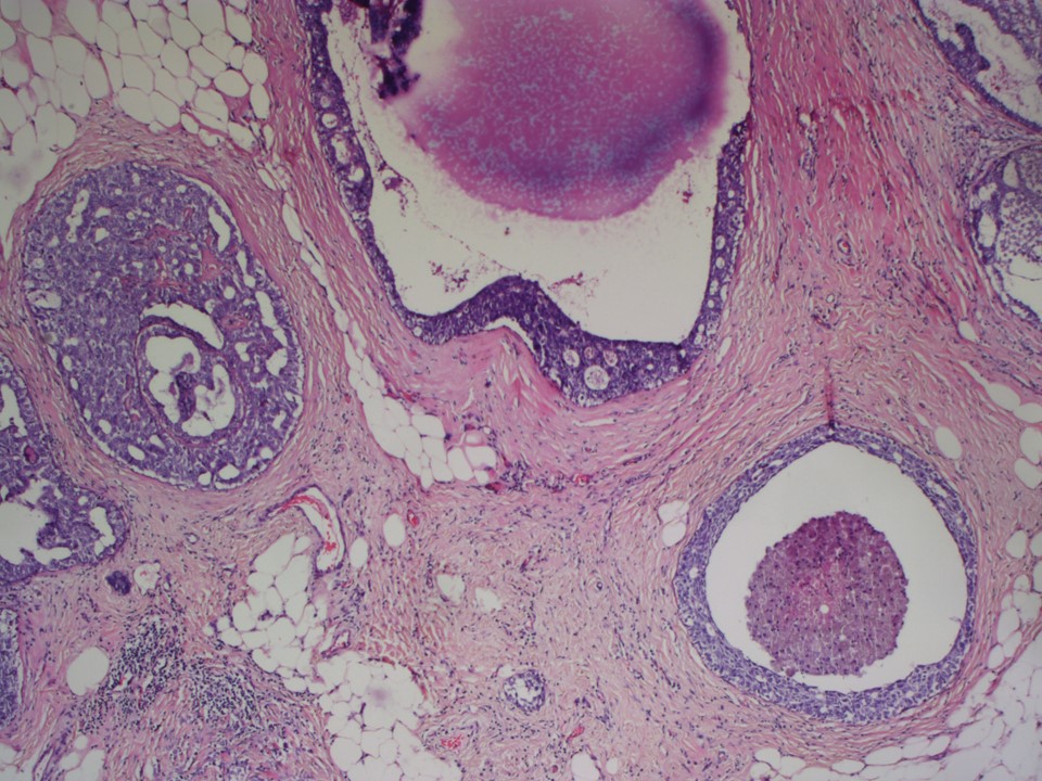 | 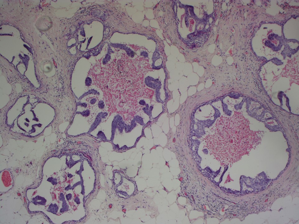 |
| Histopathology features: | |
| ‣ Specimen type: | MRM |
| ‣ Laterality: | Left |
| ‣ Macroscopy: | Left MRM specimen (24.0 × 16.0 × 7.5 cm) with skin flap (11.0 × 6.5 cm). On serial sectioning, a firm greyish white area (6.0 × 3.0 × 3.0 cm) is seen in the upper outer quadrant and extending to the lower outer quadrant. A second firm area (1.5 × 1.0 × 1.0 cm) is seen 3 cm medial to the first firm area in the upper inner quadrant. The intervening breast parenchyma shows interspersed whitish calcified areas |
| ‣ Histological type: | Microinvasive breast carcinoma |
| ‣ Histological grade: | Grade 2 (3 + 2 + 2 = 7) |
| ‣ Mitosis: | 12 |
| ‣ Maximum invasive tumour size: | Six foci of microinvasive breast carcinoma; all < 0.1 cm in greatest dimension |
| ‣ Lymph node status: | 0/10 |
| ‣ Peritumoural lymphovascular invasion: | Absent |
| ‣ DCIS/EIC: | DCIS of cribriform, comedocarcinoma, and solid type, high grade with necrosis in both the firm areas seen grossly. EIC present. Multiple foci of DCIS extending over 6 cm in greatest dimension |
| ‣ Margins: | Free of tumour |
| ‣ Pathological stage: | pT1miNo |
| ‣ Biomarkers: | ER positive, PR positive, and HER2 negative |
| ‣ Comments: | The adjacent breast also shows lobular mastitis with a chronic inflammatory infiltrate |
Case summary:
| Postmenopausal woman presented with a left breast lump. Diagnosed as solitary dilated duct with fine pleomorphic microcalcifications in segmental distribution and in clusters and in linear branching pattern, BI-RADS 5 on imaging, as breast carcinoma on cytology, and as microinvasive breast carcinoma pT1miN0 with EIC on histopathology. |
Learning points:
|
EIC is defined by the following criteria:
|