


Home / Training / Manuals / Atlas of breast cancer early detection / Cases
Atlas of breast cancer early detection
Filter by language: English / Русский
Go back to the list of case studies
.png) Click on the pictures to magnify and display the legends
Click on the pictures to magnify and display the legends
| Case number: | 179 |
| Age: | 70 |
| Clinical presentation: | Postmenopausal woman with increased risk of developing breast cancer presented with a lump in the left axillary tail. The patient had ovarian carcinoma detected 1 year ago and had undergone a hysterectomy. |
Mammography:
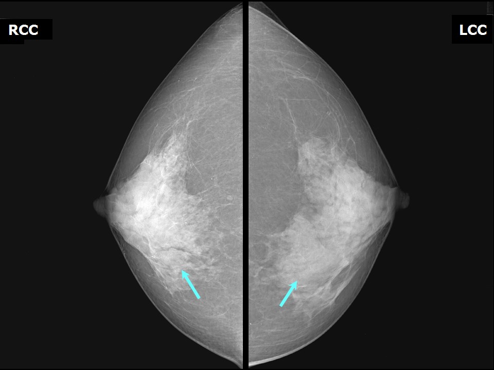 | 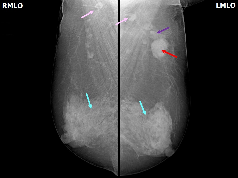 |
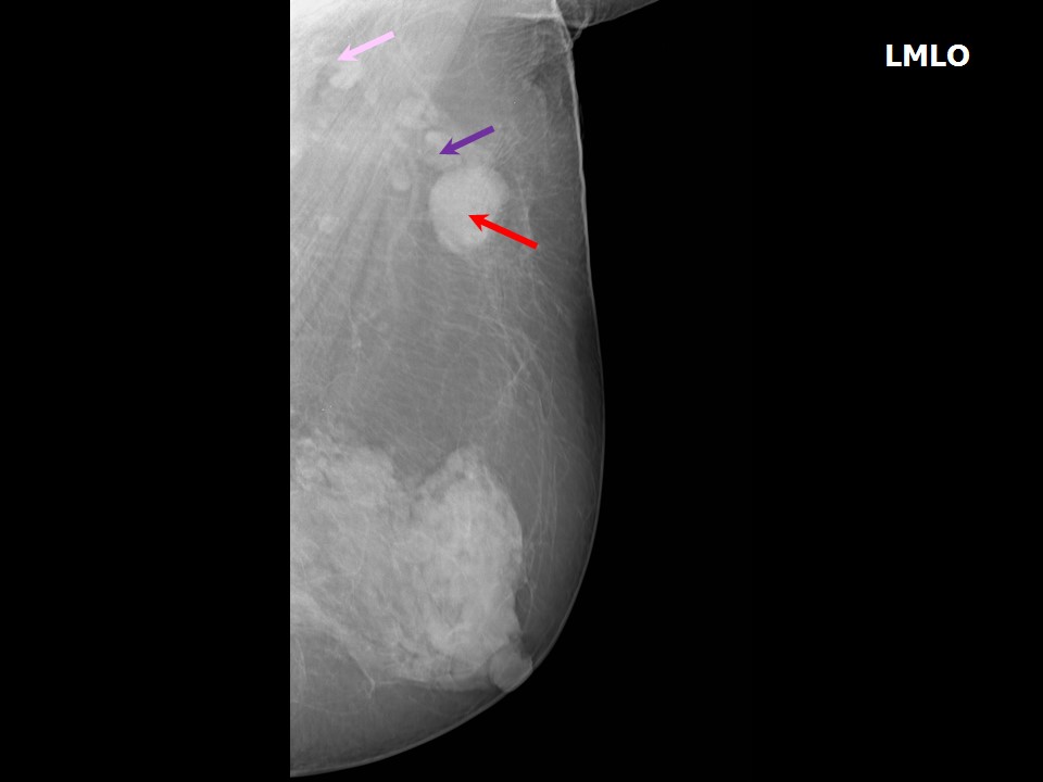 |
| Breast composition: | ACR category d (the breasts are extremely dense, which lowers the sensitivity of mammography) | Mammography features: |
| ‣ Location of the lesion: | Left axilla, enlarged axillary lymph node (2.4 × 1.4 cm) of altered morphology with loss of fatty hilum |
| ‣ Mass: | |
| • Number: | 1 |
| • Size: | None |
| • Shape: | None |
| • Margins: | None |
| • Density: | None |
| ‣ Calcifications: | |
| • Typically benign: | None |
| • Suspicious: | None |
| • Distribution: | None |
| ‣ Architectural distortion: | Present |
| ‣ Asymmetry: | None |
| ‣ Intramammary node: | None |
| ‣ Skin lesion: | None |
| ‣ Solitary dilated duct: | None |
| ‣ Associated features: | None |
Ultrasound:
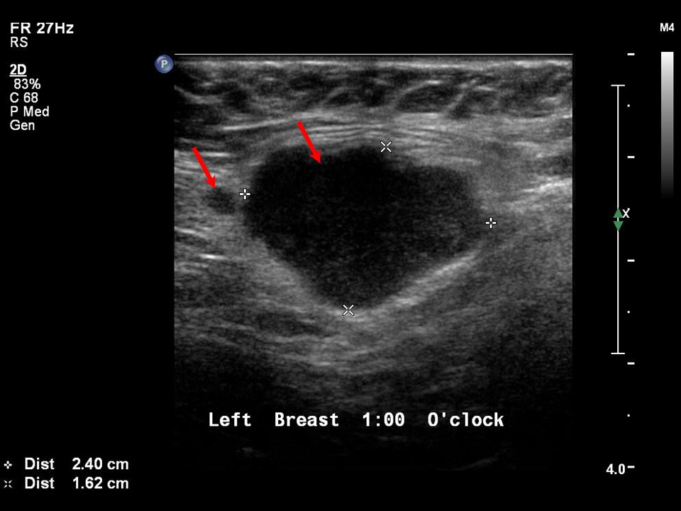 | 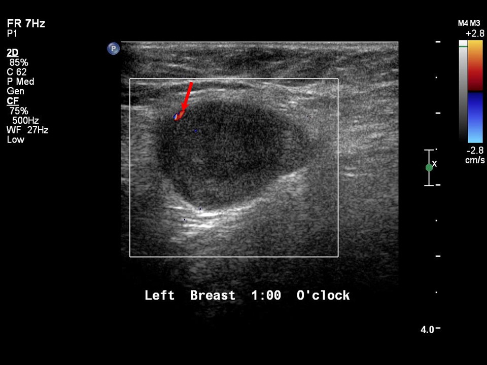 |
| Ultrasound features: None | |
| ‣ Mass | |
| • Location: | None |
| • Number: | 1 |
| • Size: | None |
| • Shape: | None |
| • Orientation: | None |
| • Margins: | None |
| • Echo pattern: | None |
| • Posterior features: | No posterior features |
| ‣ Calcifications: | None |
| ‣ Associated features: | None |
| ‣ Special cases: | Lymph nodes, axillary |
BI-RADS:
BI-RADS Category: 4C (high suspicion for malignancy)Further assessment:
Further assessment advised: Referral for cytologyCytology:
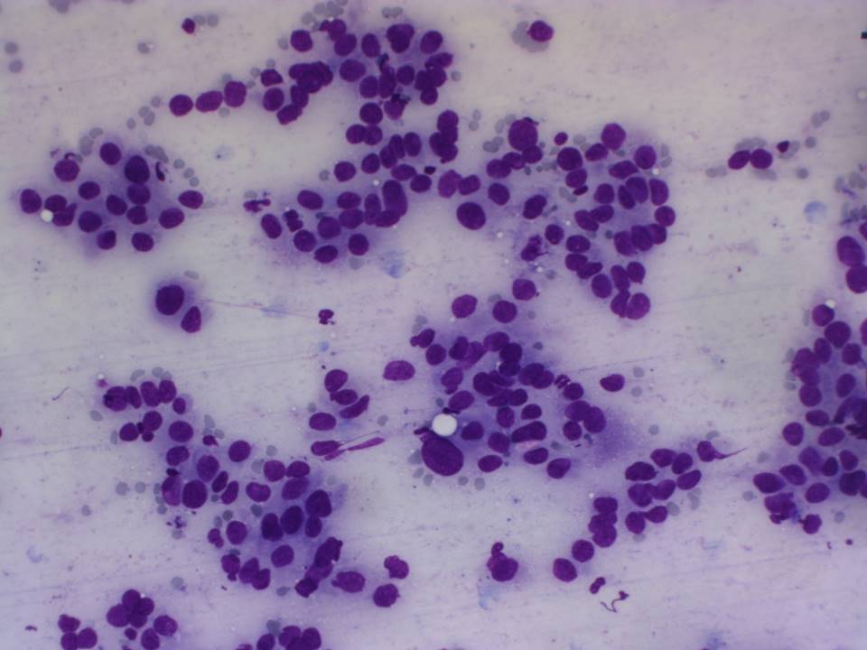 |
| Cytology features: | |
| ‣ Type of sample: | FNAC |
| ‣ Site of biopsy: | |
| • Laterality: | Left axillary node |
| • Quadrant: | No lump in breast |
| • Localization technique: | Palpation |
| • Nature of aspirate: | Whitish |
| ‣ Cytological description: | Smears are very cellular and show dyscohesive clusters of malignant epithelial cells with marked pleomorphism and nuclear hyperchromasia. A glandular arrangement of the epithelial cells is seen in some cell clusters |
| ‣ Reporting category: | Malignant |
| ‣ Diagnosis: | Distant metastases, correlating with the clinical history of ovarian carcinoma |
| ‣ Comments: | None |
Case summary:
| Postmenopausal woman, who had undergone surgery for ovarian carcinoma a year ago, presented with a lump in the left axillary tail. Diagnosed as left axillary tail enlarged dysmorphic lymph nodes, BI-RADS 4C on imaging. No suspicious finding was seen in bilateral breasts on imaging other than the left axillary node findings. On FNAC of the node, malignant cells were seen. |
Learning points:
|