


Home / Training / Manuals / Atlas of breast cancer early detection / Cases
Atlas of breast cancer early detection
Filter by language: English / Русский
Go back to the list of case studies
.png) Click on the pictures to magnify and display the legends
Click on the pictures to magnify and display the legends
| Case number: | 087 |
| Age: | 42 |
| Clinical presentation: | Premenopausal woman with average risk of developing breast cancer presented with a lump in the left breast noticed 1 month ago. On clinical examination, a single left upper quadrant lump 3 cm in diameter was found. |
Mammography:
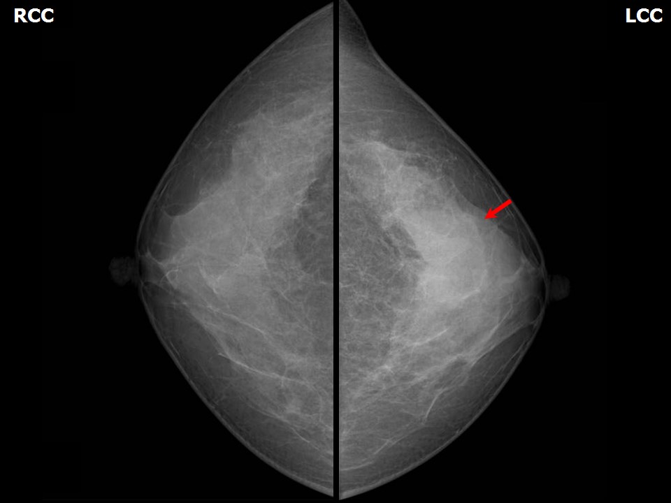 | 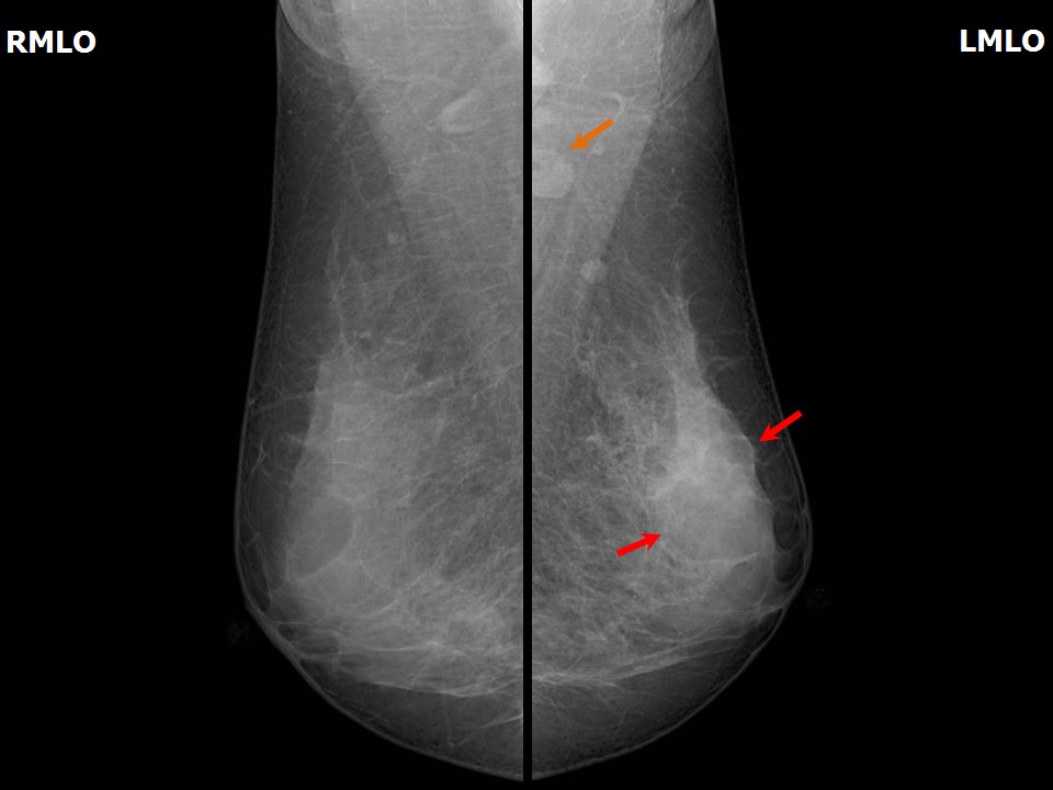 |
| Breast composition: | ACR category c (the breasts are heterogeneously dense, which may obscure small masses) | Mammography features: |
| ‣ Location of the lesion: | Left breast, upper outer quadrant at 121 oclock, middle third |
| ‣ Mass: | |
| • Number: | 1 |
| • Size: | Not measurable in the dense breast parenchyma |
| • Shape: | Irregular |
| • Margins: | Indistinct |
| • Density: | High |
| ‣ Calcifications: | |
| • Typically benign: | None |
| • Suspicious: | None |
| • Distribution: | None |
| ‣ Architectural distortion: | Present |
| ‣ Asymmetry: | Focal |
| ‣ Intramammary node: | None |
| ‣ Skin lesion: | None |
| ‣ Solitary dilated duct: | None |
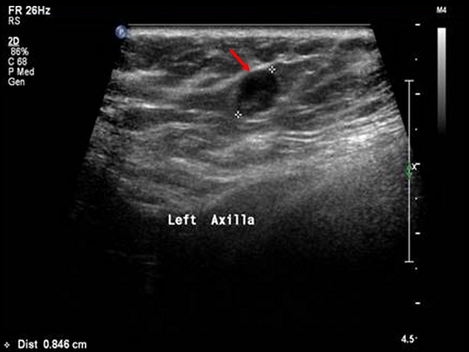
| ‣ Associated features: | Architectural distortion and axillary lymphadenopathy |
Ultrasound:
 |  |
 |
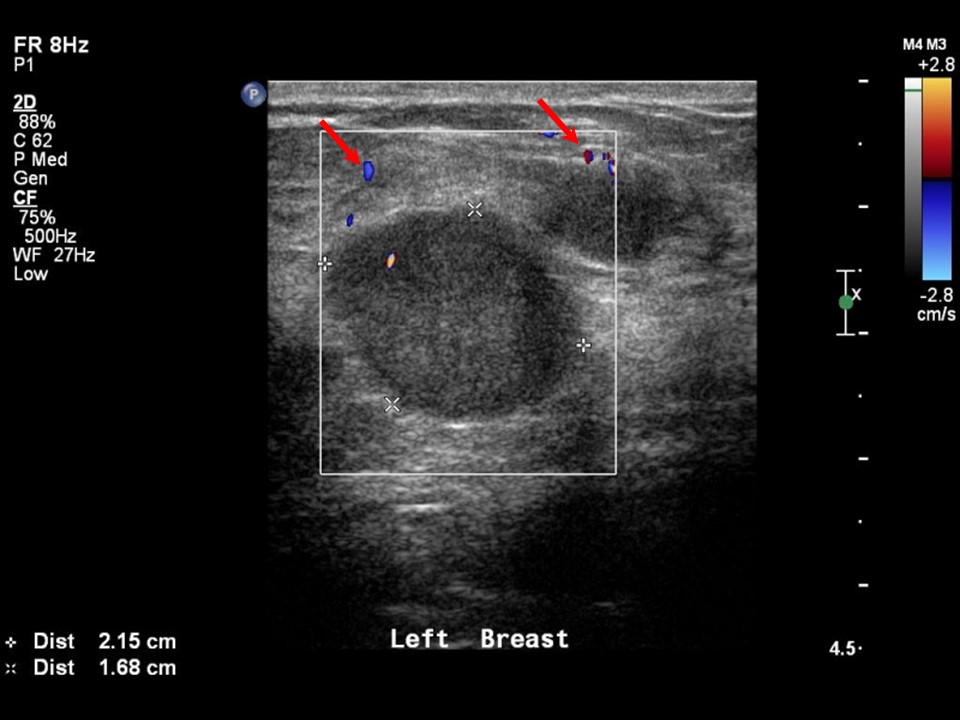
| Ultrasound features: Left breast, central portion of the breast, central quadrant at 121 oclock | |
| ‣ Mass | |
| • Location: | Left breast, central portion of the breast, central quadrant at 121 oclock |
| • Number: | 3 |
| • Size: | Largest 2.2 × 1.7 cm |
| • Shape: | Irregular |
| • Orientation: | Not parallel |
| • Margins: | Indistinct |
| • Echo pattern: | Hypoechoic |
| • Posterior features: | No posterior features |
| ‣ Calcifications: | None |
| ‣ Associated features: | Internal vascularity |
| ‣ Special cases: | None |
BI-RADS:
BI-RADS Category: 4C (high suspicion for malignancy)Further assessment:
Further assessment advised: Referral for core biopsyCytology:
 |
| Cytology features: | |
| ‣ Type of sample: | FNAC (solid lesion) |
| ‣ Site of biopsy: | |
| • Laterality: | Left |
| • Quadrant: | Upper outer |
| • Localization technique: | Ultrasound-guided |
| • Nature of aspirate: | Whitish |
| ‣ Cytological description: | Smears show dyscohesive clusters of pleomorphic malignant cells |
| ‣ Reporting category: | Malignant |
| ‣ Diagnosis: | Carcinoma high grade |
| ‣ Comments: | None |
Histopathology:
Core needle biopsy
 |
| Histopathology features: | |
| ‣ Specimen type: | Core needle biopsy |
| ‣ Laterality: | Left |
| ‣ Macroscopy: | 3 cores |
| ‣ Histological type: | Invasive carcinoma of no special type |
| ‣ Histological grade: | Grade 3 (3 + 2 + 2 = 7) |
| ‣ Mitosis: | 11 |
| ‣ Maximum invasive tumour size: | |
| ‣ Lymph node status: | |
| ‣ Peritumoural lymphovascular invasion: | |
| ‣ DCIS/EIC: | |
| ‣ Margins: | |
| ‣ Pathological stage: | |
| ‣ Biomarkers: | Triple negative: ER negative (internal control positive), PR negative (internal control positive), and HER2/neu negative (score 0) |
| ‣ Comments: |
Case summary:
| Premenopausal woman presented with left breast lump. Diagnosed as retroareolar high-density focal asymmetry in left breast, BI-RADS 4C on imaging, as left breast carcinoma, high grade on cytology, and as invasive carcinoma of no special type, triple negative on histopathology. |
Learning points:
|