


Home / Training / Manuals / Atlas of breast cancer early detection / Cases
Atlas of breast cancer early detection
Filter by language: English / Русский
Go back to the list of case studies
.png) Click on the pictures to magnify and display the legends
Click on the pictures to magnify and display the legends
| Case number: | 092 |
| Age: | 68 |
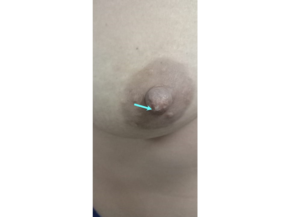
| Clinical presentation: | Postmenopausal woman with average risk of developing breast cancer presented with nipple discharge from the right nipple of duration 1 year. Examination revealed right breast nodularity with tenderness felt along the axillary tail. There was clear watery discharge form the nipple. |
 |
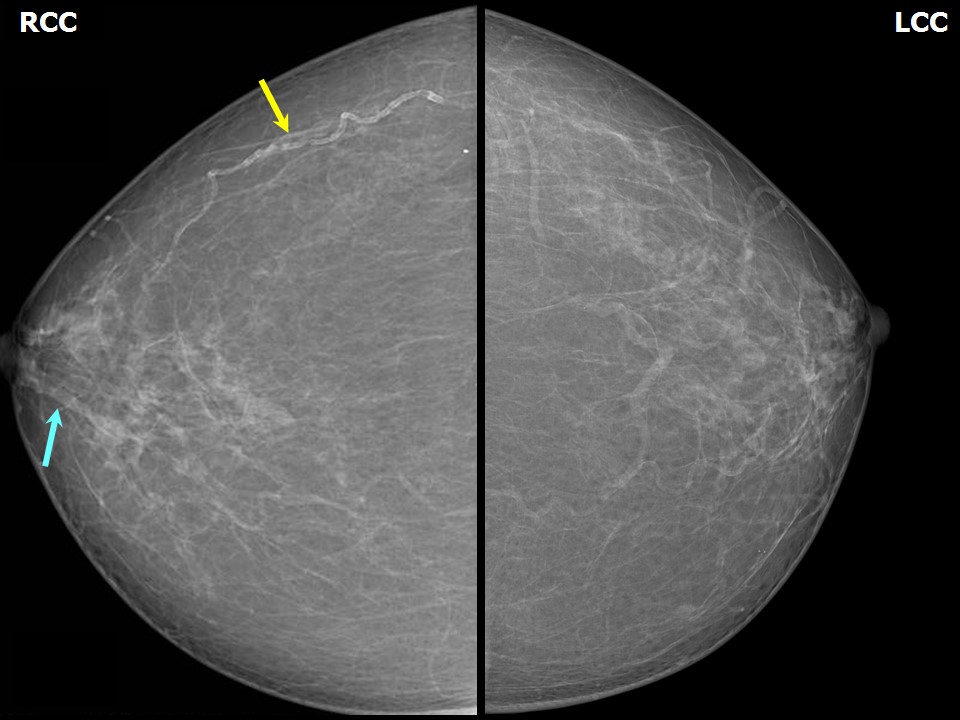
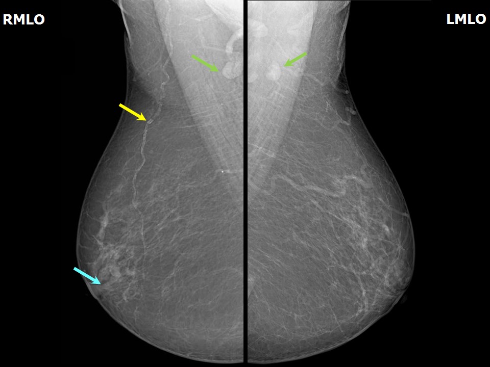
Mammography:
 |  |
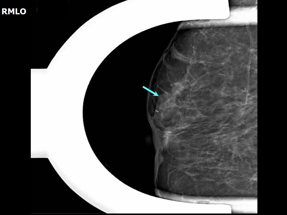 |
| Breast composition: | ACR category a (the breasts are almost entirely fatty) | Mammography features: |
| ‣ Location of the lesion: | Right breast, central portion of the breast, upper inner quadrant at 121 oclock, anterior third |
| ‣ Mass: | |
| • Number: | 1 |
| • Size: | None |
| • Shape: | Linear dilated duct |
| • Margins: | Circumscribed |
| • Density: | Equal |
| ‣ Calcifications: | |
| • Typically benign: | None |
| • Suspicious: | None |
| • Distribution: | None |
| ‣ Architectural distortion: | None |
| ‣ Asymmetry: | None |
| ‣ Intramammary node: | None |
| ‣ Skin lesion: | None |
| ‣ Solitary dilated duct: | None |
| ‣ Associated features: | Dilated duct in subareolar region |
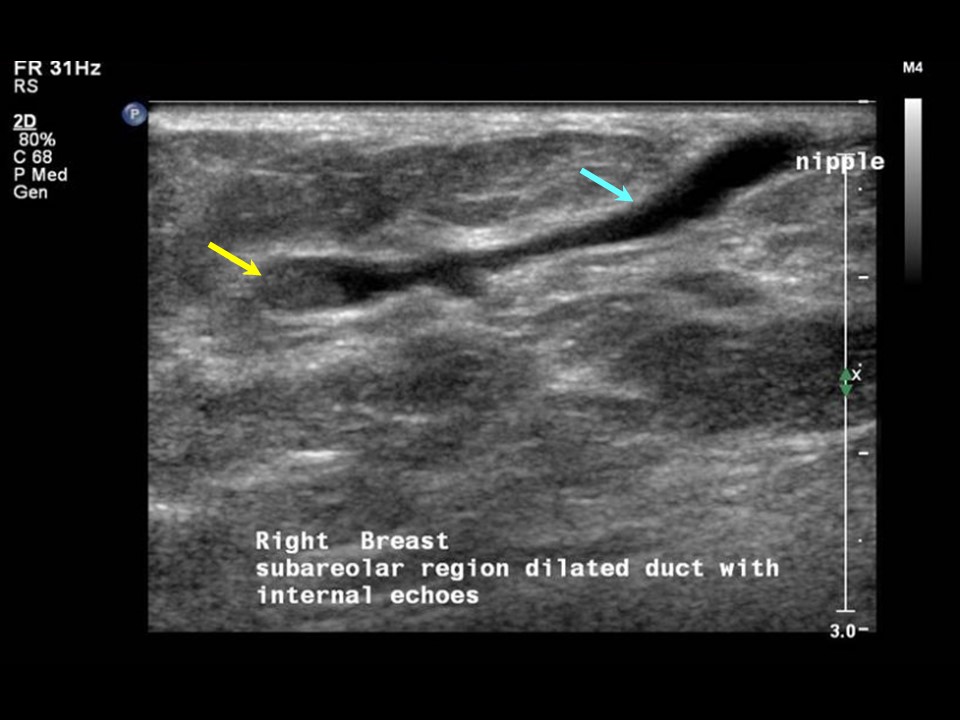
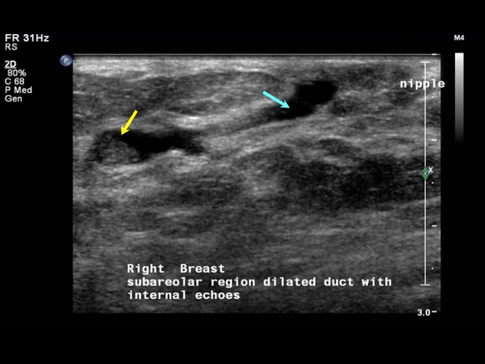
Ultrasound:
 |  |
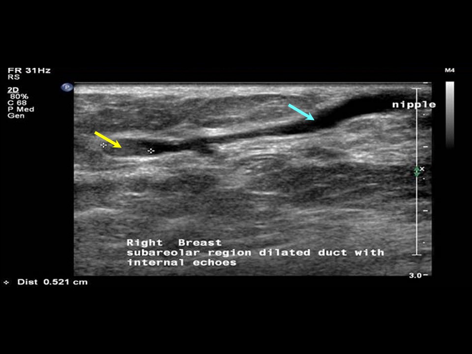 |
| Ultrasound features: Right breast, central portion of the breast | |
| ‣ Mass | |
| • Location: | Right breast, central portion of the breast |
| • Number: | 1 |
| • Size: | 0.5 cm in the dilated duct |
| • Shape: | Irregular |
| • Orientation: | None |
| • Margins: | None |
| • Echo pattern: | Isoechoic |
| • Posterior features: | No posterior features |
| ‣ Calcifications: | None |
| ‣ Associated features: | Duct changes |
| ‣ Special cases: | Solitary dilated duct |
BI-RADS:
BI-RADS Category: 4A (low level of suspicion for malignancy)Further assessment:
Further assessment advised: Referral for cytologyCytology:
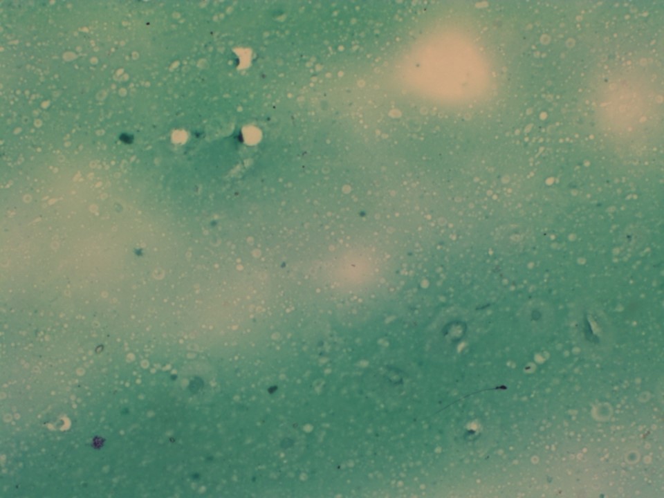 |
| Cytology features: | |
| ‣ Type of sample: | Nipple discharge |
| ‣ Site of biopsy: | |
| • Laterality: | Right |
| • Quadrant: | |
| • Localization technique: | |
| • Nature of aspirate: | Clear serous fluid discharge from the nipple |
| ‣ Cytological description: | Smears show only thin proteinaceous material. Cellular material not seen |
| ‣ Reporting category: | Benign |
| ‣ Diagnosis: | Benign, negative for malignant cells |
| ‣ Comments: | None |
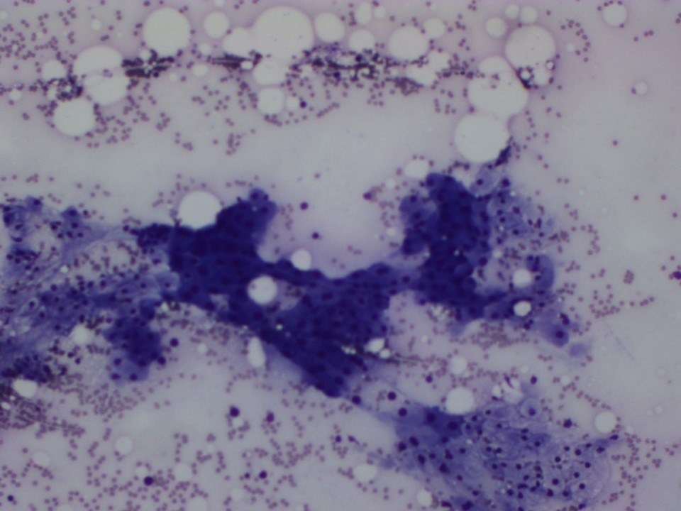 |
| Cytology features: | |
| ‣ Type of sample: | FNAC (solid lesion) |
| ‣ Site of biopsy: | |
| • Laterality: | Right |
| • Quadrant: | Subareolar |
| • Localization technique: | Ultrasound-guided FNAC |
| • Nature of aspirate: | Scant brownish fluid |
| ‣ Cytological description: | Smears show plenty of foamy histiocytes and a few ductal epithelial cells, some of which show apocrine change |
| ‣ Reporting category: | Benign |
| ‣ Diagnosis: | Benign, proliferative fibrocystic change |
| ‣ Comments: | None |
Case summary:
| Postmenopausal woman presented with right breast clear watery nipple discharge. Diagnosed as right breast subareolar solitary dilated duct with intraductal echoes, BI-RADS 4A on imaging and as benign proliferative fibrocystic change on cytology. |
Learning points:
|