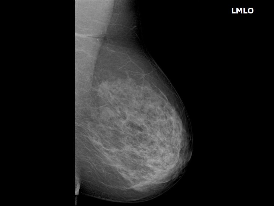
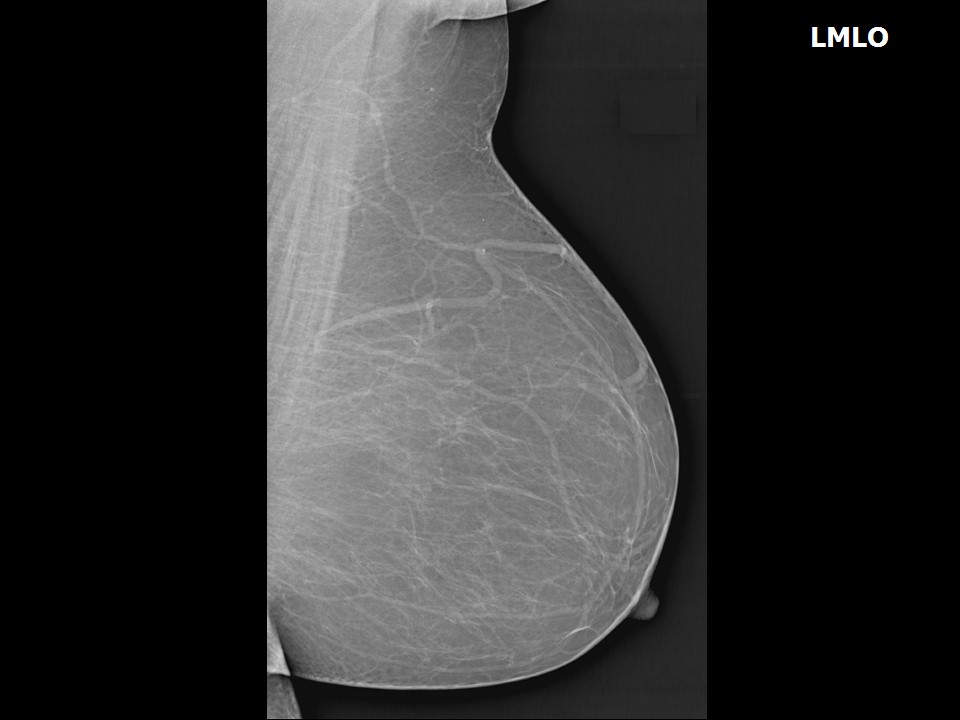
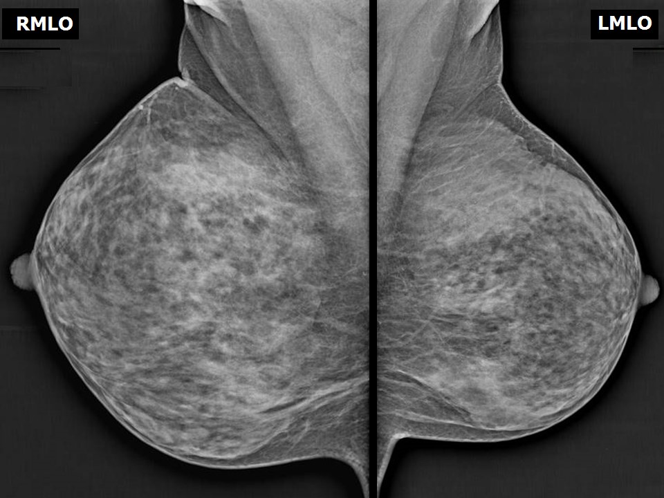
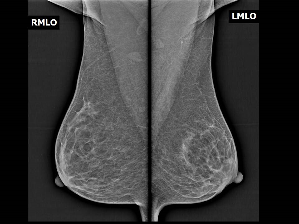
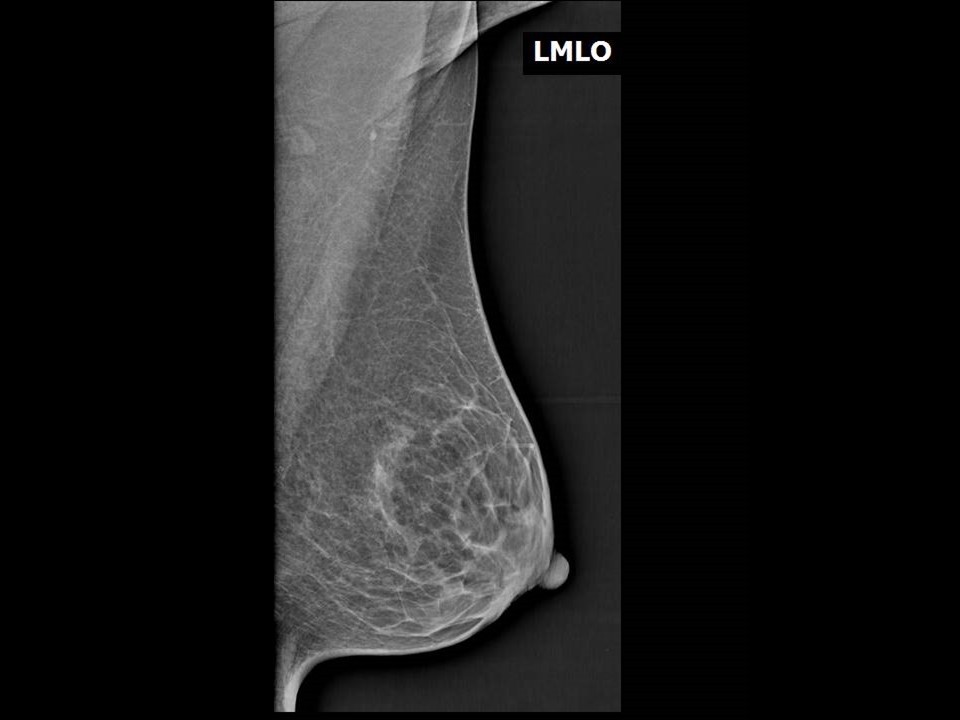
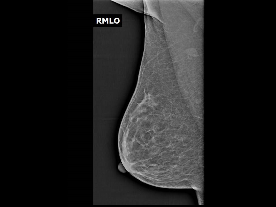
The mammographic images obtained should be checked to ensure that the images are satisfactory for interpretation. The MLO view is the single most important mammographic projection of the breast. The following criteria should be fulfilled to ensure that the MLO image obtained is accurate and technically adequate to enable a diagnosis to be made:
- In a single projection the image should enable visualization of as much breast tissue as possible, from the axilla to the inframammary fold.
- The image should include the free margin of the pectoralis muscle and the inframammary fold.
- The pectoralis muscle should be visible down to the axis of the nipple with bilaterally symmetrical images.
- Bilateral MLO views should show the pectoralis muscles forming a V shape, viewed as mirror images.
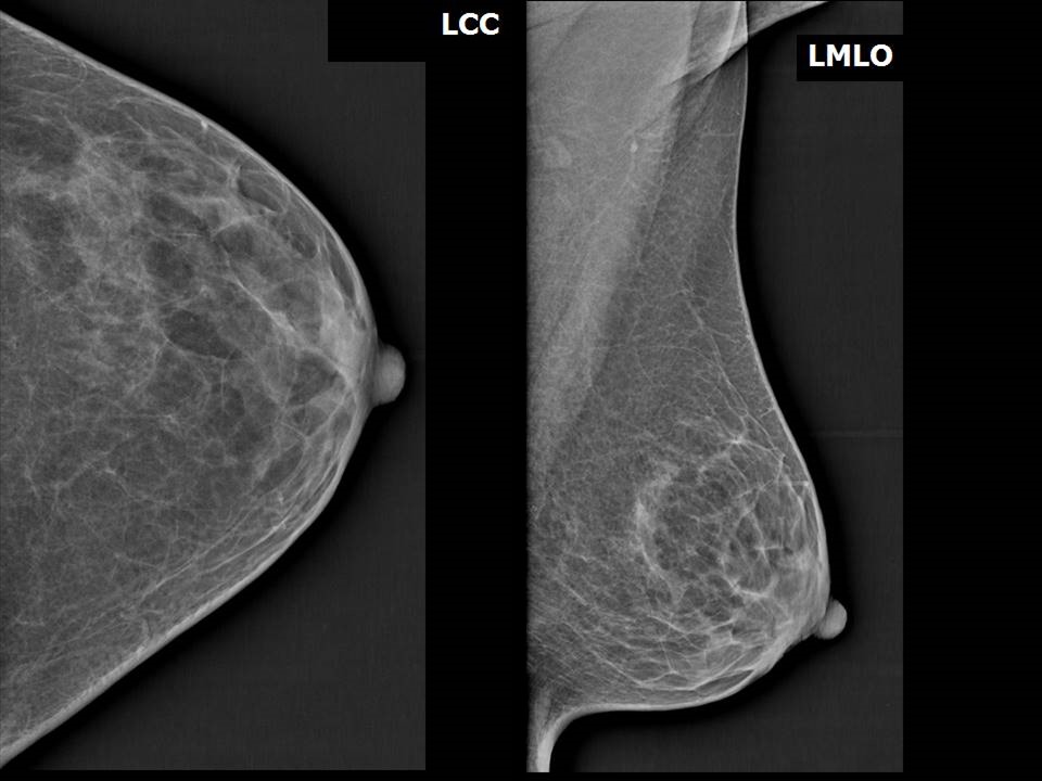
- The distance back from the nipple to the free lower edge of the pectoralis major muscle is called the nipple axis line (NAL) or the posterior nipple line (PNL). PNL-MLO should be no more than 1 cm longer than PNL-CC of the same breast.
- The PNL should be below the level of the inferior aspect of the pectoral muscle, and the distance between the PNL and the inferior aspect of the pectoralis muscle should not exceed 10 mm.
Normal anatomical landmarks on MLO view
Bilateral symmetrical MLO – V-shaped pectoralis shadow
View of entire breast tissue from axilla to inframammary fold
Technical adequacy of MLO views The PNL
Lower end of pectoralis muscle to PNL
Relationship between PNL-MLO and PNL-CC
|