|
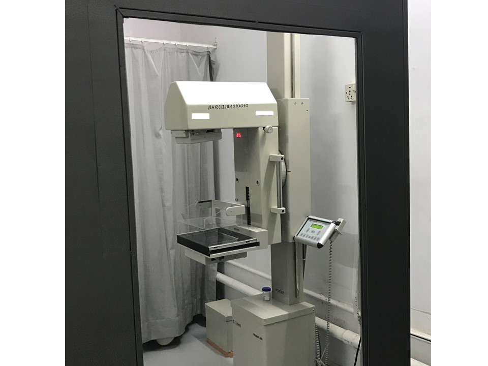
Breast imaging – Mammography technique – Mammography procedure – Obtaining mammographic images – Right CC view |   |
Steps to obtain the right CC view
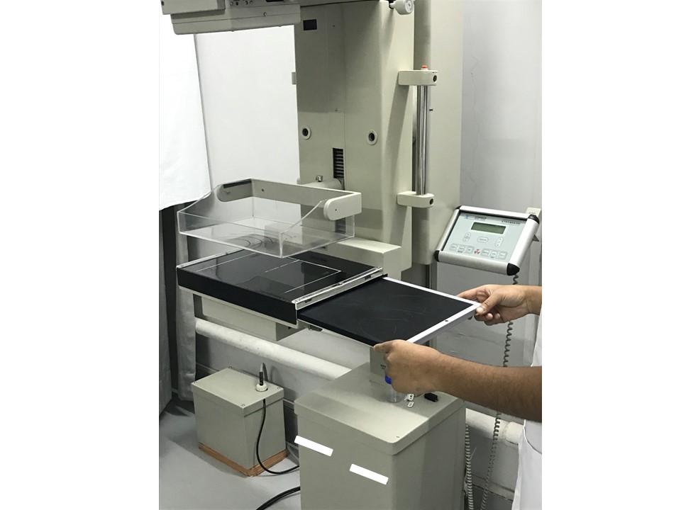
- Rotate the mammography unit gantry and bring it back to vertical (0 degrees) so that the beam is perpendicular to the floor. Place the image receptor in the detector.
- Place the image receptor in the detector.
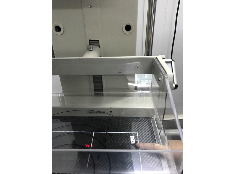
- Place a lead identification marker “RCC” on the breast support in the region of the outer quadrant, along the axillary side of the right breast.
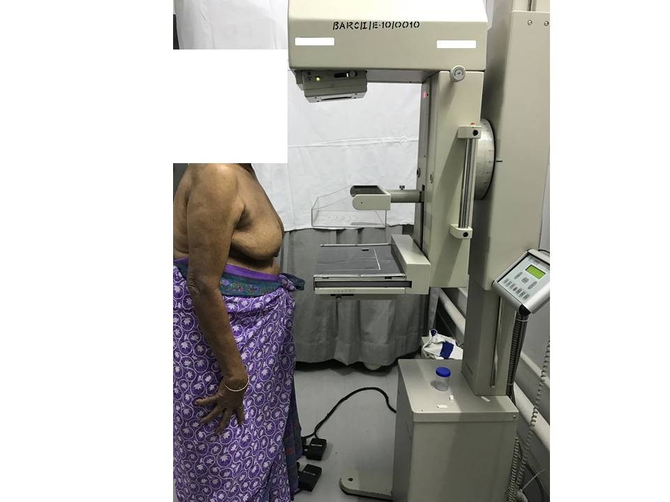
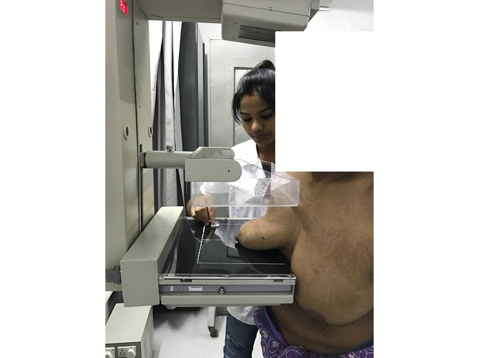
- Ask the patient to stand straight in front and close to the mammography unit with no body tilt/angulation; both hands should be relaxed by the sides of the body.
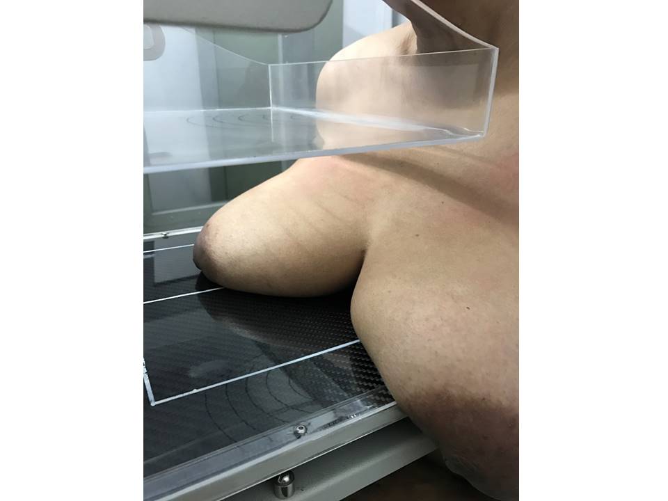
- Project the right nipple in profile so that it lies approximately in the middle of the detector.
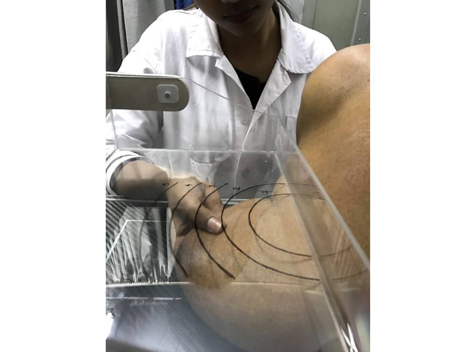
- Hold the right breast gently in your palm so that the inframammary fold is straight and the upper tissues are relaxed. Adjust the detector height to this level.
- Raise the right breast and pull it away from the chest wall into the machine using two hands by gathering the tissues from below and pulling the breast upwards.
- Guide the patient into the machine so that the edge of the detector is against her ribs, ensuring that all deeper structures of the breast are included in the image detector.
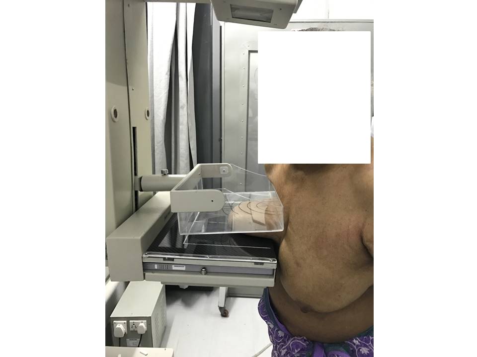
- The left breast is moved away from the detector either by the patient's left hand or it is gently raised and placed on the breast support.
- Ask the patient to turn her face to the left with her neck tilted backwards; ensure that no body parts overlap the breast being radiographed.
- Lower the compression paddle gently, maintaining the position of the breast and ensuring that the nipple is in profile. Ensure that the breast does not slip out.
- When adequate compression has been achieved, step behind the protective lead shield and take the X-ray.
- The exposure is completed controlled by the AEC; a latent image is formed on the image receptor and compression releases automatically.
- Ask the patient to step aside from the mammography unit. Prepare the unit for the next exposure.
|   |
|
.png)
Click on the pictures to magnify and display the legends

Click on this icon to display a case study
25 avenue Tony Garnier CS 90627 69366, LYON CEDEX 07 France - Tel: +33 (0)4 72 73 84 85 © IARC 2026 - Terms of use - Privacy Policy.
| .png)


