Home / Training / Manuals / Atlas of breast cancer early detection / Learning

Atlas of breast cancer early detection

Filter by language: English / Русский

Breast imaging – Mammography technique – Mammography procedure – Additional mammographic views – Lumpectomy specimen mammography


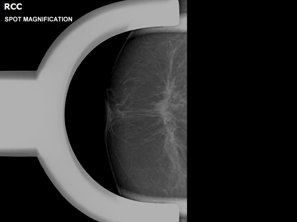
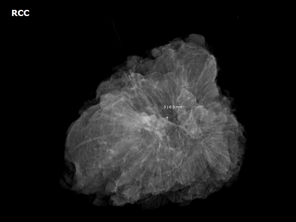
Preclinical cancers or suspicious clusters of microcalcifications detected on screening mammography are further evaluated with image-guided biopsy. If carcinoma in situ or malignancy is detected, most patients undergo wide excision of the lesion as a breast-conserving procedure. Hookwire localization is used for the excision and biopsy of clinically non-palpable breast lesions. Mammography of the lumpectomy specimen is an important intraoperative step to ensure complete excision.

Advantages of specimen mammography
  • Specimen mammography can confirm complete excision of a small, clinically non-palpable mass or cluster of microcalcifications.
  • It can help to ascertain margin clearance macroscopically, although histopathology of the specimen remains the reference standard for involvement or clearance of the margins of the excised tissue. Frozen sections can be viewed intraoperatively and decisions to revise margin clearance can be taken if needed.

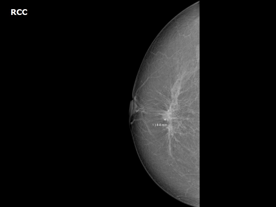
Mammogram right breast shows developing asymmetry


Lumpectomy specimen mammogram


Click on the pictures to magnify and display the legends


Click on this icon to display a case study

25 avenue Tony Garnier CS 90627 69366, LYON CEDEX 07 France - Tel: +33 (0)4 72 73 84 85
© IARC 2024 - Terms of use  -  Privacy Policy.