|
Breast imaging – Mammography technique – Mammography procedure – Obtaining mammographic images – Right MLO view |   |
Steps to obtain the right MLO view
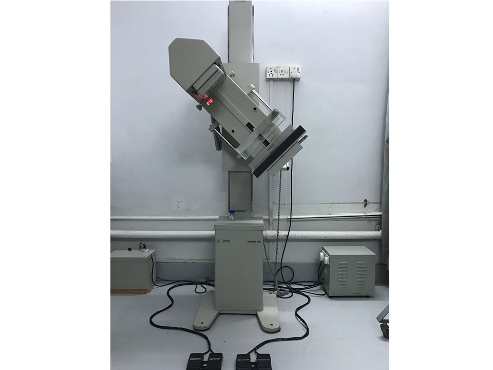
- Adjust the angle of the mammography unit gantry to 45 degrees so that the plane of the detector is parallel to the right pectoralis muscle.
- Place the image receptor in the detector.
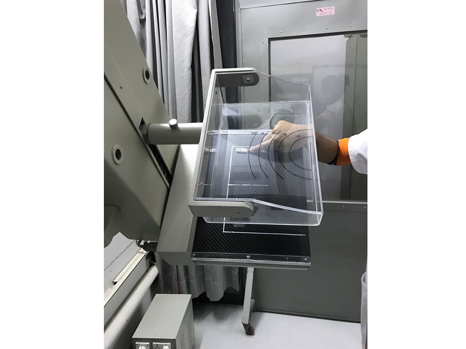
- Place a lead identification marker “RMLO” on the breast support in the region of the upper axillary portion of the right breast.
- Adjust the height of the breast support to suit the woman’s habitus.
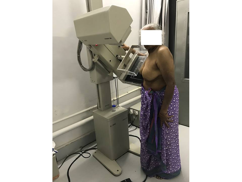
- Elevate the woman’s right arm and place it across the breast support, taking care that the arm is never higher than the right shoulder.
- Place the corner of the detector high in the right axilla and the compression paddle at the level of the right infraclavicular region.
- Raise the right breast and pull it forward, trying to gather all the deep lateral tissues. Make sure the woman’s shoulder is relaxed at all times.
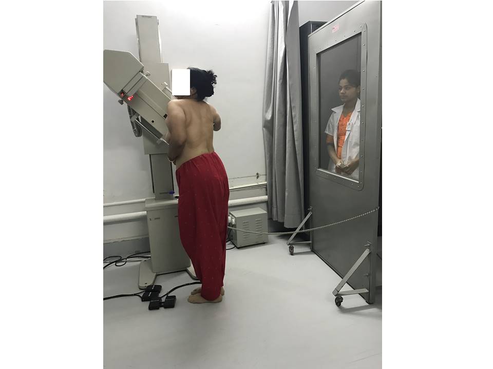
- Ask the woman to turn her face to the left with her neck tilted backwards; ensure that no body parts overlap the breast being radiographed.
- Lower the compression paddle gently, maintaining the position of the breast, and ensuring that the nipple is in profile. Ensure that the breast does not slip out.
- When adequate compression (8–12 kg) has been achieved, step behind the protective lead shield and take the X-ray.
- The exposure is completed controlled by the AEC; a latent image is formed on the image receptor and compression releases automatically.
- Ask the patient to step aside from the mammography unit. Prepare the unit for the next exposure.
Gantry position and placement of marker
Patient positioning
|
.png)
Click on the pictures to magnify and display the legends

Click on this icon to display a case study
25 avenue Tony Garnier CS 90627 69366, LYON CEDEX 07 France - Tel: +33 (0)4 72 73 84 85 © IARC 2026 - Terms of use - Privacy Policy.
| .png)


