|
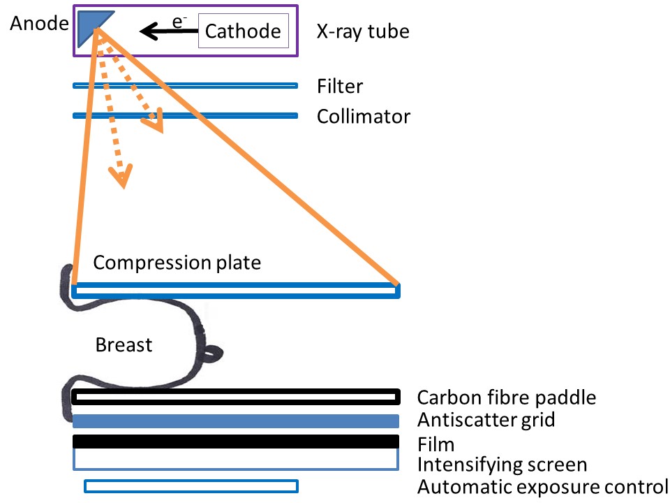
Breast imaging – Mammography technique – Basic functioning of mammography unit |   |
- The breast to be examined is placed on the breast support and held in position with the compression plate.
- A high-voltage supply of electrons from the cathode collides with the rotating molybdenum anode to produce a characteristic radiation spectrum optimal for breast imaging.
- The X-ray photons exit the tube through a port window of beryllium and additional molybdenum filters to modify the spectrum.
- A molybdenum filter is used to remove the undesirable high-energy photons (more than 20 keV) from the spectrum. Only radiation with photon energies less than 20 keV reaches the breast tissue.
- These X-rays are then directed through the compressed breast tissue by a collimator.
- The X-ray beam passing through the breast is attenuated depending on the density of the tissues it encounters in the breast.
- The attenuated beam passes through the anti-scatter grid to a detector located on the opposite side. The detector is either a photographic film plate, which captures the X-ray image on film, or a solid-state detector, which transmits electronic signals to a computer to form a digital image of the breast.
- The AEC sensor at the base of the image receptor cassette measures the radiation passing through the breast and cassette. When a sufficient number of X-ray photons have reached the sensor, exposure is automatically terminated by the sensor.
|   |
|
.png)
Click on the pictures to magnify and display the legends

Click on this icon to display a case study
25 avenue Tony Garnier CS 90627 69366, LYON CEDEX 07 France - Tel: +33 (0)4 72 73 84 85 © IARC 2026 - Terms of use - Privacy Policy.
| .png)


