English
Français
Home
Research projects
Breast cancer
Cancer control
Cervical cancer
Colorectal cancer
Oral cancer
Training
Manuals
eLearning courses
Digital learning series
Video tutorials
Other useful screening videos
Audio presentations
Quick clinical reference charts
Online library
SCR publications
Scientific papers
Collaborators
About the group
Visitors and news
Menu
Home
Research projects
Cervical cancer
Breast cancer
Oral cancer
HPV vaccine
Training materials
Manuals
eLearning courses
Digital learning series
Video tutorials
Other useful screening videos
Audio presentations
Quick clinical reference charts
Online Library
SCR publications
Scientific papers
Collaborators
About the group
Visitors and news
Chapters
Introduction
Introduction
Visual inspection after application of acetic acid (VIA)
VIA as a screening test for cervical cancer
Advantages of VIA
Limitations of VIA
VIA to triage women positive on the HPV test
Determining eligibility for ablative treatment after application of acetic acid
In the context of VIA screening
In the context of HPV screening
Anatomical considerations
Gross anatomy of female genital organs
External genitalia
Internal genitalia
Gross anatomy of cervix
Cervical epithelium
Cervical epithelium
Squamous epithelium
Columnar epithelium
Squamocolumnar junction
Physiological changes of cervical epithelium
Ectropion
Squamous metaplasia
Transformation zone of the cervix
Pregnancy
Atrophic changes
Congenital transformation zone (CTZ)
Neoplastic changes of the cervical epithelium
Genesis of cervical cancer
Cervical intraepithelial neoplasia (CIN)
Adenocarcinoma in situ (AIS)
Cervical cancers
Changes in the cervical epithelium after application of acetic acid
Pathophysiological basis
Instruments, consumables, and setup required for examination after application of acetic acid
Instruments and consumables required
Preparation of 5% dilute acetic acid
Setup for the procedures
VIA procedure
As a screening test or a triage test
Counselling
History taking
Preparation for VIA procedure
Steps for inspection of the external genitalia
Steps for insertion of the speculum and exposure of the cervix
Steps for examination before application of acetic acid
Steps
Normal findings
Normal cervicovaginal discharge
Bleeding through the external os
Nabothian cyst
Ectropion
Abnormal findings
Cervicovaginal inflammation
Cervical Polyp
Leukoplakia
Cervical condyloma
Growth or ulcer
Examination after application of acetic acid
Steps for applying acetic acid to the cervix
Detecting changes in the cervical epithelium after application of acetic acid
Normal findings
Squamous epithelium
Columnar epithelium
Squamocolumnar junction (SCJ)
Transformation zone (TZ)
Features during pregnancy
Abnormal features
Abnormal features
Rapidity of onset of acetowhitening
Intensity of acetowhitening
Margin of acetowhite area
Location of acetowhite area
Surface of acetowhite area
Size of acetowhite area
Conclusion of examination
Post-VIA counselling
Documentation of findings
Interpretation of VIA test results
VIA Negative
No acetowhite area
Columnar epithelium appearing white
Polyps with faint acetowhite change
Nabothian cysts appearing white
Line-like acetowhitening at the SCJ
Patchy acetowhite areas without any definite margin
Thin acetowhite areas with tongue-shaped projections towards the external os
Acetowhite areas away from the SCJ
VIA Positive
Dense acetowhite areas in the TZ with well-defined margins
Dense acetowhite area on columnar epithelium
Entire cervix appearing dense acetowhite
Leukoplakia becoming acetowhite
CTZ appearing acetowhite
Suspicious of invasive cancer
Preventing errors in VIA
Inadequate exposure of the cervix
Inadequate visualization of the transformation zone
Inadequate visualization of the margins of acetowhite areas
Errors in assessment
Errors in assessment of acetowhite areas
Errors in recognizing cervical cancer
Errors in assessment of the postmenopausal cervix
Management of women with an abnormal VIA test
Steps to determine eligibility for ablative treatment
Principles
Principles of treating cervical precancerous lesions
Criteria used to determine eligibility for ablative treatment
Steps to determine eligibility for ablative treatment
Role of Lugol’s iodine in identifying the transformation zone for treatment
Steps for applying Lugol’s iodine to the cervix
Lugol’s iodine for pre-treatment assessment
Preparation of Lugol’s iodine
Treatment by cryotherapy
Principles
Cryosurgical unit
Ancillary instruments and consumables
Steps
Side-effects and complications
Post-treatment advice and follow-up
Video
Treatment by thermal ablation
Principles
The benchtop model
The battery-operated model
Complications
Video
Videos
Steps of applying acetic acid for VIA and identifying the transformation zone (VIA negative)
Steps of applying acetic acid for VIA, determining eligibility for ablation and treatment with thermal ablation
Preparation of Monsel’s solution
Infection prevention
Standard procedures for protection from infection
Essential steps for infection prevention in VIA screening or triage
Preparation of 0.5% chlorine solution
Case study
VIA-negative cases
VIA-positive cases
VIA-suspicious of cancer cases
Quiz
Acknowledgement
Suggested citation
Copyright
Home
/
Training
/
Manuals
/
Atlas of visual inspection of the cervix with acetic acid for screening, triage, and assessment for treatment
/ Cases
Atlas of visual inspection of the cervix with acetic acid for screening, triage, and assessment for treatment
Filter by language:
English
/
Français
/
Español
/
Русский
/
українська
Click on the pictures to magnify and display the legends
Case:
157
Age:
32
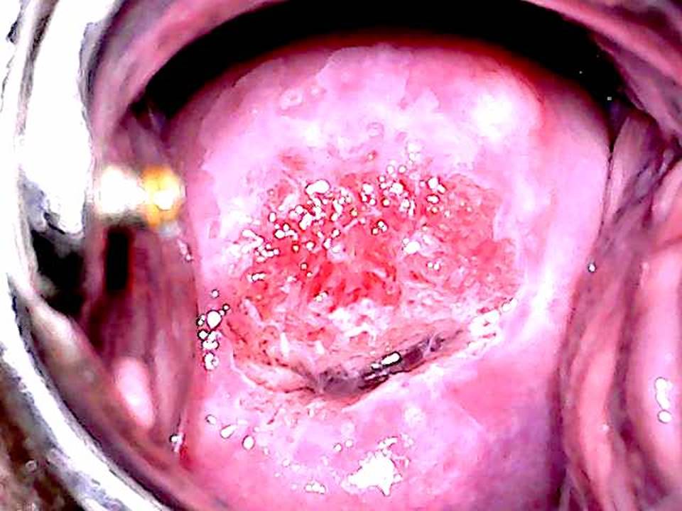
Before application of acetic acid
: The cervix appears normal. The SCJ is fully visible on the ectocervix.
Cervix before application of acetic acid:
Normal
Abnormal
After application of acetic acid:
Thin acetowhite areas with geographical margins and attached to the SCJ are seen at the 11–1 o’clock position.
Cervix after application of acetic acid:
Squamocolumnar junction visibility:
Fully visible
Not visible
Partially visible
Squamocolumnar junction location:
On ectocervix
Partly on ectocervix and partly on endocervix
On endocervix
Inside canal
Acetowhite area:
Absent
Present
If present, description of acetowhite area:
Colour:
Dense
Thin
Margin:
Diffuse
Sharp but irregular
Sharp and regular
Sharp and raised
Surface:
Smooth
Irregular
Location:
Within TZ or close to the external os (if SCJ is not visible)
Outside TZ
Size:
Covering less than 75% of ectocervix
Covering more than 75% of ectocervix
VIA category
:
Negative
Positive
Suspicious of cancer
After application of Lugol’s iodine
: The lesions appear patchy yellow, with well-demarcated margins.
Determined eligibility for ablative treatment
:
Eligible for ablation
Not eligible
Histopathology
: LSIL-CIN1
Remarks
: None.
IARC, 150 Cours Albert Thomas, 69372 Lyon CEDEX 08, France - Tel: +33 (0)4 72 73 84 85 - Fax: +33 (0)4 72 73 85 75
© IARC 2024 -
All Rights Reserved
.