


Chapters
Introduction
Visual inspection after application of acetic acid (VIA)
Determining eligibility for ablative treatment after application of acetic acid
Anatomical considerations
Cervical epithelium
Physiological changes of cervical epithelium
Neoplastic changes of the cervical epithelium
Changes in the cervical epithelium after application of acetic acid
Instruments, consumables, and setup required for examination after application of acetic acid
VIA procedure
Interpretation of VIA test results
Preventing errors in VIA
Management of women with an abnormal VIA test
Steps to determine eligibility for ablative treatment
Role of Lugols iodine in identifying the transformation zone for treatment
Treatment by cryotherapy
Treatment by thermal ablation
Videos
Preparation of Monsels solution
Infection prevention
Case study
Quiz
Acknowledgement
Suggested citation
Copyright
Home / Training / Manuals / Atlas of visual inspection of the cervix with acetic acid for screening, triage, and assessment for treatment
.png)
Click on the pictures to magnify and display the legends
Atlas of visual inspection of the cervix with acetic acid for screening, triage, and assessment for treatment
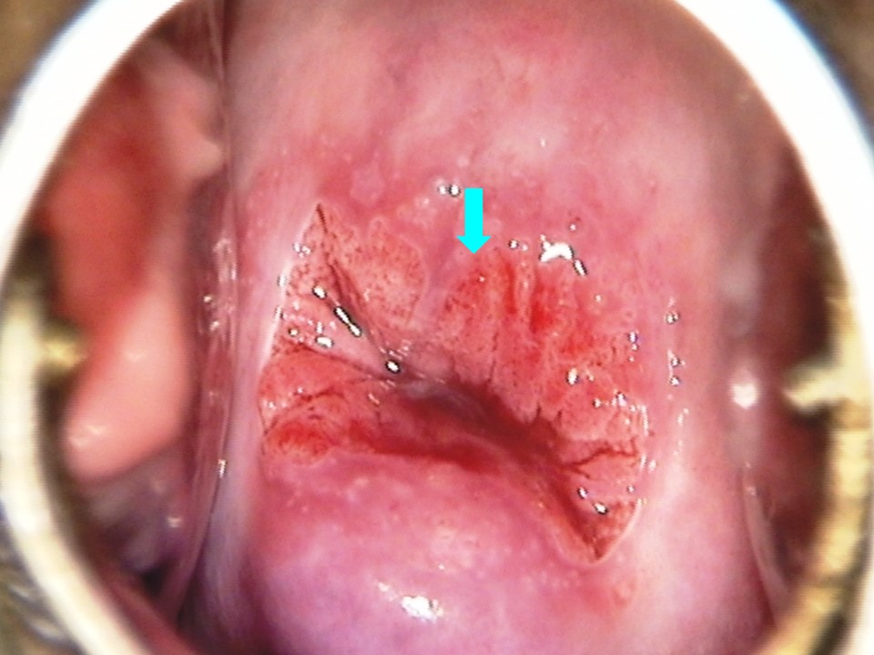
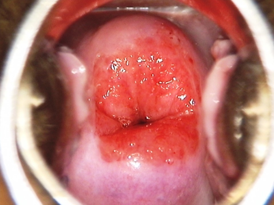
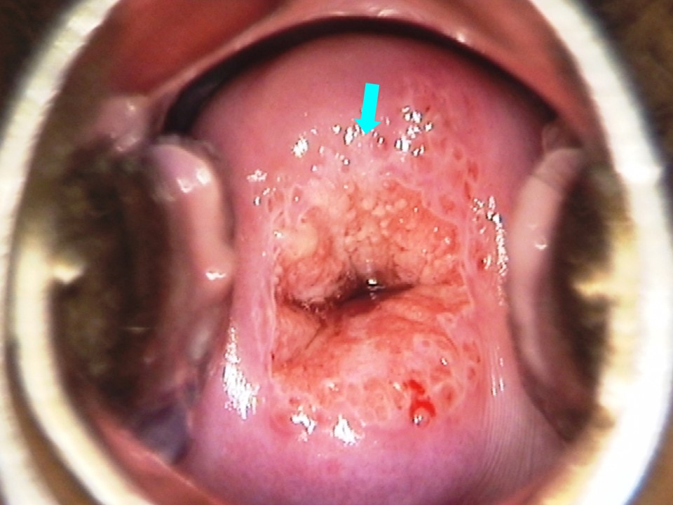
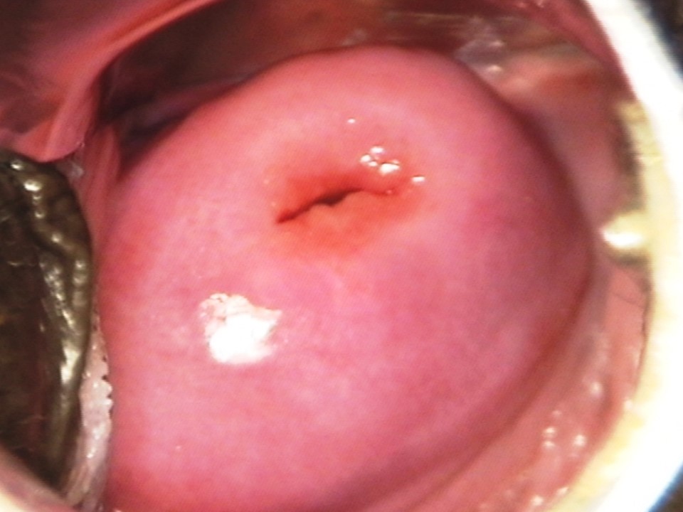
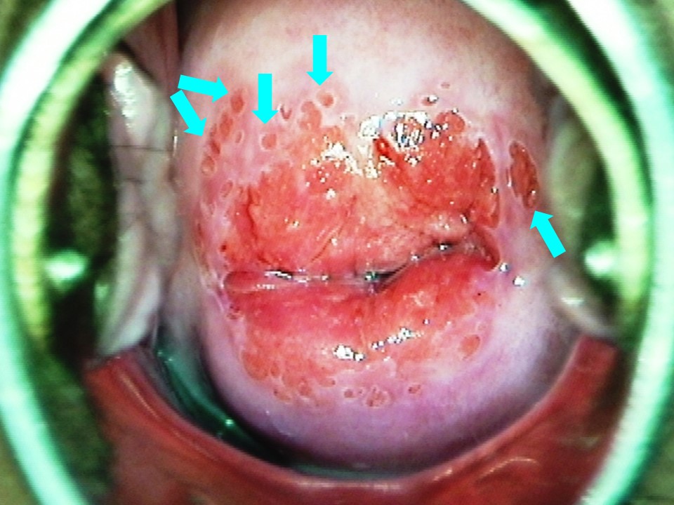
Filter by language: English / Français / Español / Русский / українськаVIA procedure Examination after application of 5% acetic acid Normal features Transformation zone (TZ) |
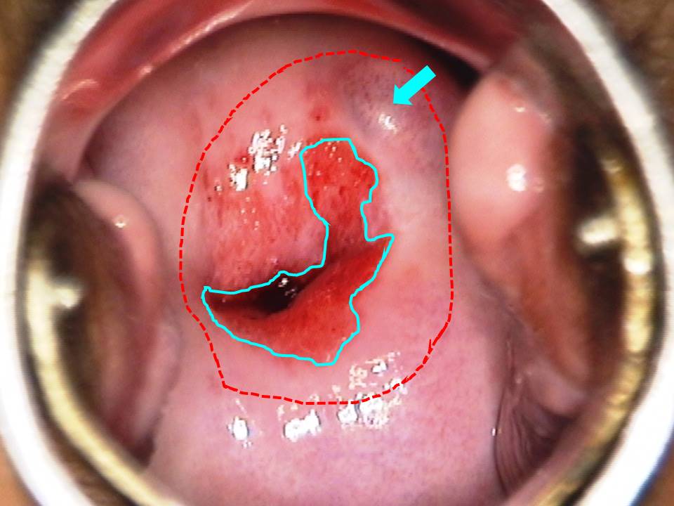
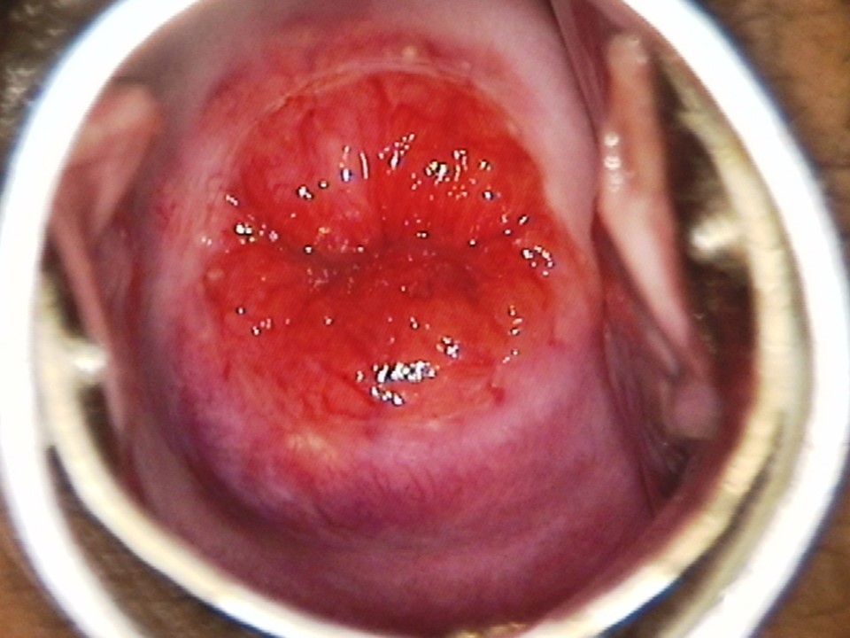
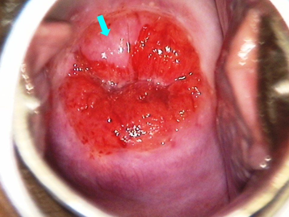
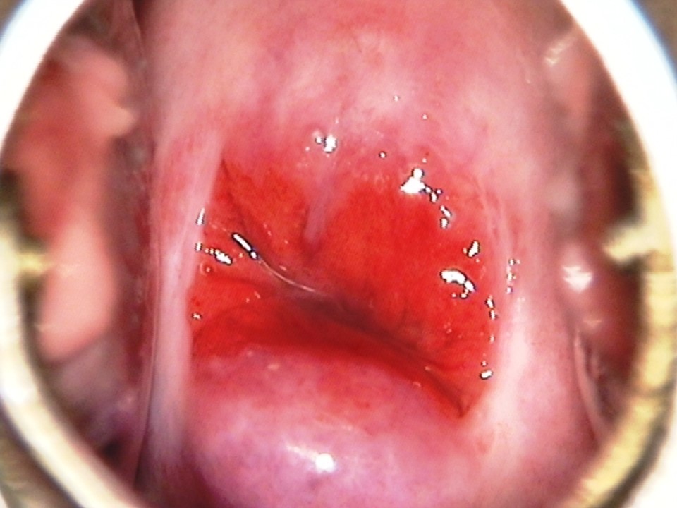
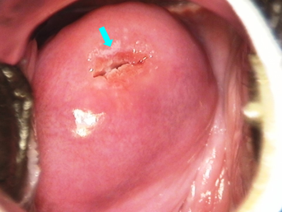
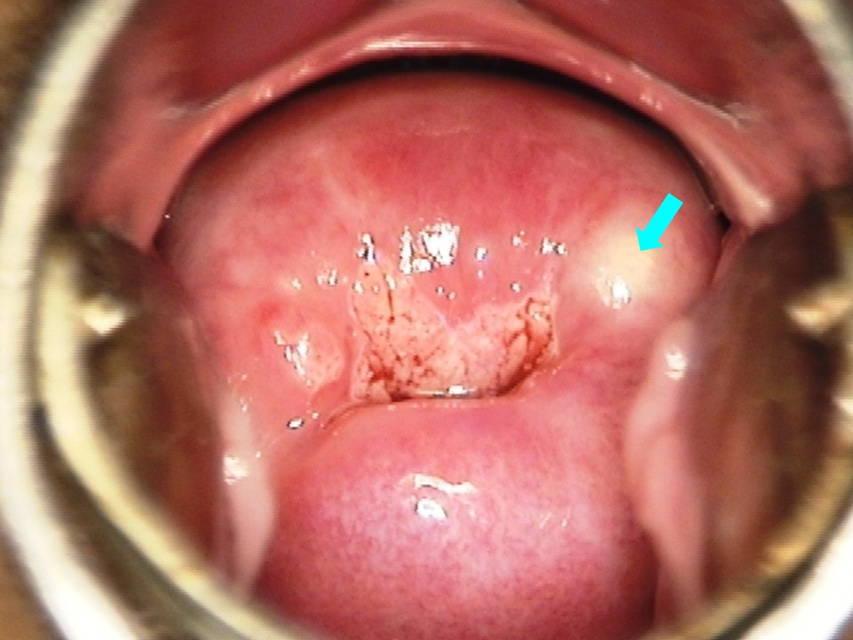
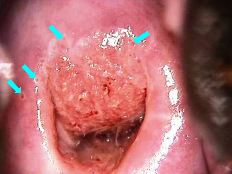
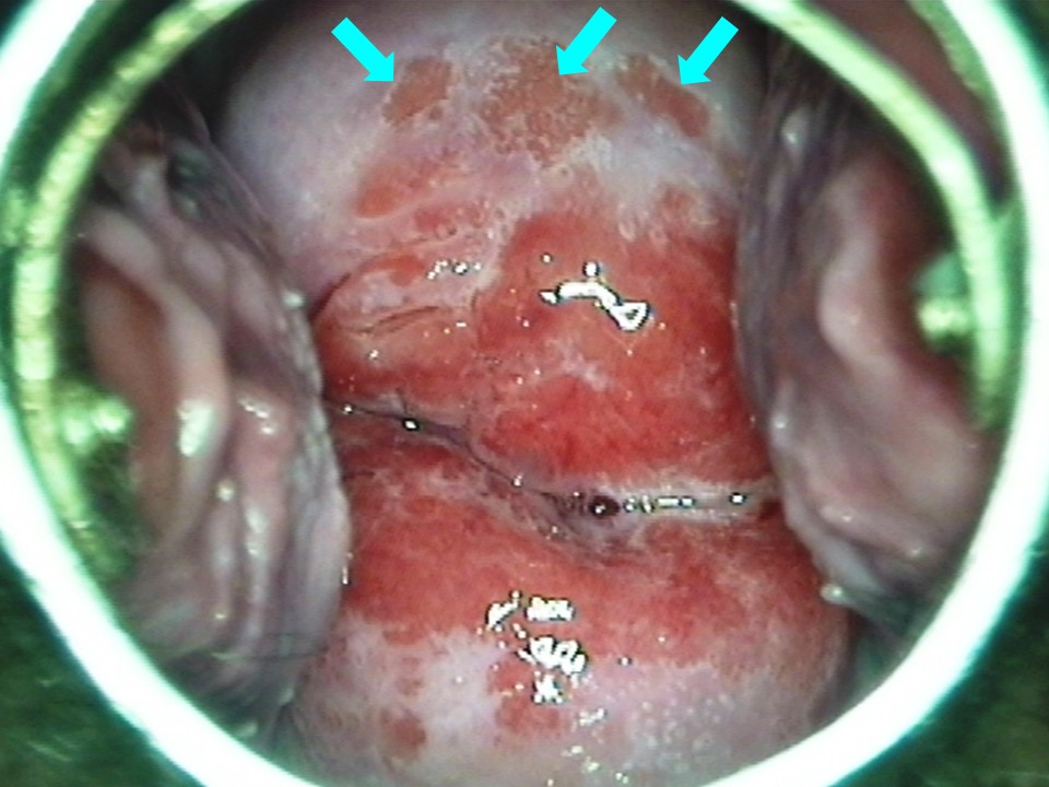
The transformation zone (TZ) is the area on the cervix where metaplastic changes take place. The TZ can be identified by the presence of the metaplastic epithelium, nabothian cysts, crypt openings, and islands of columnar epithelium. In the fully mature squamous epithelium, none of the features of metaplasia may be visible. In the presence of a CIN lesion, extent of TZ is determined by the extent of the lesion. The TZ may be completely or partially inside the endocervical canal, if the SCJ moves inside the canal. This is frequently seen in postmenopausal women.
Squamous metaplastic epithelium appears pale pinkish and is present towards the external os. It is often visible as tongue-shaped projections of pinkish epithelium projecting and coalescing towards the external os. Sometimes the metaplastic epithelium may be faintly acetowhite without any distinct outside border. Metaplastic epithelium may also appear as a thin transparent acetowhite patch extending outwards from the external os with a diffuse or feathery margin. Nabothian cysts are a feature of the TZ. A nabothian cyst looks like a raised pearly white or ivory white pimple on the squamous epithelium, and it becomes prominent after application of acetic acid. Crypt openings (usually visible with magnification) are present only in the TZ. After application of acetic acid, the tiny crypt opening appears as a small black dot surrounded by a thin acetowhite rim. During VIA, crypt openings may look like small white spots on the squamous epithelium. A skip area or an island of columnar epithelium is visible as a small pocket of red columnar epithelium on the metaplastic squamous epithelium. The process of replacement of columnar epithelium by metaplastic epithelium is not always uniform, and small islands of red columnar epithelium may be left behind on the original ectropion. |
Click on the pictures to magnify and display the legends
IARC, 150 Cours Albert Thomas, 69372 Lyon CEDEX 08, France - Tel: +33 (0)4 72 73 84 85 - Fax: +33 (0)4 72 73 85 75
© IARC 2025 - All Rights Reserved.
© IARC 2025 - All Rights Reserved.