


Chapters
Introduction
Visual inspection after application of acetic acid (VIA)
Determining eligibility for ablative treatment after application of acetic acid
Anatomical considerations
Cervical epithelium
Physiological changes of cervical epithelium
Neoplastic changes of the cervical epithelium
Changes in the cervical epithelium after application of acetic acid
Instruments, consumables, and setup required for examination after application of acetic acid
VIA procedure
Interpretation of VIA test results
Preventing errors in VIA
Management of women with an abnormal VIA test
Steps to determine eligibility for ablative treatment
Role of Lugols iodine in identifying the transformation zone for treatment
Treatment by cryotherapy
Treatment by thermal ablation
Videos
Preparation of Monsels solution
Infection prevention
Case study
Quiz
Acknowledgement
Suggested citation
Copyright
Home / Training / Manuals / Atlas of visual inspection of the cervix with acetic acid for screening, triage, and assessment for treatment
.png)
Click on the pictures to magnify and display the legends
Atlas of visual inspection of the cervix with acetic acid for screening, triage, and assessment for treatment
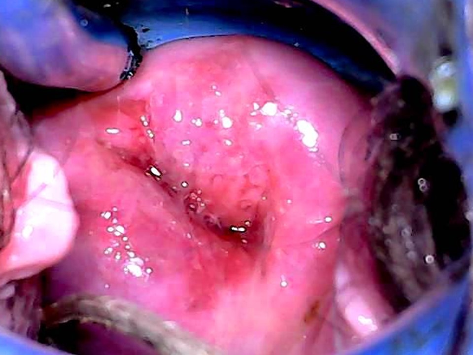
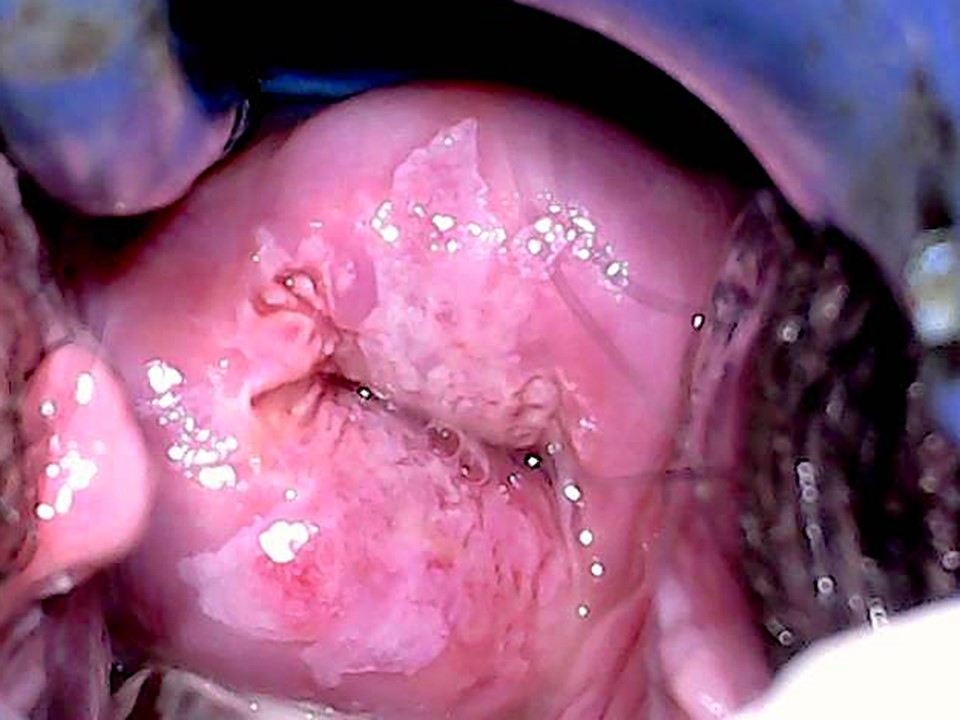
Filter by language: English / Français / Español / Русский / українськаChanges in the cervical epithelium after application of acetic acid Pathophysiological basis |
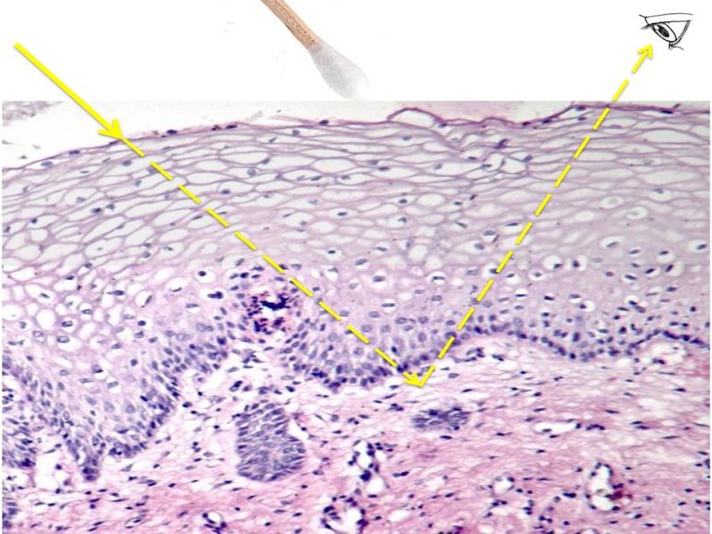
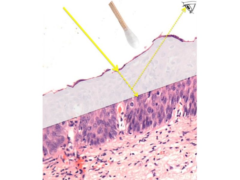
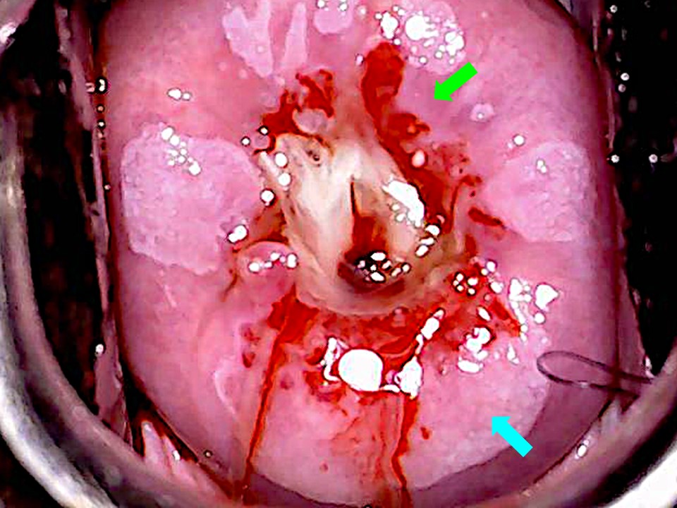
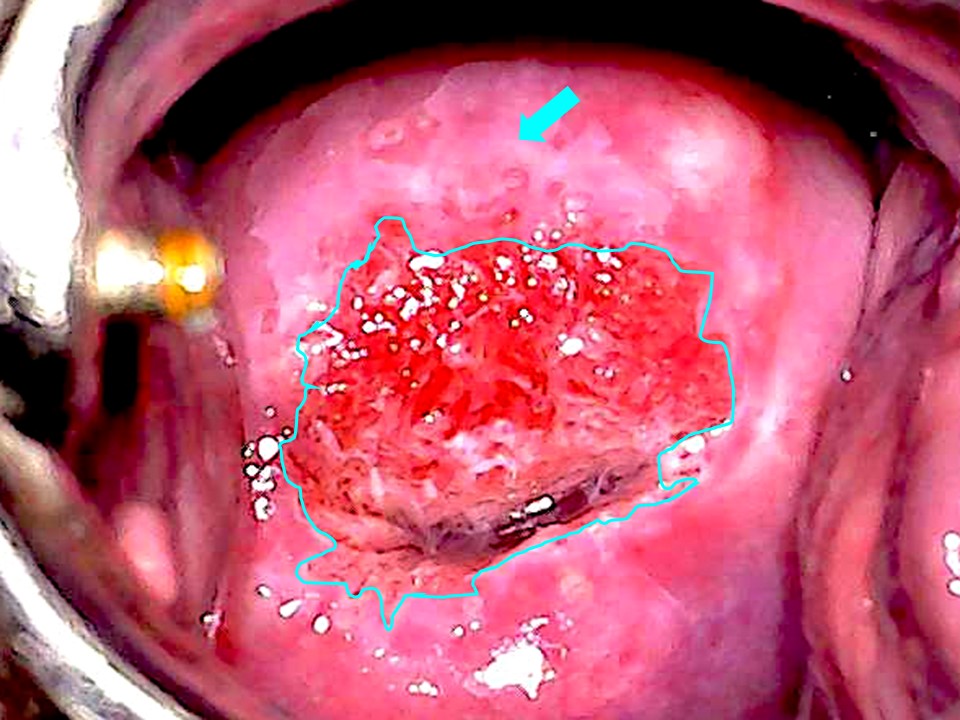
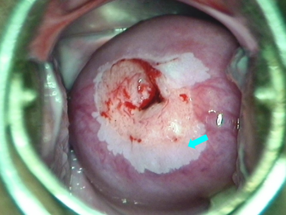
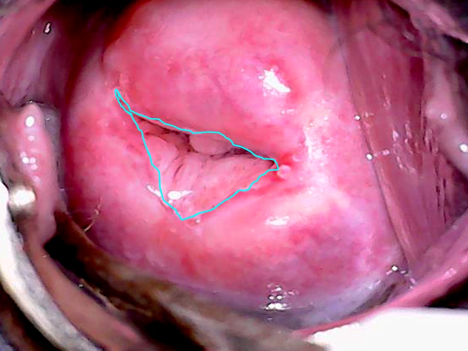
A cervix with a precancerous lesion looks healthy to the naked eye. Even early invasive cancers are not easily recognizable with the naked eye. To make these neoplastic lesions visible, 35% acetic acid is liberally applied to the cervix for 1 minute. Neoplastic lesions on the ectocervix become visible as white patches after application of acetic acid. By detecting the white patches that appear after application of acetic acid and identifying certain characteristics, it is possible to detect cervical precancer and cancer. This is the basis of the VIA test, whether it is used as a primary screening test or as a triage test. Acetic acid also helps in determining eligibility for treatment, by making the SCJ much more easily recognizable and enabling the size of the lesion to be measured. How does acetic acid change the colour of abnormal epithelium? Dilute acetic acid causes transient and reversible coagulation and precipitation of the cellular proteins of the cervical epithelium. Application of acetic acid also induces dehydration of the cells, resulting in the collapse of the cell membranes around the nucleus. Normal cervical epithelium is transparent, but it appears pink because of reflection of light from the underlying stroma, which is rich in blood vessels. There is very little cellular protein in the superficial layers of the normal mature squamous epithelium (the nuclei are very small or absent, and the cytoplasm is replaced by glycogen). Thus, there is very little protein to coagulate when acetic acid is applied to normal epithelium, and the normal pink colour of the mature squamous epithelium remains unchanged. Cervical epithelium with high-grade CIN or invasive cancer contains cells with large nuclei with abundant nuclear proteins. The rapidly proliferating cells do not accumulate glycogen, and they have intact cytoplasm. Acetic acid induces cellular dehydration and coagulation of the cellular proteins, causing the epithelium to appear white because light cannot pass through the layers of coagulated proteins. Coagulation of the protein contained in the neoplastic lesions makes the epithelium appear opaque white, and thus the lesions can be distinctly seen on naked-eye examination. This is the basis of the VIA test. The density of the acetowhitening is directly proportional to the grade of the cellular abnormality. High-grade abnormalities (CIN2 or CIN3) have more cellular proteins and appear densely acetowhite. Most of the cells in a CIN1 lesion are normal, especially in the superficial and intermediate layers, and as a result the epithelium appears thinly acetowhite. Invasive cancers have a high amount of cellular protein, and the number of abnormal cells is also high; after application of acetic acid, they form a dense, chalky white plaque, which may be partially obliterated by bleeding on contact. Identifying the SCJ and its localization is crucial to decide on the type of treatment. SCJ is much better visualized after application of acetic acid. Acetic acid cleans the mucus and causes the columnar epithelium to be oedematous (swollen) and prominent. The SCJ is often seen as a distinct white line after application of acetic acid. |
Click on the pictures to magnify and display the legends
IARC, 150 Cours Albert Thomas, 69372 Lyon CEDEX 08, France - Tel: +33 (0)4 72 73 84 85 - Fax: +33 (0)4 72 73 85 75
© IARC 2025 - All Rights Reserved.
© IARC 2025 - All Rights Reserved.