Chapters
Introduction
Visual inspection after application of acetic acid (VIA)
Determining eligibility for ablative treatment after application of acetic acid
Anatomical considerations
Cervical epithelium
Physiological changes of cervical epithelium
Neoplastic changes of the cervical epithelium
Changes in the cervical epithelium after application of acetic acid
Instruments, consumables, and setup required for examination after application of acetic acid
VIA procedure
Interpretation of VIA test results
Preventing errors in VIA
Management of women with an abnormal VIA test
Steps to determine eligibility for ablative treatment
Role of Lugols iodine in identifying the transformation zone for treatment
Treatment by cryotherapy
Treatment by thermal ablation
Videos
Preparation of Monsels solution
Infection prevention
Case study
Quiz
Acknowledgement
Suggested citation
Copyright
Home / Training / Manuals / Atlas of visual inspection of the cervix with acetic acid for screening, triage, and assessment for treatment / Cases
Atlas of visual inspection of the cervix with acetic acid for screening, triage, and assessment for treatment
Filter by language: English / Français / Español / Русский / українська
VIA-positive cases  Click to return to the atlas
.png) Click on the pictures to magnify and display the legends
Click on the pictures to magnify and display the legends
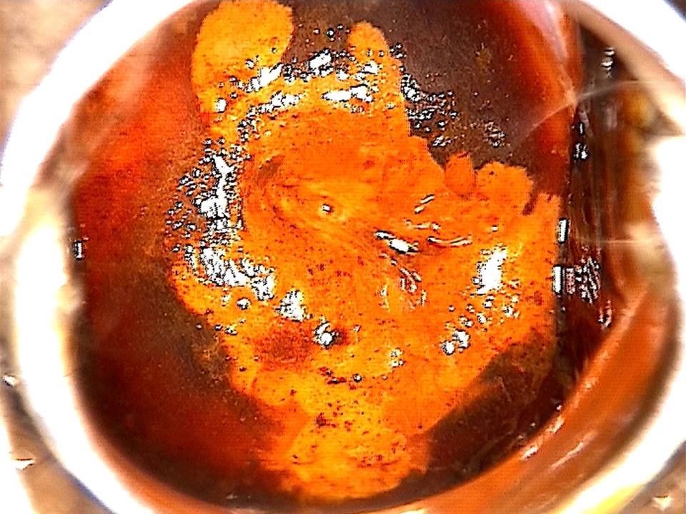
Before application of acetic acid: Scanty thin curdy white discharge is present on the cervix. Note that a red patch around the external os is not always columnar epithelium. Wait until after application of acetic acid before trying to locate the SCJ.
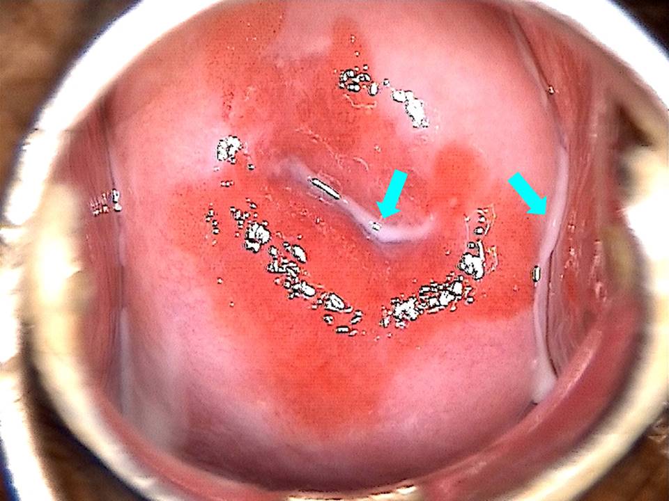
After application of acetic acid: Large dense acetowhite areas are seen around the external os occupying more than 75% of the ectocervix. The acetowhite areas extend into the endocervical canal. The SCJ is not visible. Multiple areas of peeled-off epithelium (at the 10, 12, and 6 oclock positions) can be seen as red patches within the dense acetowhite lesion.
 |
Strawberry appearance of cervix Cervicitis Polyp Bleeding on contact White patch Growth Ulcer Erosion |
 |
|