Chapters
Introduction
Visual inspection after application of acetic acid (VIA)
Determining eligibility for ablative treatment after application of acetic acid
Anatomical considerations
Cervical epithelium
Physiological changes of cervical epithelium
Neoplastic changes of the cervical epithelium
Changes in the cervical epithelium after application of acetic acid
Instruments, consumables, and setup required for examination after application of acetic acid
VIA procedure
Interpretation of VIA test results
Preventing errors in VIA
Management of women with an abnormal VIA test
Steps to determine eligibility for ablative treatment
Role of Lugolís iodine in identifying the transformation zone for treatment
Treatment by cryotherapy
Treatment by thermal ablation
Videos
Preparation of Monselís solution
Infection prevention
Case study
Quiz
Acknowledgement
Suggested citation
Copyright
Home / Training / Manuals / Atlas of visual inspection of the cervix with acetic acid for screening, triage, and assessment for treatment
.png)
Click on the pictures to magnify and display the legends
Atlas of visual inspection of the cervix with acetic acid for screening, triage, and assessment for treatment
Filter by language: English / FranÁais / EspaŮol / Русский / українськаVIA procedure Ė Examination before application of acetic acid Ė Abnormal findings on speculum examination Ė Cervical polyp |
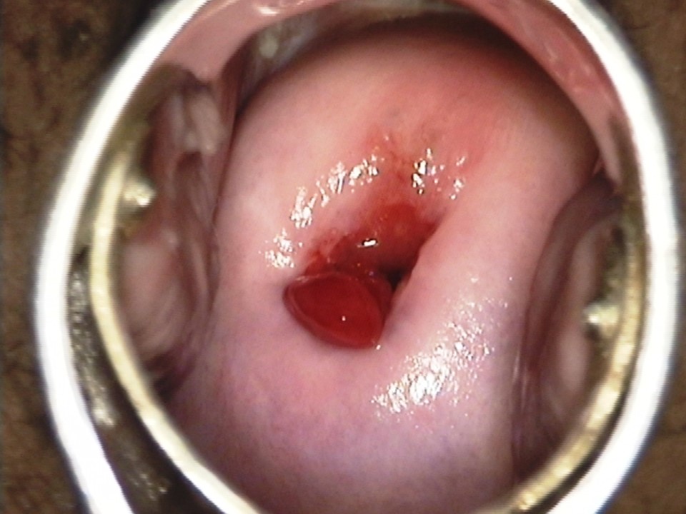
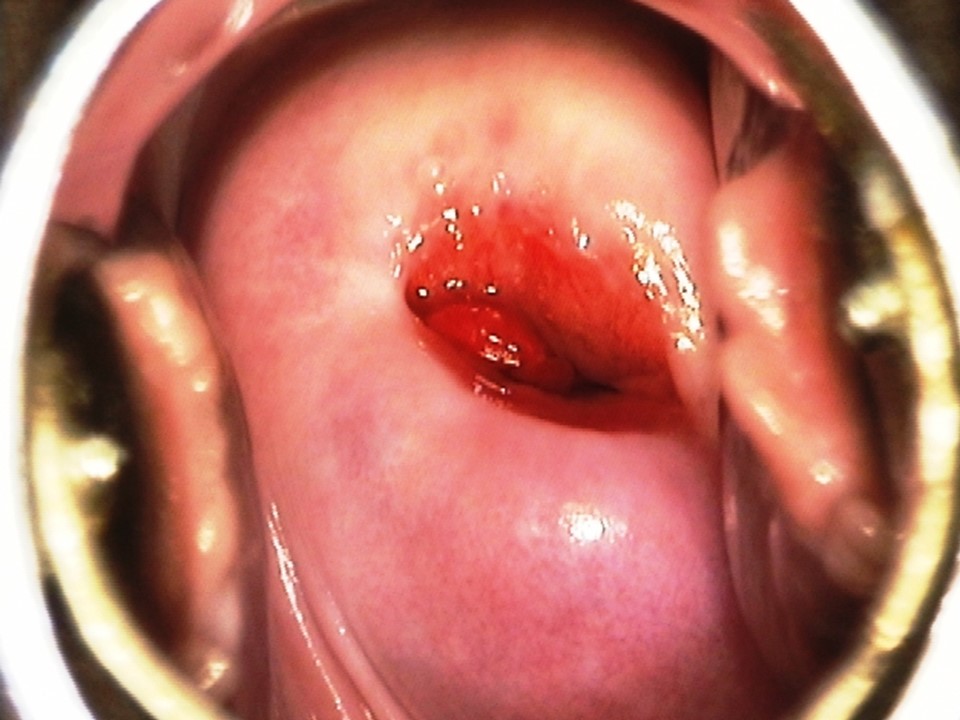
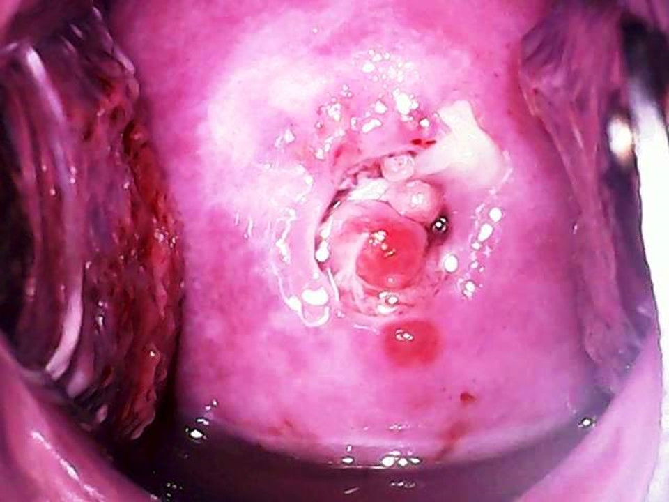
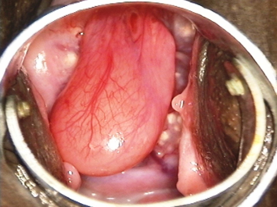
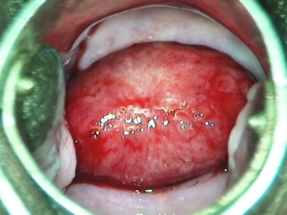
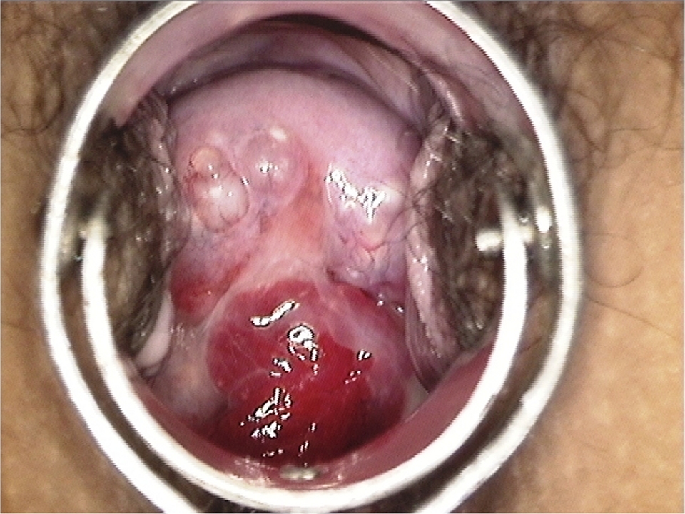
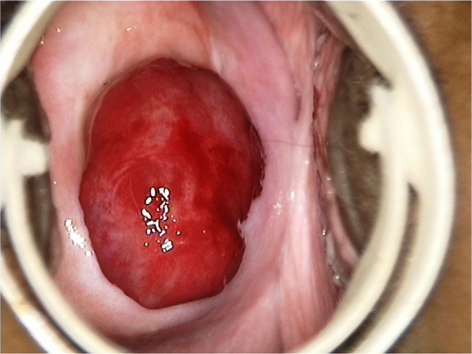
Cervical mucous polyps are overgrowths of the endocervical columnar epithelium and appear as single or multiple smooth reddish or pinkish soft masses protruding through the external os. The presence of multiple polyps or large polyps at the external os often interferes with proper visualization of the SCJ and the TZ. Because most polyps are mobile, they can be gently pushed in different directions with the help of a small cotton swab in an attempt to visualize the SCJ in its entirety. If the polyps are very large, it may not be possible to visualize the SCJ completely even after adequate manipulation. Most mucous polyps remain small, do not cause any symptoms, and regress after menopause. Some larger polyps may cause symptoms, such as vaginal discharge or postcoital bleeding. Such polyps that are pedunculated can be easily removed by holding the polyp with a sponge-holding forceps and twisting the polyp at the base repeatedly until the polyp comes out. No anaesthesia is required, and the small amount of bleeding from the attachment of the polyp can be easily stopped with gentle pressure. Sometimes a polyp may not be mobile, because of the presence of a thick and short stalk, and is firm in consistency. Such a polyp, which is usually due to a benign tumour originating from the muscle and fibres of the cervix or even the body of the uterus, is known as a fibroid polyp. A fibroid polyp may be located inside the endocervical canal and bulge through the external os. In such a situation, it may become difficult to see the SCJ or even the cervix. Such polyps require surgical excision. A polyp with necrosis may sometimes be misdiagnosed as a cancer. The next section describes a condition called leukoplakia. |
Click on the pictures to magnify and display the legends
IARC, 150 Cours Albert Thomas, 69372 Lyon CEDEX 08, France - Tel: +33 (0)4 72 73 84 85 - Fax: +33 (0)4 72 73 85 75
© IARC 2026 - All Rights Reserved.
© IARC 2026 - All Rights Reserved.


