


Home / Training / Manuals / Histopathology of the uterine cervix - digital atlas / Glossary Definitions
Histopathology of the uterine cervix - digital atlas
Glossary Definitions
Filter by language: English / Français / Portugues / 中文|
|
 |
|
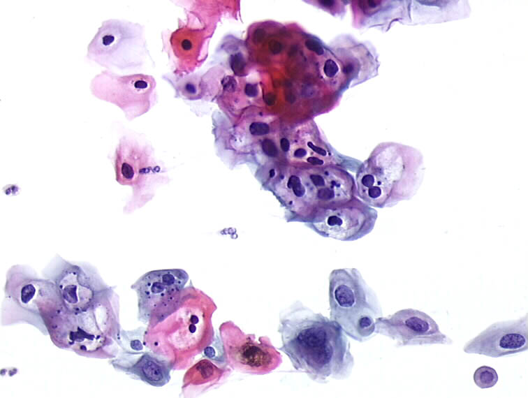
Koilocyte The koilocyte is pathognomonic for HPV infection. They occur singly, but may be in small clusters. It is generally a superficial or large intermediate cell (never parabasal) with a very large, clear (empty) and irregular perinuclear zone (to distinguish them from clear halos as seen in Trichomonas infection for example). The halo shows clear-cut borders and the periperal rim of cytoplasm is usually thickened. The cytoplasm is dense and may be basophilic or eosinophilic. The nucleus is enlarged. The nucleus is two to three times the size of the nucleus of the normal cell of the corresponding type. At times binucleation and irregular nuclear borders are noted. The chromatin is smudged and homogenous or exhibits varying degrees of density. Many pseudokoilocyte may be found in different situations (inflammation, metaplastic cells, post coital smears, post menopausal, etc.). Differential diagnosis of koilocytotic atypias: * Mild reactive epithelial changes in glycogenated cells (slightly hyperchromatic nuclei without significant enlargement in normal cells) * Prominent cytoplasmic halos, without nuclear atypia * Mild nuclear enlargement (x2) and discrete concentric halos in inflammatory conditions (trichomoniasis) * Conditions of maturation disturbance during pregnancy, postpartum, or menopausal conditions. Histopathology atlas Cytopathology atlas |
25 avenue Tony Garnier CS 90627 69366, LYON CEDEX 07 France - Tel: +33 (0)4 72 73 84 85
© IARC 2026 - Terms of use - Privacy Policy.
© IARC 2026 - Terms of use - Privacy Policy.